Summary

Atomic Force Microscopy Cantilever-Based Nanoindentation: Mechanische eigenschapsmetingen op nanoschaal in lucht en vloeistof

Published: December 02, 2022
doi:

Summary

Het kwantificeren van het contactoppervlak en de kracht die door een atoomkrachtmicroscoop (AFM) sondepunt op een monsteroppervlak wordt uitgeoefend, maakt mechanische eigenschapsbepaling op nanoschaal mogelijk. Best practices voor het implementeren van AFM cantilever-gebaseerde nano-indentatie in lucht of vloeistof op zachte en harde monsters om elastische modulus of andere nanomechanische eigenschappen te meten, worden besproken.

Abstract

Een atoomkrachtmicroscoop (AFM) meet fundamenteel de interactie tussen een AFM-sondepunt op nanoschaal en het monsteroppervlak. Als de kracht die door de sondepunt wordt uitgeoefend en het contactgebied met het monster kunnen worden gekwantificeerd, is het mogelijk om de mechanische eigenschappen op nanoschaal (bijvoorbeeld elastiek of de modulus van Young) van het te onderzoeken oppervlak te bepalen. Een gedetailleerde procedure voor het uitvoeren van kwantitatieve AFM cantilever-gebaseerde nano-indentatie-experimenten wordt hier gegeven, met representatieve voorbeelden van hoe de techniek kan worden toegepast om de elastische moduli van een breed scala aan monstertypen te bepalen, variërend van kPa tot GPa. Deze omvatten levende mesenchymale stamcellen (MSC’s) en kernen in fysiologische buffer, hars-ingebedde gedehydrateerde loblolly pine doorsneden en Bakken leisteen van verschillende samenstelling.

Daarnaast wordt AFM cantilever-gebaseerde nano-indentatie gebruikt om de breuksterkte (d.w.z. doorbraakkracht) van fosfolipide bilayers te onderzoeken. Belangrijke praktische overwegingen zoals methodekeuze en -ontwikkeling, sondeselectie en kalibratie, identificatie van interessegebieden, heterogeniteit van monsters, functiegrootte en beeldverhouding, tipslijtage, oppervlakteruwheid en gegevensanalyse en meetstatistieken worden besproken om een goede implementatie van de techniek te bevorderen. Ten slotte wordt co-lokalisatie van AFM-afgeleide nanomechanische kaarten met elektronenmicroscopietechnieken aangetoond die aanvullende informatie geven over de elementaire samenstelling.

Introduction

Het begrijpen van de mechanische eigenschappen van materialen is een van de meest fundamentele en essentiële taken in engineering. Voor de analyse van de eigenschappen van bulkmateriaal zijn er tal van methoden beschikbaar om de mechanische eigenschappen van materiaalsystemen te karakteriseren, waaronder trekproeven1, compressietests2 en drie- of vierpuntsbuigproeven (buigproeven)3. Hoewel deze tests op microschaal waardevolle informatie kunnen opleveren over de eigenschappen van bulkmateriaal, worden ze over het algemeen uitgevoerd om te falen en zijn ze daarom destructief. Bovendien missen ze de ruimtelijke resolutie die nodig is om de micro- en nanoschaaleigenschappen van veel materiaalsystemen die vandaag de dag van belang zijn, zoals dunne films, biologische materialen en nanocomposieten, nauwkeurig te onderzoeken. Om te beginnen met het aanpakken van enkele van de problemen met grootschalige mechanische tests, voornamelijk de destructieve aard ervan, werden microhardheidstests overgenomen van mineralogie. Hardheid is een maat voor de weerstand van een materiaal tegen plastische vervorming onder specifieke omstandigheden. Over het algemeen gebruiken microhardheidstests een stijve sonde, meestal gemaakt van gehard staal of diamant, om in een materiaal te springen. De resulterende inkepingsdiepte en/of -oppervlakte kan vervolgens worden gebruikt om de hardheid te bepalen. Er zijn verschillende methoden ontwikkeld, waaronder Vickers4, Knoop5 en Brinell6 hardheid; Elk biedt een maat voor de materiaalhardheid op microschaal, maar onder verschillende omstandigheden en definities, en produceert als zodanig alleen gegevens die kunnen worden vergeleken met tests die onder dezelfde omstandigheden worden uitgevoerd.

Geïnstrumenteerde nano-indentatie werd ontwikkeld om de relatieve waarden verkregen via de verschillende microhardheidstestmethoden te verbeteren, de ruimtelijke resolutie te verbeteren die mogelijk is voor de analyse van mechanische eigenschappen en de analyse van dunne films mogelijk te maken. Belangrijk is dat door gebruik te maken van de methode die voor het eerst is ontwikkeld door Oliver en Pharr7, het elastische of Young’s modulus, E, van een monstermateriaal kan worden bepaald via geïnstrumenteerde nano-indentatie. Bovendien kan, door gebruik te maken van een Berkovich driezijdige piramidale nanoindenter-sonde (waarvan de ideale tipgebiedfunctie overeenkomt met die van de Vickers vierzijdige piramidale sonde)8, een directe vergelijking tussen nanoschaal en meer traditionele microschaalhardheidsmetingen worden gemaakt. Met de groeiende populariteit van de AFM begon ook AFM cantilever-gebaseerde nano-indentatie aandacht te krijgen, met name voor het meten van de mechanische eigenschappen van zachtere materialen. Als gevolg hiervan, zoals schematisch weergegeven in figuur 1, zijn de twee meest gebruikte technieken om mechanische eigenschappen op nanoschaal te ondervragen en te kwantificeren geïnstrumenteerde nano-indentatie (figuur 1A) en AFM cantilever-gebaseerde nano-indentatie (figuur 1B)9, waarvan de laatste de focus van dit werk is.

Figure 1
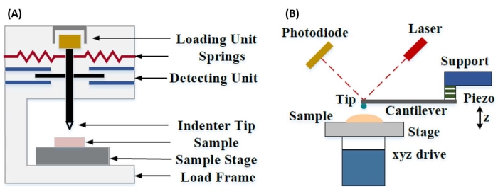
Figuur 1: Vergelijking van geïnstrumenteerde en AFM cantilever-gebaseerde nano-indentatiesystemen. Schematische diagrammen met typische systemen voor het uitvoeren van (A) geïnstrumenteerde nano-indentatie en (B) AFM cantilever-gebaseerde nano-indentatie. Dit cijfer is gewijzigd ten opzichte van Qian et al.51. Afkorting: AFM = atoomkrachtmicroscopie. Klik hier om een grotere versie van deze figuur te bekijken.

Zowel geïnstrumenteerde als AFM cantilever-gebaseerde nano-indentatie maken gebruik van een stijve sonde om een monsteroppervlak van belang te vervormen en de resulterende kracht en verplaatsing te bewaken als een functie van de tijd. Doorgaans wordt het gewenste belastings- (d.w.z. kracht) of (Z-piëzo) verplaatsingsprofiel door de gebruiker gespecificeerd via de software-interface en rechtstreeks door het instrument geregeld, terwijl de andere parameter wordt gemeten. De mechanische eigenschap die het vaakst wordt verkregen uit nano-indentatie-experimenten is de elastische modulus (E), ook wel de Young-modulus genoemd, die drukeenheden heeft. De elastische modulus van een materiaal is een fundamentele eigenschap met betrekking tot de bindingsstijfheid en wordt gedefinieerd als de verhouding tussen trek- of drukspanning (σ, de uitgeoefende kracht per oppervlakte-eenheid) tot axiale spanning (ε, de proportionele vervorming langs de inkepingsas) tijdens elastische (d.w.z. omkeerbare of tijdelijke) vervorming voorafgaand aan het begin van plastische vervorming (vergelijking [1]):

Equation 1(1)

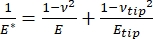
Opgemerkt moet worden dat, omdat veel materialen (vooral biologische weefsels) in feite visco-elastisch zijn, de (dynamische of complexe) modulus in werkelijkheid bestaat uit zowel elastische (opslag, in fase) als viskeuze (verlies, uit fase) componenten. In de praktijk wordt in een nano-indentatie-experiment de gereduceerde modulus, E *, gemeten, die gerelateerd is aan de werkelijke monstermodulus van belang, E, zoals weergegeven in vergelijking (2):

Equation 2(2)

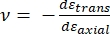
Waarbij E-tip en ν-tip respectievelijk de elastische modulus en Poisson-verhouding van de nano-indenterpunt zijn, en ν de geschatte Poisson-verhouding van het monster. De Poisson-verhouding is de negatieve verhouding van de transversale tot axiale spanning en geeft dus de mate van transversale verlenging van een monster aan bij blootstelling aan axiale spanning (bijvoorbeeld tijdens nano-indentatiebelasting), zoals weergegeven in vergelijking (3):

Equation 3(3)

De omzetting van gereduceerde naar werkelijke modulus is noodzakelijk omdat a) een deel van de axiale spanning die door de indrukkerijpunt wordt gegeven, kan worden omgezet in dwarsspanning (d.w.z. het monster kan vervormen door uitzetting of samentrekking loodrecht op de belastingsrichting), en b) de indrukkerijpunt niet oneindig hard is, en dus resulteert de handeling van het inspringen van het monster in een (kleine) hoeveelheid vervorming van de punt. Merk op dat in het geval dat E-tip >> E (d.w.z. de indrukpunt is veel harder dan het monster, wat vaak het geval is bij het gebruik van een diamantsonde), de relatie tussen de gereduceerde en werkelijke monstermodulus aanzienlijk vereenvoudigt tot E ≈ E * (1 – v2). Terwijl geïnstrumenteerde nano-indentatie superieur is in termen van nauwkeurige krachtkarakterisering en dynamisch bereik, is AFM cantilever-gebaseerde nano-indentatie sneller, biedt ordes van grootte grotere kracht- en verplaatsingsgevoeligheid, maakt beeldvorming met een hogere resolutie en verbeterde inspringing lokalisatie mogelijk en kan tegelijkertijd magnetische en elektrische eigenschappen op nanoschaal onderzoeken9. In het bijzonder is AFM cantilever-gebaseerde nano-indentatie superieur voor de kwantificering van mechanische eigenschappen op nanoschaal van zachte materialen (bijv. polymeren, gels, lipide bilayers en cellen of andere biologische materialen), extreem dunne (sub-μm) films (waarbij substraateffecten een rol kunnen spelen afhankelijk van de inkepingsdiepte)10,11, en gesuspendeerde tweedimensionale (2D) materialen12,13,14 zoals grafeen 15,16, mica17, hexagonaal boornitride (h-BN)18, of overgangsmetaaldichalcogeniden (TMDC’s; bijv. MoS2)19. Dit komt door de voortreffelijke kracht (sub-nN) en verplaatsingsgevoeligheid (sub-nm), die belangrijk is voor het nauwkeurig bepalen van het begincontactpunt en het blijven binnen het elastische vervormingsgebied.

Bij AFM cantilever-gebaseerde nano-indentatie wordt de verplaatsing van een AFM-sonde naar het monsteroppervlak geactiveerd door een gekalibreerd piëzo-elektrisch element (figuur 1B), waarbij de flexibele cantilever uiteindelijk buigt als gevolg van de weerstandskracht die wordt ervaren bij contact met het monsteroppervlak. Deze buiging of afbuiging van de cantilever wordt meestal bewaakt door een laser van de achterkant van de cantilever te reflecteren en in een fotodetector (positiegevoelige detector [PSD]). In combinatie met de kennis van de cantileverstijfheid (in nN/nm) en doorbuigingsgevoeligheid (in nm/V) is het mogelijk om deze gemeten cantileverafbuiging (in V) om te zetten in de kracht (in nN) die op het monster wordt uitgeoefend. Na contact levert het verschil tussen de Z-piëzobeweging en de cantileverafbuiging de inkepingdiepte van het monster op. In combinatie met de kennis van de tipgebiedfunctie maakt dit de berekening van het tip-monster contactgebied mogelijk. De helling van de contactgedeelten van de resulterende krachtafstands- of krachtverplaatsingscurven (F-D) kan vervolgens worden aangepast met behulp van een geschikt contactmechanicamodel (zie het gedeelte Gegevensanalyse van de discussie) om de nanomechanische eigenschappen van het monster te bepalen. Hoewel AFM cantilever-gebaseerde nano-indentatie een aantal duidelijke voordelen heeft ten opzichte van geïnstrumenteerde nano-indentatie zoals hierboven beschreven, brengt het ook verschillende praktische implementatie-uitdagingen met zich mee, zoals kalibratie, tipslijtage en data-analyse, die hier zullen worden besproken. Een ander potentieel nadeel van AFM cantilever-gebaseerde nano-indentatie is de aanname van lineaire elasticiteit, omdat de contactradius en indrukkingsdiepten veel kleiner moeten zijn dan de indrukkerijradius, wat moeilijk te bereiken kan zijn bij het werken met AFM-sondes op nanoschaal en / of monsters met een aanzienlijke oppervlakteruwheid.

Traditioneel is nano-indentatie beperkt tot individuele locaties of kleine rasterinspringingsexperimenten, waarbij een gewenste locatie (d.w.z. regio van belang [ROI]) wordt geselecteerd en een enkele gecontroleerde inspringing, meerdere inspringingen op een enkele locatie gescheiden door enige wachttijd, en / of een grof raster van inspringingen worden uitgevoerd met een snelheid in de orde van Hz. Recente ontwikkelingen in de AFM maken het echter mogelijk om mechanische eigenschappen en topografie gelijktijdig te verwerven door het gebruik van op hoge snelheid op krachtcurve gebaseerde beeldvormingsmodi (aangeduid met verschillende handelsnamen, afhankelijk van de systeemfabrikant), waarbij krachtcurven worden uitgevoerd met een kHz-snelheid onder belastingscontrole, waarbij de maximale tip-samplekracht wordt gebruikt als het beeldvormingsinstelpunt. Er zijn ook point-and-shoot-methoden ontwikkeld, die het mogelijk maken om een AFM-topografiebeeld te verkrijgen, gevolgd door daaropvolgende selectieve nano-indentatie op interessante punten in het beeld, waardoor ruimtelijke controle op nanoschaal over de locatie van nano-indentatie mogelijk is. Hoewel dit niet de primaire focus van dit werk is, worden specifieke geselecteerde toepassingsvoorbeelden van zowel op krachtcurve gebaseerde beeldvorming als op point-and-shoot cantilever-gebaseerde nano-indentatie gepresenteerd in de representatieve resultaten en kunnen worden gebruikt in combinatie met het onderstaande protocol, indien beschikbaar op het specifieke AFM-platform dat wordt gebruikt. In het bijzonder schetst dit werk een algemeen protocol voor de praktische implementatie van AFM cantilever-gebaseerde nano-indentatie op elk capabel AFM-systeem en biedt vier use case-voorbeelden (twee in lucht, twee in vloeistof) van de techniek, inclusief representatieve resultaten en een diepgaande bespreking van de nuances, uitdagingen en belangrijke overwegingen om de techniek met succes toe te passen.

Protocol

OPMERKING: Vanwege de grote verscheidenheid aan commercieel verkrijgbare AFM’s en de diversiteit van monstertypen en toepassingen die bestaan voor op cantilever gebaseerde nano-indentatie, is het volgende protocol opzettelijk ontworpen om relatief algemeen van aard te zijn, met de nadruk op de gedeelde stappen die nodig zijn voor alle op cantilever gebaseerde nano-indentatie-experimenten, ongeacht het instrument of de fabrikant. Daarom gaan de auteurs ervan uit dat de lezer op zijn minst basiskennis bezit van het bediene…

Representative Results

Kracht-verplaatsingscurvenFiguur 7 toont representatieve, bijna ideale F-D-curven verkregen uit nano-indentatie-experimenten uitgevoerd in lucht op in hars ingebedde loblolly-dennenmonsters (figuur 7A) en in vloeistof (fosfaat-gebufferde zoutoplossing [PBS]) op mesenchymale stamcelkernen (MSC) (figuur 7B). Het gebruik van elk contactmechanicamodel is afhankelijk van de nauwkeurige en betrouwbare bepaling van het…

Discussion

Monstervoorbereiding
Voor nano-indentatie in lucht omvatten gebruikelijke bereidingsmethoden cryosectie (bijv. weefselmonsters), slijpen en / of polijsten gevolgd door ultramicrotoming (bijv. In hars ingebedde biologische monsters), ionenfrezen of voorbereiding van gerichte ionenbundels (bijv. halfgeleider-, poreuze of gemengde hardheidsmonsters die niet geschikt zijn voor polijsten), mechanisch of elektrochemisch polijsten (bijv. Metaallegeringen) of dunnefilmafzetting (bijv. Atomaire laag of chemisc…

Disclosures

The authors have nothing to disclose.

Acknowledgements

Alle AFM-experimenten zijn uitgevoerd in het Boise State University Surface Science Laboratory (SSL). SEM-karakterisering werd uitgevoerd in het Boise State Center for Materials Characterization (BSCMC). Onderzoek gerapporteerd in deze publicatie met betrekking tot grondstoffen voor biobrandstoffen werd gedeeltelijk ondersteund door het Amerikaanse ministerie van Energie, Office of Energy Efficiency and Renewable Energy, Bioenergy Technologies Office als onderdeel van het Feedstock Conversion Interface Consortium (FCIC) en onder DOE Idaho Operations Office Contract DE-AC07-051ID14517. Celmechanicastudies werden ondersteund door de National Institutes of Health (VS) onder subsidies AG059923, AR075803 en P20GM109095, en door subsidies van de National Science Foundation (VS) 1929188 en 2025505. Het model lipide bilayer systemen werk werd ondersteund door de National Institutes of Health (USA) onder subsidie R01 EY030067. De auteurs bedanken Dr. Elton Graugnard voor het produceren van de samengestelde afbeelding in figuur 11.

Materials

Atomic force microscope Bruker Dimension Icon Uses Nanoscope control software, including PeakForce Quantitative Nanomechanical Mapping (PF-QNM), FastForce Volume (FFV), and Point-and-Shoot Ramping experimental workspaces
AtomicJ American Institute of Physics https://doi.org/10.1063/1.4881683 Flexible, powerful, free open source Java-based force curve analysis software package. Supports numerous contact mechanic models, such as Hertz, Sneddon DMT, JKR, Maugis, and cone or pyramid (including blunt and truncated). Also includes a variety of initial contact point estimation methods to choose from. Supports batch processing of data and subsequent statistical analysis (e.g., averages, standard deviations, histograms, goodness of fit, etc.). Literature citation is: P. Hermanowicz, M. Sarna, K. Burda, and H. GabryEquation 1, “AtomicJ: An open source software for analysis of force curves” Rev. Sci. Instrum. 85: 063703 (2014), https://doi.org/10.1063/1.4881683
Buffer solution (PBS) Fisher Chemical (NaCl), Sigma Aldrich (KCl), Fisher BioReagents (Na2HPO4 and KH2PO4) S271 (>99% purity NaCl), P9541 (>99% purity KCl), BP332(>99% purity Na2HPO4), BP362 (>99% purity KH2PO4) Phosphate buffered saline (PBS) was prepared in the laboratory as an aqueous solution consisting of 137 mM NaCl, 2.7 mM KCl, 10 mM Na2HPO4, and 1.8 mM KH2PO4 dissolved in ultrapure water. Reagents were measured out using an analytical balance, and glassware was cleaned with soap and water followed by autoclaving immediately prior to use.
Chloroform
Diamond tip AFM probe Bruker PDNISP Pre-mounted factory-calibrated cube corner diamond (E = 1140 GPa) tip AFM probe (nominal R = 40 nm) with a stainless steel cantilever (nominal k = 225 N/m, f0 = 50 kHz). Spring constant is measured at the factory (k = 256 N/m for the probe, Serial #13435414, used here) and calibration data (including AFM images of indents showing probe geometry) is provided with the probe.
Diamond ultramicrotome blade Diatome Ultra 35° 2.1 mm width. Also used a standard glass blade for intial rough cut of sample surface before transitioning to diamond blade for final surface preparation
Epoxy Gorilla Glue 26853-31-6 Epoxy resin and hardner were mixed in a 1:1 ratio, a small drop was placed on a stainless steel sample puck (Ted Pella), and V1 grade muscovite mica (Ted Pella) was attached to create an atomically flat surface for preparation of phospholipid membranes.
Ethanol
LR white resin, medium grade (catalyzed) Electron Microscopy Sciences 14381 500 mL bottle, Lot #150629
Mesenchymal stem cells (MSCs) N/A N/A MSCs for nanomechanical studies were primary cells harvested from 8-10 week old male C57BL/6 mice as described in Goelzer, M. et al. "Lamin A/C Is Dispensable to Mechanical Repression of Adipogenesis" Int J Mol Sci 22: 6580 (2021) doi:10.3390/ijms22126580 and Peister, A. et al. "Adult stem cells from bone marrow (MSCs) isolated from different strains of inbred mice vary in surface epitopes, rates of proliferation, and differentiation potential" Blood 103: 1662-1668 (2004), doi:10.1182/blood-2003-09-3070.
Modulus standards Bruker PFQNM-SMPKIT-12M Used HOPG (E = 18 GPa) and PS (E = 2.7 GPa). Also contains 2x PDMS (Tack 0, E = 2.5 MPa; Tack 4, E = 3.5 MPa), PS-LDPE (E = 2.0/0.2 GPa), fused silica (E = 72.9 GPa), sapphire (E – 345 GPa), and tip characterization (titanium roughness) sample. All samples come pre-mounted on a 12 mm diameter steel disc (sample puck).
Muscovite mica Ted Pella 50-12 12 mm diameter, V1 grade muscovite mica
Nanscope Analysis Bruker Version 2.0 Free AFM image processing and analysis software package, but designed for, and proprietary/limited to Bruker AFMs; similar functionality is available from free, platform-independent AFM image processing and analysis software packages such as Gwyddion, WSxM, and others. Has built-in capabilities for force curve analysis, but AtomicJ is more flexible/full featured (e.g., more built-in contact mechanics models to choose from, statistical analysis of force curve fitting results, etc.) for force curve analysis and handles batch processing of force curves.
Phospholipids: POPC, Cholesterol (ovine) Avanti Polar Lipids POPC: CAS # 26853-31-6, Cholesterol: CAS # 57-88-5 POPC lipid dissolved in chloroform (25 mg/mL) was obtained from vendor and used without further purification. Cholesterol powder from the same vendor was dissolved in chloroform (20 mg/mL). 
Probe holder (fluid, lipid bilayers) Bruker MTFML-V2 Specific to the particular AFM used; MTFML-V2 is a glass probe holder for scanning in fluid on a MultiMode AFM.
Probe holder (fluid, MSCs) Bruker FastScan Bio Z-scanner Used with Dimension FastScan head (XY flexure scanners). Serial number MXYPOM5-1B154.
Probe holder (standard, ambient) Bruker DAFMCH Specific to the particular AFM used; DAFMCH is the standard contact and tapping mode probe holder for the Dimension Icon AFM, suitable for nanoindentation (PF-QNM, FFV, and point-and-shoot ramping)
Sample Puck Ted Pella 16218 Product number is for 15 mm diameter stainless steel sample puck. Also available in 6 mm, 10 mm, 12 mm, and 20 mm diameters at https://www.tedpella.com/AFM_html/AFM.aspx#anchor842459
Sapphire substrate Bruker PFQNM-SMPKIT-12M Extremely hard surface (E = 345 GPa) for measuring deflection sensitivity of probes (want all of the deflection to come from the probe, not the substrate). Part of the PF-QNM/modulus standards kit.
Scanning electron microscope Hitachi S-3400N-II Located at Boise State. Used to perform co-localized SEM/EDS on all samples except additively manufactured (AM) Ti-6Al-4V.
Silicon AFM probes (standard) NuNano Scout 350 Standard tapping mode silicon probe with reflective aluminum backside coating; k = 42 N/m (nominal), f0 = 350 kHz. Nominal R = 5 nm. Also available uncoated or with reflective gold backside coating. Probes with similar specifications are available from other manufacturers (e.g., Bruker TESPA-V2).
Silicon AFM probes (stiff) Bruker RTESPA-525, RTESPA-525-30  Rotated tip etched silicon probes with reflective aluminum backside coating; k = 200 N/m (nominal), f0 = 525 kHz. Nominal R = 8 nm for RTESPA-525, R = 30 nm for RTESPA-525-30. Spring constant of each RTESPA-525-30 is measured individually at the factory via laser Doppler vibrometry and supplied with the probe.
Silicon carbide grit paper (abrasive discs) Allied 50-10005 120 grit
Silicon nitride AFM probes (soft, large radius hemispherical tip) Bruker MLCT-SPH-5UM, MLCT-SPH-5UM-DC Also MLCT-SPH-1UM-DC. New product line of factory-calibrated (probe radius and spring constants of all cantilevers) large radius (R = 1 or 5 mm) hemispherical tip (at the end of a 23 mm long cylindrical shaft) probes. DC = drift compensation coating. 6 cantilevers/probe (A-F). Nominal spring constants: A, k = 0.07 N/m; B, k = 0.02 N/m; C, k = 0.01 N/m; D, k = 0.03 N/m; E, k = 0.1 N/m; F, k = 0.6 N/m.
Silicon nitride AFM probes (soft, medium sharp tip) Bruker DNP 4 cantilevers/probe (A-d). Nominal spring constants: A, k = 0.35 N/m; B, k = 0.12 N/m; C, k = 0.24 N/m; D, k = 0.06 N/m. Nominal radii of curvature, R = 10 nm.
Silicon nitride AFM probes (soft, sharp tip) Bruker ScanAsyst-Air Nominal values: resonance frequency, f0 = 70 kHz; spring constant, k = 0.4 N/m; radius of curvature, R = 2 nm. Designed for force curve based AFM imaging.
Superglue Henkel Loctite 495 Cyanoacrylate based instant adhesive. Lots of roughly equivalent products are readily available.
Syringe pump New Era Pump Systems NE1000US One channel syringe pump system with infusion and withdrawal capacity
Tip characterization standard Bruker PFQNM-SMPKIT-12M Titanium (Ti) roughness standard. Part of the PF-QNM/modulus standards kit.
Ultrahigh purity nitrogen (UHP N2), 99.999% Norco SPG TUHPNI – T T size compressed gas cylinder of ultrahigh purity (99.999%) nitrogen for drying samples
Ultramicrotome Leica EM UC6 Equipped with a glass blade (standard, for intial sample preparation) and a diamond blade (for final preparation)
Ultrapure water Thermo Fisher Barnstead Nanopure Model 7146 Model has been discontinued, but equivalent products are available. Produces ≥18.2 MΩ*cm ultrapure water with 1-5 ppb TOC (total organic content), per inline UV monitoring. Includes 0.2 µm particulate filter, ion exchange columns, and UV oxidation chamber.
Variable Speed Grinder Buehler EcoMet 3000 Used with silicon carbide grit papers during hand polishing.
Vibration isolation table (active) Herzan TS-140 Used with Bruker MultiMode AFM. Sits on a TMC 65-531 vibration isolation table. Bruker Dimension Icon AFM utilizes strictly passive vibration isolation (comes from manufacturer with custom acoustic hood, air table, and granite slab).
Vibration isolation table (passive) TMC 65-531 35" x 30" vibration isolation table with optional air damping (disabled). Used with Bruker MultiMode AFM. Herzan TS-140 "Table Stable" active vibration control table is located on top.

References

  1. Hart, E. W. Theory of the tensile test. Acta Metallurgica. 15 (2), 351-355 (1967).
  2. Fell, J. T., Newton, J. M. Determination of tablet strength by the diametral-compression test. Journal of Pharmaceutical Sciences. 59 (5), 688-691 (1970).
  3. Babiak, M., Gaff, M., Sikora, A., Hysek, &. #. 3. 5. 2. ;. Modulus of elasticity in three- and four-point bending of wood. Composite Structures. 204, 454-465 (2018).
  4. Song, S., Yovanovich, M. M. Relative contact pressure-Dependence on surface roughness and Vickers microhardness. Journal of Thermophysics and Heat Transfer. 2 (1), 43-47 (1988).
  5. Hays, C., Kendall, E. G. An analysis of Knoop microhardness. Metallography. 6 (4), 275-282 (1973).
  6. Hill, R., Storåkers, B., Zdunek, A. B. A theoretical study of the Brinell hardness test. Proceedings of the Royal Society of London. A. Mathematical and Physical Sciences. 423 (1865), 301-330 (1989).
  7. Oliver, W. C., Pharr, G. M. An improved technique for determining hardness and elastic modulus using load and displacement sensing indentation experiments. Journal of Materials Research. 7 (6), 1564-1583 (1992).
  8. Sakharova, N. A., Fernandes, J. V., Antunes, J. M., Oliveira, M. C. Comparison between Berkovich, Vickers and conical indentation tests: A three-dimensional numerical simulation study. International Journal of Solids and Structures. 46 (5), 1095-1104 (2009).
  9. Cohen, S. R., Kalfon-Cohen, E. Dynamic nanoindentation by instrumented nanoindentation and force microscopy: a comparative review. Beilstein Journal of Nanotechnology. 4 (1), 815-833 (2013).
  10. Saha, R., Nix, W. D. Effects of the substrate on the determination of thin film mechanical properties by nanoindentation. Acta Materialia. 50 (1), 23-38 (2002).
  11. Tsui, T. Y., Pharr, G. M. Substrate effects on nanoindentation mechanical property measurement of soft films on hard substrates. Journal of Materials Research. 14 (1), 292-301 (1999).
  12. Cao, G., Gao, H. Mechanical properties characterization of two-dimensional materials via nanoindentation experiments. Progress in Materials Science. 103, 558-595 (2019).
  13. Castellanos-Gomez, A., Singh, V., vander Zant, H. S. J., Steele, G. A. Mechanics of freely-suspended ultrathin layered materials. Annalen der Physik. 527 (1-2), 27-44 (2015).
  14. Cao, C., Sun, Y., Filleter, T. Characterizing mechanical behavior of atomically thin films: A review. Journal of Materials Research. 29 (3), 338-347 (2014).
  15. Lee, C., Wei, X., Kysar, J. W., Hone, J. Measurement of the elastic properties and intrinsic strength of monolayer graphene. Science. 321 (5887), 385-388 (2008).
  16. Elibol, K., et al. Visualising the strain distribution in suspended two-dimensional materials under local deformation. Scientific Reports. 6 (1), 28485 (2016).
  17. Castellanos-Gomez, A., et al. Mechanical properties of freely suspended atomically thin dielectric layers of mica. Nano Research. 5 (8), 550-557 (2012).
  18. Song, L., et al. Large scale growth and characterization of atomic hexagonal boron nitride layers. Nano Letters. 10 (8), 3209-3215 (2010).
  19. Castellanos-Gomez, A., et al. Elastic properties of freely suspended MoS2 nanosheets. Advanced Materials. 24 (6), 772-775 (2012).
  20. D’Costa, N. P., Hoh, J. H. Calibration of optical lever sensitivity for atomic force microscopy. Review of Scientific Instruments. 66 (10), 5096-5097 (1995).
  21. Wu, Y., et al. Evaluation of elastic modulus and hardness of crop stalks cell walls by nano-indentation. Bioresource Technology. 101 (8), 2867-2871 (2010).
  22. Barns, S., et al. Investigation of red blood cell mechanical properties using AFM indentation and coarse-grained particle method. BioMedical Engineering OnLine. 16 (1), 140 (2017).
  23. Hermanowicz, P., Sarna, M., Burda, K., Gabryś, H. AtomicJ: An open source software for analysis of force curves. Review of Scientific Instruments. 85 (6), 063703 (2014).
  24. Broitman, E. Indentation hardness measurements at macro-, micro-, and nanoscale: a critical overview. Tribology Letters. 65 (1), 23 (2016).
  25. Tiwari, A. . Nanomechanical Analysis of High Performance Materials. , (2015).
  26. Aggarwal, R. L., Ramdas, A. K. . Physical Properties of Diamond and Sapphire. , (2019).
  27. Boyd, E. J., Uttamchandani, D. Measurement of the anisotropy of Young’s modulus in single-crystal silicon. Journal of Microelectromechanical Systems. 21 (1), 243-249 (2012).
  28. Harding, J. W., Sneddon, I. N. The elastic stresses produced by the indentation of the plane surface of a semi-infinite elastic solid by a rigid punch. Mathematical Proceedings of the Cambridge Philosophical Society. 41 (1), 16-26 (2008).
  29. Lin, D. C., Dimitriadis, E. K., Horkay, F. Robust strategies for automated AFM force curve analysis-I. Non-adhesive indentation of soft, inhomogeneous materials. Journal of Biomechanical Engineering. 129 (3), 430-440 (2006).
  30. Lin, D. C., Dimitriadis, E. K., Horkay, F. Robust strategies for automated AFM force curve analysis-II: Adhesion-influenced indentation of soft, elastic materials. Journal of Biomechanical Engineering. 129 (6), 904-912 (2007).
  31. Haile, S., Palmer, M., Otey, A. Potential of loblolly pine: switchgrass alley cropping for provision of biofuel feedstock. Agroforestry Systems. 90 (5), 763-771 (2016).
  32. Lu, X., et al. Biomass logistics analysis for large scale biofuel production: Case study of loblolly pine and switchgrass. Bioresource Technology. 183, 1-9 (2015).
  33. Susaeta, A., Lal, P., Alavalapati, J., Mercer, E., Carter, D. Economics of intercropping loblolly pine and switchgrass for bioenergy markets in the southeastern United States. Agroforestry Systems. 86 (2), 287-298 (2012).
  34. Garcia, R. Nanomechanical mapping of soft materials with the atomic force microscope: methods, theory and applications. Chemical Society Reviews. 49 (16), 5850-5884 (2020).
  35. Derjaguin, B. V., Muller, V. M., Toporov, Y. P. Effect of contact deformations on the adhesion of particles. Journal of Colloid and Interface Science. 53 (2), 314-326 (1975).
  36. Ciesielski, P. N., et al. Engineering plant cell walls: tuning lignin monomer composition for deconstructable biofuel feedstocks or resilient biomaterials. Green Chemistry. 16 (5), 2627-2635 (2014).
  37. Liu, K., Ostadhassan, M., Zhou, J., Gentzis, T., Rezaee, R. Nanoscale pore structure characterization of the Bakken shale in the USA. Fuel. 209, 567-578 (2017).
  38. Maryon, O. O., et al. Co-localizing Kelvin probe force microscopy with other microscopies and spectroscopies: selected applications in corrosion characterization of alloys. JoVE. (184), e64102 (2022).
  39. Eliyahu, M., Emmanuel, S., Day-Stirrat, R. J., Macaulay, C. I. Mechanical properties of organic matter in shales mapped at the nanometer scale. Marine and Petroleum Geology. 59, 294-304 (2015).
  40. Li, C., et al. Nanomechanical characterization of organic matter in the Bakken formation by microscopy-based method. Marine and Petroleum Geology. 96, 128-138 (2018).
  41. Bouzid, T., et al. The LINC complex, mechanotransduction, and mesenchymal stem cell function and fate. Journal of Biological Engineering. 13 (1), 68 (2019).
  42. Dupont, S., et al. Role of YAP/TAZ in mechanotransduction. Nature. 474 (7350), 179-183 (2011).
  43. Wang, S., et al. CCM3 is a gatekeeper in focal adhesions regulating mechanotransduction and YAP/TAZ signalling. Nature Cell Biology. 23 (7), 758-770 (2021).
  44. Sen, B., et al. Mechanical strain inhibits adipogenesis in mesenchymal stem cells by stimulating a durable β-catenin signal. Endocrinology. 149 (12), 6065-6075 (2008).
  45. Sen, B., et al. mTORC2 regulates mechanically induced cytoskeletal reorganization and lineage selection in marrow-derived mesenchymal stem cells. Journal of Bone and Mineral Research. 29 (1), 78-89 (2014).
  46. Sen, B., et al. Mechanically induced nuclear shuttling of β-catenin requires co-transfer of actin. Stem Cells. 40 (4), 423-434 (2022).
  47. Newberg, J., et al. Isolated nuclei stiffen in response to low intensity vibration. Journal of Biomechanics. 111, 110012 (2020).
  48. Ding, Y., Xu, G. -. K., Wang, G. -. F. On the determination of elastic moduli of cells by AFM based indentation. Scientific Reports. 7 (1), 45575 (2017).
  49. Khadka, N. K., Timsina, R., Rowe, E., O’Dell, M., Mainali, L. Mechanical properties of the high cholesterol-containing membrane: An AFM study. Biochimica et Biophysica Acta. Biomembranes. 1863 (8), 183625 (2021).
  50. Castellana, E. T., Cremer, P. S. Solid supported lipid bilayers: From biophysical studies to sensor design. Surface Science Reports. 61 (10), 429-444 (2006).
  51. Qian, L., Zhao, H. Nanoindentation of soft biological materials. Micromachines. 9 (12), 654 (2018).
  52. Pittenger, B., Yablon, D. Improving the accuracy of nanomechanical measurements with force-curve-based AFM techniques. Bruker Application Notes. 149, (2017).
  53. Vorselen, D., Kooreman, E. S., Wuite, G. J. L., Roos, W. H. Controlled tip wear on high roughness surfaces yields gradual broadening and rounding of cantilever tips. Scientific Reports. 6 (1), 36972 (2016).
  54. Bhaskaran, H., et al. Ultralow nanoscale wear through atom-by-atom attrition in silicon-containing diamond-like carbon. Nature Nanotechnology. 5 (3), 181-185 (2010).
  55. Giannazzo, F., Schilirò, E., Greco, G., Roccaforte, F. Conductive atomic force microscopy of semiconducting transition metal dichalcogenides and heterostructures. Nanomaterials. 10 (4), 803 (2020).
  56. Melitz, W., Shen, J., Kummel, A. C., Lee, S. Kelvin probe force microscopy and its application. Surface Science Reports. 66 (1), 1-27 (2011).
  57. Kazakova, O., et al. Frontiers of magnetic force microscopy. Journal of Applied Physics. 125 (6), 060901 (2019).
  58. Kim, H. -. J., Yoo, S. -. S., Kim, D. -. E. Nano-scale wear: A review. International Journal of Precision Engineering and Manufacturing. 13 (9), 1709-1718 (2012).
  59. Heath, G. R., et al. Localization atomic force microscopy. Nature. 594 (7863), 385-390 (2021).
  60. Strahlendorff, T., Dai, G., Bergmann, D., Tutsch, R. Tip wear and tip breakage in high-speed atomic force microscopes. Ultramicroscopy. 201, 28-37 (2019).
  61. Lantz, M. A., et al. Wear-resistant nanoscale silicon carbide tips for scanning probe applications. Advanced Functional Materials. 22 (8), 1639-1645 (2012).
  62. Khurshudov, A. G., Kato, K., Koide, H. Wear of the AFM diamond tip sliding against silicon. Wear. 203, 22-27 (1997).
  63. Villarrubia, J. S. Algorithms for scanned probe microscope image simulation, surface reconstruction, and tip estimation. Journal of Research of the National Institute of Standards and Technology. 102 (4), 425 (1997).
  64. Kain, L., et al. Calibration of colloidal probes with atomic force microscopy for micromechanical assessment. Journal of the Mechanical Behavior of Biomedical Materials. 85, 225-236 (2018).
  65. Slattery, A. D., Blanch, A. J., Quinton, J. S., Gibson, C. T. Accurate measurement of Atomic Force Microscope cantilever deflection excluding tip-surface contact with application to force calibration. Ultramicroscopy. 131, 46-55 (2013).
  66. Dobrovinskaya, E. R., Lytvynov, L. A., Pishchik, V. . Sapphire: Material, Manufacturing, Applications. , (2009).
  67. te Riet, J., et al. Interlaboratory round robin on cantilever calibration for AFM force spectroscopy. Ultramicroscopy. 111 (12), 1659-1669 (2011).
  68. Pratt, J. R., Shaw, G. A., Kumanchik, L., Burnham, N. A. Quantitative assessment of sample stiffness and sliding friction from force curves in atomic force microscopy. Journal of Applied Physics. 107 (4), 044305 (2010).
  69. Slattery, A. D., Blanch, A. J., Quinton, J. S., Gibson, C. T. Calibration of atomic force microscope cantilevers using standard and inverted static methods assisted by FIB-milled spatial markers. Nanotechnology. 24 (1), 015710 (2012).
  70. Higgins, M. J., et al. Noninvasive determination of optical lever sensitivity in atomic force microscopy. Review of Scientific Instruments. 77 (1), 013701 (2006).
  71. Lévy, R., Maaloum, M. Measuring the spring constant of atomic force microscope cantilevers: thermal fluctuations and other methods. Nanotechnology. 13 (1), 33-37 (2001).
  72. Sikora, A. Quantitative normal force measurements by means of atomic force microscopy towards the accurate and easy spring constant determination. Nanoscience and Nanometrology. 2 (1), 8-29 (2016).
  73. Ohler, B. Cantilever spring constant calibration using laser Doppler vibrometry. Review of Scientific Instruments. 78 (6), 063701 (2007).
  74. Gates, R. S., Pratt, J. R. Accurate and precise calibration of AFM cantilever spring constants using laser Doppler vibrometry. Nanotechnology. 23 (37), 375702 (2012).
  75. Cleveland, J. P., Manne, S., Bocek, D., Hansma, P. K. A nondestructive method for determining the spring constant of cantilevers for scanning force microscopy. Review of Scientific Instruments. 64 (2), 403-405 (1993).
  76. Sader, J. E., Chon, J. W. M., Mulvaney, P. Calibration of rectangular atomic force microscope cantilevers. Review of Scientific Instruments. 70 (10), 3967-3969 (1999).
  77. Sader, J. E., et al. Spring constant calibration of atomic force microscope cantilevers of arbitrary shape. Review of Scientific Instruments. 83 (10), 103705 (2012).
  78. Sader, J. E. Frequency response of cantilever beams immersed in viscous fluids with applications to the atomic force microscope. Journal of Applied Physics. 84 (1), 64-76 (1998).
  79. Sader, J. E., Pacifico, J., Green, C. P., Mulvaney, P. General scaling law for stiffness measurement of small bodies with applications to the atomic force microscope. Journal of Applied Physics. 97 (12), 124903 (2005).
  80. Mendels, D. -. A., et al. Dynamic properties of AFM cantilevers and the calibration of their spring constants. Journal of Micromechanics and Microengineering. 16 (8), 1720-1733 (2006).
  81. Gao, S., Brand, U. In-situ nondestructive characterization of the normal spring constant of AFM cantilevers. Measurement Science and Technology. 25 (4), 044014 (2014).
  82. Gibson, C. T., Watson, G. S., Myhra, S. Determination of the spring constants of probes for force microscopy/spectroscopy. Nanotechnology. 7 (3), 259-262 (1996).
  83. Gates, R. S., Pratt, J. R. Prototype cantilevers for SI-traceable nanonewton force calibration. Measurement Science and Technology. 17 (10), 2852-2860 (2006).
  84. Neumeister, J. M., Ducker, W. A. Lateral, normal, and longitudinal spring constants of atomic force microscopy cantilevers. Review of Scientific Instruments. 65 (8), 2527-2531 (1994).
  85. Kim, M. S., Choi, I. M., Park, Y. K., Kang, D. I. Atomic force microscope probe calibration by use of a commercial precision balance. Measurement. 40 (7), 741-745 (2007).
  86. Kim, M. -. S., Choi, J. -. H., Park, Y. -. K., Kim, J. -. H. Atomic force microscope cantilever calibration device for quantified force metrology at micro- or nano-scale regime: the nano force calibrator (NFC). Metrologia. 43 (5), 389-395 (2006).
  87. Tian, Y., et al. A novel method and system for calibrating the spring constant of atomic force microscope cantilever based on electromagnetic actuation. Review of Scientific Instruments. 89 (12), 125119 (2018).
  88. Clifford, C. A., Seah, M. P. The determination of atomic force microscope cantilever spring constants via dimensional methods for nanomechanical analysis. Nanotechnology. 16 (9), 1666-1680 (2005).
  89. Chen, B. -. Y., Yeh, M. -. K., Tai, N. -. H. Accuracy of the spring constant of atomic force microscopy cantilevers by finite element method. Analytical Chemistry. 79 (4), 1333-1338 (2007).
  90. Mick, U., Eichhorn, V., Wortmann, T., Diederichs, C., Fatikow, S. Combined nanorobotic AFM/SEM system as novel toolbox for automated hybrid analysis and manipulation of nanoscale objects. 2010 IEEE International Conference on Robotics and Automation. , 4088-4093 (2010).
  91. Kim, M. -. S., Choi, J. -. H., Kim, J. -. H., Park, Y. -. K. Accurate determination of spring constant of atomic force microscope cantilevers and comparison with other methods. Measurement. 43 (4), 520 (2010).
  92. Zhang, G., Wei, Z., Ferrell, R. E. Elastic modulus and hardness of muscovite and rectorite determined by nanoindentation. Applied Clay Science. 43 (2), 271-281 (2009).
  93. Bobko, C. P., Ortega, J. A., Ulm, F. -. J. Comment on "Elastic modulus and hardness of muscovite and rectorite determined by nanoindentation by G. Zhang, Z. Wei and R.E. Ferrell. Applied Clay Science. 46 (4), 425-428 (2009).
  94. Zhang, G., Wei, Z., Ferrell, R. E. Reply to the Comment on "Elastic modulus and hardness of muscovite and rectorite determined by nanoindentation" by G. Zhang, Z. Wei and R. E. Ferrell. Applied Clay Science. 46 (4), 429-432 (2009).
  95. Jin, D. W., et al. Thermal stability and Young’s modulus of mechanically exfoliated flexible mica. Current Applied Physics. 18 (12), 1486-1491 (2018).
  96. Xiao, J., et al. Anisotropic friction behaviour of highly oriented pyrolytic graphite. Carbon. 65, 53-62 (2013).
  97. Hertz, H. Ueber die Berührung fester elastischer Körper. Journal für die reine und angewandte Mathematik. 1882 (92), 156-171 (1882).
  98. Johnson, K. L., Kendall, K., Roberts, A. D., Tabor, D. Surface energy and the contact of elastic solids. Proceedings of the Royal Society of London. A. Mathematical and Physical Sciences. 324 (1558), 301-313 (1971).
  99. Muller, V. M., Derjaguin, B. V., Toporov, Y. P. On two methods of calculation of the force of sticking of an elastic sphere to a rigid plane. Colloids and Surfaces. 7 (3), 251-259 (1983).
  100. Maugis, D. Adhesion of spheres: The JKR-DMT transition using a dugdale model. Journal of Colloid and Interface Science. 150 (1), 243-269 (1992).
  101. Muller, V. M., Yushchenko, V. S., Derjaguin, B. V. On the influence of molecular forces on the deformation of an elastic sphere and its sticking to a rigid plane. Journal of Colloid and Interface Science. 77 (1), 91-101 (1980).
  102. Muller, V. M., Yushchenko, V. S., Derjaguin, B. V. General theoretical consideration of the influence of surface forces on contact deformations and the reciprocal adhesion of elastic spherical particles. Journal of Colloid and Interface Science. 92 (1), 92-101 (1983).
  103. Johnson, K. L., Greenwood, J. A. An adhesion map for the contact of elastic spheres. Journal of Colloid and Interface Science. 192 (2), 326-333 (1997).
  104. Shi, X., Zhao, Y. -. P. Comparison of various adhesion contact theories and the influence of dimensionless load parameter. Journal of Adhesion Science and Technology. 18 (1), 55-68 (2004).
check_url/kr/64497?article_type=t

Play Video

Cite This Article
Enrriques, A. E., Howard, S., Timsina, R., Khadka, N. K., Hoover, A. N., Ray, A. E., Ding, L., Onwumelu, C., Nordeng, S., Mainali, L., Uzer, G., Davis, P. H. Atomic Force Microscopy Cantilever-Based Nanoindentation: Mechanical Property Measurements at the Nanoscale in Air and Fluid. J. Vis. Exp. (190), e64497, doi:10.3791/64497 (2022).

View Video