सामग्री के यांत्रिक गुणों को समझना इंजीनियरिंग में सबसे मौलिक और आवश्यक कार्यों में से एक है। थोक सामग्री गुणों के विश्लेषण के लिए, सामग्री प्रणालियों के यांत्रिक गुणों को चिह्नित करने के लिए कई तरीके उपलब्ध हैं, जिनमें तन्यता परीक्षण1, संपीड़न परीक्षण2, और तीन- या चार-बिंदु झुकने (लचीले) परीक्षण3 शामिल हैं। जबकि ये माइक्रोस्केल परीक्षण थोक सामग्री गुणों के बारे में अमूल्य जानकारी प्रदान कर सकते हैं, वे आम तौर पर विफलता के लिए आयोजित किए जाते हैं, और इसलिए विनाशकारी होते हैं। इसके अतिरिक्त, उनके पास कई भौतिक प्रणालियों के सूक्ष्म और नैनोस्केल गुणों की सटीक जांच करने के लिए आवश्यक स्थानिक संकल्प की कमी है, जो आज रुचि रखते हैं, जैसे पतली फिल्में, जैविक सामग्री और नैनोकम्पोजिट्स। बड़े पैमाने पर यांत्रिक परीक्षण के साथ कुछ समस्याओं को संबोधित करना शुरू करने के लिए, मुख्य रूप से इसकी विनाशकारी प्रकृति, खनिज विज्ञान से माइक्रोहार्डनेस परीक्षणों को अपनाया गया था। कठोरता विशिष्ट परिस्थितियों में प्लास्टिक विरूपण के लिए एक सामग्री के प्रतिरोध का एक उपाय है। सामान्य तौर पर, माइक्रोहार्डनेस परीक्षण एक कठोर जांच का उपयोग करते हैं, जो आमतौर पर कठोर स्टील या हीरे से बना होता है, ताकि किसी सामग्री में प्रवेश किया जा सके। परिणामस्वरूप इंडेंटेशन गहराई और / या क्षेत्र का उपयोग कठोरता निर्धारित करने के लिए किया जा सकता है। विकर्स4, नोप5 और ब्रिनेल6 कठोरता सहित कई तरीकों को विकसित किया गया है; प्रत्येक माइक्रोस्केल सामग्री कठोरता का एक उपाय प्रदान करता है, लेकिन विभिन्न स्थितियों और परिभाषाओं के तहत, और इस तरह केवल डेटा का उत्पादन करता है जिसकी तुलना समान परिस्थितियों में किए गए परीक्षणों से की जा सकती है।
विभिन्न माइक्रोहार्डनेस परीक्षण विधियों के माध्यम से प्राप्त सापेक्ष मूल्यों में सुधार करने, यांत्रिक गुणों के विश्लेषण के लिए संभव स्थानिक रिज़ॉल्यूशन में सुधार करने और पतली फिल्मों के विश्लेषण को सक्षम करने के लिए इंस्ट्रूमेंटेड नैनोइंडेंटेशन विकसित किया गया था। महत्वपूर्ण रूप से, ओलिवर और फार7 द्वारा पहली बार विकसित विधि का उपयोग करके, एक नमूना सामग्री के लोचदार या यंग के मापांक, ई, को यंत्रीकृत नैनोइंडेंटेशन के माध्यम से निर्धारित किया जा सकता है। इसके अलावा, एक बर्कोविच तीन-तरफा पिरामिड नैनोइंडेंटर जांच (जिसका आदर्श टिप क्षेत्र फ़ंक्शन विकर्स चार-तरफा पिरामिड जांच से मेल खाता है) को नियोजित करके, नैनोस्केल और अधिक पारंपरिक माइक्रोस्केल कठोरता माप के बीच सीधी तुलना की जा सकती है। एएफएम की लोकप्रियता में वृद्धि के साथ, एएफएम कैंटिलीवर-आधारित नैनोइंडेंटेशन ने विशेष रूप से नरम सामग्री के यांत्रिक गुणों को मापने के लिए भी ध्यान आकर्षित करना शुरू कर दिया। नतीजतन, जैसा कि चित्र 1 में योजनाबद्ध रूप से दर्शाया गया है, नैनोस्केल यांत्रिक गुणों से पूछताछ और मात्रा निर्धारित करने के लिए आज दो सबसे अधिक नियोजित तकनीकें उपकरण नैनोइंडेंटेशन (चित्रा 1 ए) और एएफएम कैंटिलीवर-आधारित नैनोइंडेंटेशन (चित्रा 1 बी) 9 हैं, जिनमें से उत्तरार्द्ध इस काम का फोकस है।
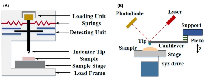
चित्रा 1: उपकरण और एएफएम कैंटिलीवर आधारित नैनोइंडेंटेशन सिस्टम की तुलना। (ए) यंत्रीकृत नैनोइंडेंटेशन और (बी) एएफएम कैंटिलीवर-आधारित नैनोइंडेंटेशन के संचालन के लिए विशिष्ट प्रणालियों को दर्शाने वाले योजनाबद्ध आरेख। यह आंकड़ा कियान एट अल.51 से संशोधित किया गया था। संक्षिप्त नाम: एएफएम = परमाणु बल माइक्रोस्कोपी। कृपया इस आंकड़े का एक बड़ा संस्करण देखने के लिए यहाँ क्लिक करें.
उपकरण और एएफएम कैंटिलीवर-आधारित नैनोइंडेंटेशन दोनों रुचि की नमूना सतह को विकृत करने और समय के कार्य के रूप में परिणामी बल और विस्थापन की निगरानी करने के लिए एक कठोर जांच का उपयोग करते हैं। आमतौर पर, या तो वांछित लोड (यानी, बल) या (जेड-पीज़ो) विस्थापन प्रोफ़ाइल उपयोगकर्ता द्वारा सॉफ्टवेयर इंटरफ़ेस के माध्यम से निर्दिष्ट की जाती है और सीधे उपकरण द्वारा नियंत्रित की जाती है, जबकि अन्य पैरामीटर मापा जाता है। नैनोइंडेंटेशन प्रयोगों से प्राप्त यांत्रिक गुण अक्सर लोचदार मापांक (ई) है, जिसे यंग के मापांक के रूप में भी जाना जाता है, जिसमें दबाव की इकाइयाँ होती हैं। किसी सामग्री का लोचदार मापांक बंधन कठोरता से संबंधित एक मौलिक गुण है और इसे प्लास्टिक विरूपण की शुरुआत से पहले लोचदार (यानी, प्रतिवर्ती या अस्थायी) विरूपण के दौरान अक्षीय तनाव (ε, इंडेंटेशन अक्ष के साथ आनुपातिक विरूपण) के लिए तन्यता या संपीड़ित तनाव (σ, प्रति इकाई क्षेत्र में लागू बल) के अनुपात के रूप में परिभाषित किया गया है।
 (1)
(1)
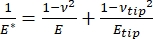
यह ध्यान दिया जाना चाहिए कि, क्योंकि कई सामग्री (विशेष रूप से जैविक ऊतक) वास्तव में विस्कोस्टिक हैं, वास्तव में, (गतिशील या जटिल) मापांक में लोचदार (भंडारण, चरण में) और चिपचिपा (हानि, चरण से बाहर) घटक दोनों होते हैं। वास्तविक व्यवहार में, नैनोइंडेंटेशन प्रयोग में जो मापा जाता है वह कम मापांक, ई * है, जो ब्याज के सही नमूना मापांक से संबंधित है, ई, जैसा कि समीकरण (2) में दिखाया गया है:
 (2)
(2)
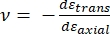
जहां ईटिप और त्रिकोण क्रमशः नैनोइंडेंट टिप के लोचदार मापांक और पॉइसन अनुपात हैं, और नमूने का अनुमानित पॉइसन अनुपात है। पॉइसन का अनुपात अनुप्रस्थ से अक्षीय तनाव का नकारात्मक अनुपात है, और इसलिए अक्षीय तनाव (जैसे, नैनोइंडेंटेशन लोडिंग के दौरान) के अधीन होने पर एक नमूने के अनुप्रस्थ बढ़ाव की डिग्री को इंगित करता है, जैसा कि समीकरण (3) में दिखाया गया है:
 (3)
(3)
कम से वास्तविक मापांक में रूपांतरण आवश्यक है क्योंकि ए) इंडेंटर टिप द्वारा प्रदान किए गए कुछ अक्षीय तनाव को अनुप्रस्थ तनाव में परिवर्तित किया जा सकता है (यानी, नमूना लोडिंग की दिशा के लंबवत विस्तार या संकुचन के माध्यम से विकृत हो सकता है), और बी) इंडेंटर टिप असीम रूप से कठिन नहीं है, और इस प्रकार नमूना को इंडेंट करने के कार्य के परिणामस्वरूप नोक की कुछ (छोटी) मात्रा में विरूपण होता है। ध्यान दें कि ऐसे मामले में जहां ई टिप >> ई (यानी, इंडेंटर टिप नमूने की तुलना में बहुत कठिन है, जो अक्सर हीरे की जांच का उपयोग करते समय सच होता है), कम और वास्तविक नमूना मापांक के बीच संबंध ई ≈ ई * (1 – वी2) के लिए बहुत सरल हो जाता है। जबकि यंत्रीकृत नैनोइंडेंटेशन सटीक बल लक्षण वर्णन और गतिशील सीमा के मामले में बेहतर है, एएफएम कैंटिलीवर-आधारित नैनोइंडेंटेशन तेज है, परिमाण अधिक बल और विस्थापन संवेदनशीलता के आदेश प्रदान करता है, उच्च रिज़ॉल्यूशन इमेजिंग और बेहतर इंडेंटेशन लोकेटिंग को सक्षम बनाता है, और एक साथ नैनोस्केल चुंबकीय औरविद्युत गुणों की जांच कर सकता है।. विशेष रूप से, एएफएम कैंटिलीवर-आधारित नैनोइंडेंटेशन नरम सामग्री (जैसे, पॉलिमर, जैल, लिपिड बाइलेयर, और कोशिकाओं या अन्य जैविक सामग्री), बेहद पतली (उप-म) फिल्मों (जहां सब्सट्रेट प्रभाव इंडेंटेशन गहराई के आधार पर खेल में आ सकते हैं) 10,11, और निलंबित दो-आयामी (2 डी) सामग्री12,13,14 जैसे ग्राफीन पर यांत्रिक गुणों की मात्रा का ठहराव करने के लिए बेहतर है।15,16, अभ्रक 17, हेक्सागोनल बोरान नाइट्राइड (एच-बीएन)18, या संक्रमण धातु डाइचल्कोजेनाइड्स (टीएमडीसी; जैसे, एमओएस2)19। यह इसके उत्तम बल (उप-एनएन) और विस्थापन (उप-एनएम) संवेदनशीलता के कारण है, जो संपर्क के प्रारंभिक बिंदु को सटीक रूप से निर्धारित करने और लोचदार विरूपण क्षेत्र के भीतर रहने के लिए महत्वपूर्ण है।
एएफएम कैंटिलीवर-आधारित नैनोइंडेंटेशन में, नमूना सतह की ओर एएफएम जांच का विस्थापन एक कैलिब्रेटेड पीजोइलेक्ट्रिक तत्व (चित्रा 1 बी) द्वारा किया जाता है, जिसमें लचीला कैंटिलीवर अंततः नमूना सतह के संपर्क में आने पर अनुभव किए गए प्रतिरोधक बल के कारण झुक जाता है। कैंटिलीवर के इस झुकाव या विक्षेपण की निगरानी आमतौर पर कैंटिलीवर के पीछे से एक लेजर को प्रतिबिंबित करके और एक फोटोडिटेक्टर (स्थिति संवेदनशील डिटेक्टर [पीएसडी]) में की जाती है। कैंटिलीवर कठोरता (nN/nm में) और विक्षेपण संवेदनशीलता (nm/V में) के ज्ञान के साथ युग्मित, इस मापा कैंटिलीवर विक्षेपण (V में) को नमूने पर लागू बल (nN में) में परिवर्तित करना संभव है। संपर्क के बाद, जेड-पीज़ो आंदोलन और कैंटिलीवर विक्षेपण के बीच का अंतर नमूना इंडेंटेशन गहराई उत्पन्न करता है। टिप क्षेत्र फ़ंक्शन के ज्ञान के साथ संयुक्त, यह टिप-नमूना संपर्क क्षेत्र की गणना को सक्षम बनाता है। परिणामस्वरूप बल-दूरी या बल-विस्थापन (एफ-डी) वक्रों के इन-संपर्क भागों की ढलान को नमूने के नैनोमैकेनिकल गुणों को निर्धारित करने के लिए एक उपयुक्त संपर्क यांत्रिकी मॉडल (चर्चा का डेटा विश्लेषण अनुभाग देखें) का उपयोग करके फिट किया जा सकता है। जबकि एएफएम कैंटिलीवर-आधारित नैनोइंडेंटेशन में ऊपर वर्णित यंत्रीकृत नैनोइंडेंटेशन पर कुछ अलग फायदे हैं, यह कई व्यावहारिक कार्यान्वयन चुनौतियों को भी प्रस्तुत करता है, जैसे अंशांकन, टिप वियर और डेटा विश्लेषण, जिन पर यहां चर्चा की जाएगी। एएफएम कैंटिलीवर-आधारित नैनोइंडेंटेशन का एक और संभावित नकारात्मक पक्ष रैखिक लोच की धारणा है, क्योंकि संपर्क त्रिज्या और इंडेंटेशन गहराई को इंडेंटर त्रिज्या की तुलना में बहुत छोटा होना चाहिए, जो नैनोस्केल एएफएम जांच और / या महत्वपूर्ण सतह खुरदरापन प्रदर्शित करने वाले नमूनों के साथ काम करते समय प्राप्त करना मुश्किल हो सकता है।
परंपरागत रूप से, नैनोइंडेंटेशन को अलग-अलग स्थानों या छोटे ग्रिड इंडेंटेशन प्रयोगों तक सीमित किया गया है, जिसमें एक वांछित स्थान (यानी, रुचि का क्षेत्र [आरओआई]) का चयन किया जाता है और एक एकल नियंत्रित इंडेंट, एक ही स्थान में कई इंडेंट कुछ प्रतीक्षा समय से अलग हो जाते हैं, और / या इंडेंट का एक मोटा ग्रिड हर्ट्ज के आदेश पर एक दर पर किया जाता है। हालांकि, एएफएम में हालिया प्रगति उच्च गति बल वक्र-आधारित इमेजिंग मोड (सिस्टम निर्माता के आधार पर विभिन्न ट्रेडनामों द्वारा संदर्भित) के उपयोग के माध्यम से यांत्रिक गुणों और स्थलाकृति के एक साथ अधिग्रहण की अनुमति देती है, जिसमें बल वक्रलोड नियंत्रण के तहत एक kHz दर पर आयोजित किए जाते हैं, जिसमें अधिकतम टिप-नमूना बल इमेजिंग सेटपॉइंट के रूप में उपयोग किया जाता है। पॉइंट-एंड-शूट विधियों को भी विकसित किया गया है, जिससे एएफएम स्थलाकृति छवि के अधिग्रहण की अनुमति मिलती है, जिसके बाद छवि के भीतर रुचि के बिंदुओं पर बाद में चयनात्मक नैनोइंडेंटेशन होता है, जो नैनोइंडेंटेशन स्थान पर नैनोस्केल स्थानिक नियंत्रण प्रदान करता है। हालांकि इस काम का प्राथमिक फोकस नहीं है, बल वक्र-आधारित इमेजिंग और पॉइंट-एंड-शूट कैंटिलीवर-आधारित नैनोइंडेंटेशन दोनों के विशिष्ट चयनित अनुप्रयोग उदाहरण प्रतिनिधि परिणामों में प्रस्तुत किए जाते हैं, और यदि विशेष एएफएम प्लेटफॉर्म पर उपलब्ध हो तो नीचे उल्लिखित प्रोटोकॉल के साथ संयोजन के रूप में उपयोग किया जा सकता है। विशेष रूप से, यह काम किसी भी सक्षम एएफएम सिस्टम पर एएफएम कैंटिलीवर-आधारित नैनोइंडेंटेशन के व्यावहारिक कार्यान्वयन के लिए एक सामान्यीकृत प्रोटोकॉल की रूपरेखा तैयार करता है और तकनीक के चार उपयोग मामले उदाहरण (हवा में दो, तरल पदार्थ में दो) प्रदान करता है, जिसमें प्रतिनिधि परिणाम और तकनीक को सफलतापूर्वक नियोजित करने के लिए बारीकियों, चुनौतियों और महत्वपूर्ण विचारों की गहन चर्चा शामिल है।
| Atomic force microscope | Bruker | Dimension Icon | Uses Nanoscope control software, including PeakForce Quantitative Nanomechanical Mapping (PF-QNM), FastForce Volume (FFV), and Point-and-Shoot Ramping experimental workspaces |
| AtomicJ | American Institute of Physics | https://doi.org/10.1063/1.4881683 | Flexible, powerful, free open source Java-based force curve analysis software package. Supports numerous contact mechanic models, such as Hertz, Sneddon DMT, JKR, Maugis, and cone or pyramid (including blunt and truncated). Also includes a variety of initial contact point estimation methods to choose from. Supports batch processing of data and subsequent statistical analysis (e.g., averages, standard deviations, histograms, goodness of fit, etc.). Literature citation is: P. Hermanowicz, M. Sarna, K. Burda, and H. Gabry , “AtomicJ: An open source software for analysis of force curves” Rev. Sci. Instrum. 85: 063703 (2014), https://doi.org/10.1063/1.4881683 , “AtomicJ: An open source software for analysis of force curves” Rev. Sci. Instrum. 85: 063703 (2014), https://doi.org/10.1063/1.4881683 |
| Buffer solution (PBS) | Fisher Chemical (NaCl), Sigma Aldrich (KCl), Fisher BioReagents (Na2HPO4 and KH2PO4) | S271 (>99% purity NaCl), P9541 (>99% purity KCl), BP332(>99% purity Na2HPO4), BP362 (>99% purity KH2PO4) | Phosphate buffered saline (PBS) was prepared in the laboratory as an aqueous solution consisting of 137 mM NaCl, 2.7 mM KCl, 10 mM Na2HPO4, and 1.8 mM KH2PO4 dissolved in ultrapure water. Reagents were measured out using an analytical balance, and glassware was cleaned with soap and water followed by autoclaving immediately prior to use. |
| Chloroform | |||
| Diamond tip AFM probe | Bruker | PDNISP | Pre-mounted factory-calibrated cube corner diamond (E = 1140 GPa) tip AFM probe (nominal R = 40 nm) with a stainless steel cantilever (nominal k = 225 N/m, f0 = 50 kHz). Spring constant is measured at the factory (k = 256 N/m for the probe, Serial #13435414, used here) and calibration data (including AFM images of indents showing probe geometry) is provided with the probe. |
| Diamond ultramicrotome blade | Diatome | Ultra 35° | 2.1 mm width. Also used a standard glass blade for intial rough cut of sample surface before transitioning to diamond blade for final surface preparation |
| Epoxy | Gorilla Glue | 26853-31-6 | Epoxy resin and hardner were mixed in a 1:1 ratio, a small drop was placed on a stainless steel sample puck (Ted Pella), and V1 grade muscovite mica (Ted Pella) was attached to create an atomically flat surface for preparation of phospholipid membranes. |
| Ethanol | |||
| LR white resin, medium grade (catalyzed) | Electron Microscopy Sciences | 14381 | 500 mL bottle, Lot #150629 |
| Mesenchymal stem cells (MSCs) | N/A | N/A | MSCs for nanomechanical studies were primary cells harvested from 8-10 week old male C57BL/6 mice as described in Goelzer, M. et al. "Lamin A/C Is Dispensable to Mechanical Repression of Adipogenesis" Int J Mol Sci 22: 6580 (2021) doi:10.3390/ijms22126580 and Peister, A. et al. "Adult stem cells from bone marrow (MSCs) isolated from different strains of inbred mice vary in surface epitopes, rates of proliferation, and differentiation potential" Blood 103: 1662-1668 (2004), doi:10.1182/blood-2003-09-3070. |
| Modulus standards | Bruker | PFQNM-SMPKIT-12M | Used HOPG (E = 18 GPa) and PS (E = 2.7 GPa). Also contains 2x PDMS (Tack 0, E = 2.5 MPa; Tack 4, E = 3.5 MPa), PS-LDPE (E = 2.0/0.2 GPa), fused silica (E = 72.9 GPa), sapphire (E – 345 GPa), and tip characterization (titanium roughness) sample. All samples come pre-mounted on a 12 mm diameter steel disc (sample puck). |
| Muscovite mica | Ted Pella | 50-12 | 12 mm diameter, V1 grade muscovite mica |
| Nanscope Analysis | Bruker | Version 2.0 | Free AFM image processing and analysis software package, but designed for, and proprietary/limited to Bruker AFMs; similar functionality is available from free, platform-independent AFM image processing and analysis software packages such as Gwyddion, WSxM, and others. Has built-in capabilities for force curve analysis, but AtomicJ is more flexible/full featured (e.g., more built-in contact mechanics models to choose from, statistical analysis of force curve fitting results, etc.) for force curve analysis and handles batch processing of force curves. |
| Phospholipids: POPC, Cholesterol (ovine) | Avanti Polar Lipids | POPC: CAS # 26853-31-6, Cholesterol: CAS # 57-88-5 | POPC lipid dissolved in chloroform (25 mg/mL) was obtained from vendor and used without further purification. Cholesterol powder from the same vendor was dissolved in chloroform (20 mg/mL). |
| Probe holder (fluid, lipid bilayers) | Bruker | MTFML-V2 | Specific to the particular AFM used; MTFML-V2 is a glass probe holder for scanning in fluid on a MultiMode AFM. |
| Probe holder (fluid, MSCs) | Bruker | FastScan Bio Z-scanner | Used with Dimension FastScan head (XY flexure scanners). Serial number MXYPOM5-1B154. |
| Probe holder (standard, ambient) | Bruker | DAFMCH | Specific to the particular AFM used; DAFMCH is the standard contact and tapping mode probe holder for the Dimension Icon AFM, suitable for nanoindentation (PF-QNM, FFV, and point-and-shoot ramping) |
| Sample Puck | Ted Pella | 16218 | Product number is for 15 mm diameter stainless steel sample puck. Also available in 6 mm, 10 mm, 12 mm, and 20 mm diameters at https://www.tedpella.com/AFM_html/AFM.aspx#anchor842459 |
| Sapphire substrate | Bruker | PFQNM-SMPKIT-12M | Extremely hard surface (E = 345 GPa) for measuring deflection sensitivity of probes (want all of the deflection to come from the probe, not the substrate). Part of the PF-QNM/modulus standards kit. |
| Scanning electron microscope | Hitachi | S-3400N-II | Located at Boise State. Used to perform co-localized SEM/EDS on all samples except additively manufactured (AM) Ti-6Al-4V. |
| Silicon AFM probes (standard) | NuNano | Scout 350 | Standard tapping mode silicon probe with reflective aluminum backside coating; k = 42 N/m (nominal), f0 = 350 kHz. Nominal R = 5 nm. Also available uncoated or with reflective gold backside coating. Probes with similar specifications are available from other manufacturers (e.g., Bruker TESPA-V2). |
| Silicon AFM probes (stiff) | Bruker | RTESPA-525, RTESPA-525-30 | Rotated tip etched silicon probes with reflective aluminum backside coating; k = 200 N/m (nominal), f0 = 525 kHz. Nominal R = 8 nm for RTESPA-525, R = 30 nm for RTESPA-525-30. Spring constant of each RTESPA-525-30 is measured individually at the factory via laser Doppler vibrometry and supplied with the probe. |
| Silicon carbide grit paper (abrasive discs) | Allied | 50-10005 | 120 grit |
| Silicon nitride AFM probes (soft, large radius hemispherical tip) | Bruker | MLCT-SPH-5UM, MLCT-SPH-5UM-DC | Also MLCT-SPH-1UM-DC. New product line of factory-calibrated (probe radius and spring constants of all cantilevers) large radius (R = 1 or 5 mm) hemispherical tip (at the end of a 23 mm long cylindrical shaft) probes. DC = drift compensation coating. 6 cantilevers/probe (A-F). Nominal spring constants: A, k = 0.07 N/m; B, k = 0.02 N/m; C, k = 0.01 N/m; D, k = 0.03 N/m; E, k = 0.1 N/m; F, k = 0.6 N/m. |
| Silicon nitride AFM probes (soft, medium sharp tip) | Bruker | DNP | 4 cantilevers/probe (A-d). Nominal spring constants: A, k = 0.35 N/m; B, k = 0.12 N/m; C, k = 0.24 N/m; D, k = 0.06 N/m. Nominal radii of curvature, R = 10 nm. |
| Silicon nitride AFM probes (soft, sharp tip) | Bruker | ScanAsyst-Air | Nominal values: resonance frequency, f0 = 70 kHz; spring constant, k = 0.4 N/m; radius of curvature, R = 2 nm. Designed for force curve based AFM imaging. |
| Superglue | Henkel | Loctite 495 | Cyanoacrylate based instant adhesive. Lots of roughly equivalent products are readily available. |
| Syringe pump | New Era Pump Systems | NE1000US | One channel syringe pump system with infusion and withdrawal capacity |
| Tip characterization standard | Bruker | PFQNM-SMPKIT-12M | Titanium (Ti) roughness standard. Part of the PF-QNM/modulus standards kit. |
| Ultrahigh purity nitrogen (UHP N2), 99.999% | Norco | SPG TUHPNI – T | T size compressed gas cylinder of ultrahigh purity (99.999%) nitrogen for drying samples |
| Ultramicrotome | Leica | EM UC6 | Equipped with a glass blade (standard, for intial sample preparation) and a diamond blade (for final preparation) |
| Ultrapure water | Thermo Fisher | Barnstead Nanopure Model 7146 | Model has been discontinued, but equivalent products are available. Produces ≥18.2 MΩ*cm ultrapure water with 1-5 ppb TOC (total organic content), per inline UV monitoring. Includes 0.2 µm particulate filter, ion exchange columns, and UV oxidation chamber. |
| Variable Speed Grinder | Buehler | EcoMet 3000 | Used with silicon carbide grit papers during hand polishing. |
| Vibration isolation table (active) | Herzan | TS-140 | Used with Bruker MultiMode AFM. Sits on a TMC 65-531 vibration isolation table. Bruker Dimension Icon AFM utilizes strictly passive vibration isolation (comes from manufacturer with custom acoustic hood, air table, and granite slab). |
| Vibration isolation table (passive) | TMC | 65-531 | 35" x 30" vibration isolation table with optional air damping (disabled). Used with Bruker MultiMode AFM. Herzan TS-140 "Table Stable" active vibration control table is located on top. |
