Summary

Atomik Kuvvet Mikroskobu Konsol Tabanlı Nanogirinti: Hava ve Sıvıda Nanoölçekte Mekanik Özellik Ölçümleri

Published: December 02, 2022
doi:

Summary

Bir atomik kuvvet mikroskobu (AFM) prob ucu tarafından bir numune yüzeyine uygulanan temas alanının ve kuvvetin ölçülmesi, nano ölçekli mekanik özellik belirlemeyi mümkün kılar. Elastik modülü veya diğer nanomekanik özellikleri ölçmek için yumuşak ve sert numunelerde havada veya sıvıda AFM konsol bazlı nanogirinti uygulamak için en iyi uygulamalar tartışılmaktadır.

Abstract

Bir atomik kuvvet mikroskobu (AFM), nano ölçekli bir AFM prob ucu ile numune yüzeyi arasındaki etkileşimi temel olarak ölçer. Prob ucu ve numune ile temas alanı tarafından uygulanan kuvvet ölçülebilirse, incelenen yüzeyin nano ölçekli mekanik özelliklerini (örneğin, elastik veya Young modülü) belirlemek mümkündür. Kantitatif AFM konsol tabanlı nanogirinti deneylerinin gerçekleştirilmesi için ayrıntılı bir prosedür, kPa’dan GPa’ya kadar çok çeşitli numune tiplerinin elastik modüllerini belirlemek için tekniğin nasıl uygulanabileceğine dair temsili örneklerle birlikte burada verilmiştir. Bunlar arasında canlı mezenkimal kök hücreler (MSC’ler) ve fizyolojik tampondaki çekirdekler, reçineye gömülü kurutulmuş loblolly çam kesitleri ve çeşitli bileşimlerde Bakken şeylleri bulunur.

Ek olarak, AFM konsol bazlı nanogirinti, fosfolipid çift katmanlarının kopma mukavemetini (yani atılım kuvvetini) araştırmak için kullanılır. Tekniğin doğru uygulanmasına yardımcı olmak için metot seçimi ve geliştirme, prob seçimi ve kalibrasyonu, ilgilenilen bölgenin tanımlanması, numune heterojenliği, özellik boyutu ve en boy oranı, uç aşınması, yüzey pürüzlülüğü ve veri analizi ve ölçüm istatistikleri gibi önemli pratik hususlar tartışılmaktadır. Son olarak, AFM türevli nanomekanik haritaların element bileşimi hakkında ek bilgi sağlayan elektron mikroskobu teknikleri ile birlikte lokalizasyonu gösterilmiştir.

Introduction

Malzemelerin mekanik özelliklerini anlamak, mühendislikteki en temel ve temel görevlerden biridir. Dökme malzeme özelliklerinin analizi için, malzeme sistemlerinin mekanik özelliklerini karakterize etmek için çekme testleri1, sıkıştırma testleri2 ve üç veya dört noktalı eğilme (eğilme) testleri3 dahil olmak üzere çok sayıda yöntem mevcuttur. Bu mikro ölçekli testler, dökme malzeme özellikleri hakkında paha biçilmez bilgiler sağlayabilirken, genellikle başarısızlığa uğrar ve bu nedenle yıkıcıdır. Ek olarak, ince filmler, biyolojik malzemeler ve nanokompozitler gibi günümüzde ilgi çekici olan birçok malzeme sisteminin mikro ve nano ölçekli özelliklerini doğru bir şekilde araştırmak için gerekli mekansal çözünürlükten yoksundurlar. Büyük ölçekli mekanik testlerle ilgili bazı problemleri, özellikle de yıkıcı doğasını ele almaya başlamak için, mikrosertlik testleri mineralojiden benimsenmiştir. Sertlik, bir malzemenin belirli koşullar altında plastik deformasyona karşı direncinin bir ölçüsüdür. Genel olarak, mikrosertlik testleri, bir malzemeye girinti yapmak için genellikle sertleştirilmiş çelik veya elmastan yapılmış sert bir prob kullanır. Elde edilen girinti derinliği ve / veya alanı daha sonra sertliği belirlemek için kullanılabilir. Vickers4, Knoop5 ve Brinell6 sertliği dahil olmak üzere çeşitli yöntemler geliştirilmiştir; Her biri mikro ölçekli malzeme sertliğinin bir ölçüsünü sağlar, ancak farklı koşullar ve tanımlar altında ve bu nedenle yalnızca aynı koşullar altında gerçekleştirilen testlerle karşılaştırılabilecek veriler üretir.

Aletli nanogirinti, çeşitli mikrosertlik test yöntemleri ile elde edilen nispi değerleri iyileştirmek, mekanik özelliklerin analizi için mümkün olan uzamsal çözünürlüğü geliştirmek ve ince filmlerin analizini sağlamak için geliştirilmiştir. Önemli olarak, ilk olarak Oliver ve Pharr7 tarafından geliştirilen yöntem kullanılarak, bir numune malzemesinin elastik veya Young modülü E, aletli nanogirinti yoluyla belirlenebilir. Ayrıca, bir Berkovich üç taraflı piramidal nanoindenter probu (ideal uç alanı fonksiyonu Vickers dört taraflı piramidal probunkiyle eşleşir)8 kullanılarak, nano ölçekli ve daha geleneksel mikro ölçekli sertlik ölçümleri arasında doğrudan karşılaştırma yapılabilir. AFM’nin popülaritesinin artmasıyla birlikte, AFM konsol bazlı nanogirinti, özellikle daha yumuşak malzemelerin mekanik özelliklerini ölçmek için de dikkat çekmeye başladı. Sonuç olarak, Şekil 1’de şematik olarak gösterildiği gibi, nano ölçekli mekanik özellikleri sorgulamak ve ölçmek için günümüzde en yaygın olarak kullanılan iki teknik, bu çalışmanın odak noktası olan aletli nanogirinti (Şekil 1A) ve AFM konsol tabanlı nanogirinti (Şekil 1B)9’dur.

Figure 1
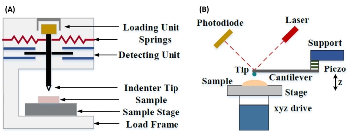
Şekil 1: Aletli ve AFM konsol bazlı nanogirinti sistemlerinin karşılaştırılması. (A) aletli nanogirinti ve (B) AFM konsol tabanlı nanogirinti iletmek için tipik sistemleri gösteren şematik diyagramlar. Bu rakam Qian ve ark.51’den değiştirilmiştir. Kısaltma: AFM = atomik kuvvet mikroskobu. Bu şeklin daha büyük bir versiyonunu görmek için lütfen buraya tıklayın.

Hem aletli hem de AFM konsol bazlı nanogirinti, ilgilenilen bir numune yüzeyini deforme etmek ve ortaya çıkan kuvveti ve yer değiştirmeyi zamanın bir fonksiyonu olarak izlemek için sert bir prob kullanır. Tipik olarak, istenen yük (yani kuvvet) veya (Z-piezo) yer değiştirme profili, kullanıcı tarafından yazılım arayüzü aracılığıyla belirlenir ve diğer parametre ölçülürken doğrudan cihaz tarafından kontrol edilir. Nanogirinti deneylerinden en sık elde edilen mekanik özellik, basınç birimlerine sahip Young modülü olarak da adlandırılan elastik modüldür (E). Bir malzemenin elastik modülü, bağ sertliği ile ilgili temel bir özelliktir ve plastik deformasyonun başlamasından önce elastik (yani tersinir veya geçici) deformasyon sırasında çekme veya basınç gerilmesinin (σ, birim alan başına uygulanan kuvvet) eksenel gerinime (ε, girinti ekseni boyunca orantılı deformasyon) oranı olarak tanımlanır (denklem [1]):

Equation 1(1)

Birçok malzemenin (özellikle biyolojik dokuların) aslında viskoelastik olması nedeniyle, gerçekte, (dinamik veya karmaşık) modülün hem elastik (depolama, faz içinde) hem de viskoz (kayıp, faz dışı) bileşenlerden oluştuğu belirtilmelidir. Gerçek uygulamada, bir nanogirinti deneyinde ölçülen şey, denklemde (2) gösterildiği gibi, ilgilenilen gerçek numune modülü E ile ilişkili olan indirgenmiş modül E * ‘dir:

Equation 2(2)

Burada E ucu ve ν ucu, nanoindenter ucunun sırasıyla elastik modülü ve Poisson oranıdır ve ν, numunenin tahmini Poisson oranıdır. Poisson oranı, enine eksenel gerinimin negatif oranıdır ve bu nedenle, denklemde (3) gösterildiği gibi, eksenel gerinimlere (örneğin, nanogirinti yüklemesi sırasında) maruz kalındığında bir numunenin enine uzama derecesini gösterir:

Equation 3(3)

İndirgenmiş modülden gerçek modüle dönüşüm gereklidir, çünkü a) girinti ucu tarafından verilen eksenel gerinimin bir kısmı enine gerinime dönüştürülebilir (yani, numune yükleme yönüne dik olarak genleşme veya büzülme yoluyla deforme olabilir) ve b) girinti ucu sonsuz derecede sert değildir ve bu nedenle numuneyi girintileme eylemi, ucun bir miktar (küçük) deformasyonuna neden olur. E ucunun E >> durumunda (yani, girinti ucu numuneden çok daha zordur, ki bu genellikle bir elmas probu kullanıldığında doğrudur), indirgenmiş ve gerçek numune modülü arasındaki ilişkinin EE * (1 – v2) için büyük ölçüde basitleştirildiğini unutmayın. Aletli nanogirinti, doğru kuvvet karakterizasyonu ve dinamik aralık açısından üstün olsa da, AFM konsol tabanlı nanogirinti daha hızlıdır, büyüklük sıralarında daha fazla kuvvet ve yer değiştirme hassasiyeti sağlar, daha yüksek çözünürlüklü görüntüleme ve gelişmiş girinti konumlandırma sağlar ve aynı anda nano ölçekli manyetik ve elektriksel özellikleri araştırabilir9. Özellikle, AFM konsol bazlı nanogirinti, yumuşak malzemelerin (örneğin, polimerler, jeller, lipit çift katmanları ve hücreler veya diğer biyolojik malzemeler), son derece ince (alt μm) filmlerin (girinti derinliğine bağlı olarak substrat etkilerinin devreye girebileceği) nano ölçekte mekanik özelliklerin nicelleştirilmesi için üstündür.10,11 ve grafen gibi asılı iki boyutlu (2D) malzemeler12,13,1415,16, mika17, altıgen bor nitrür (h-BN)18 veya geçiş metali dikalkojenitler (TMDC’ler; örneğin, MoS2)19. Bunun nedeni, ilk temas noktasını doğru bir şekilde belirlemek ve elastik deformasyon bölgesinde kalmak için önemli olan mükemmel kuvvet (sub-nN) ve yer değiştirme (sub-nm) hassasiyetidir.

AFM konsol bazlı nanogirintide, bir AFM probunun numune yüzeyine doğru yer değiştirmesi, kalibre edilmiş bir piezoelektrik eleman (Şekil 1B) tarafından harekete geçirilir ve esnek konsol, numune yüzeyiyle temas ettiğinde yaşanan dirençli kuvvet nedeniyle sonunda bükülür. Konsoldaki bu bükülme veya sapma tipik olarak bir lazeri konsol arkasından ve bir fotodetektöre (konuma duyarlı dedektör [PSD]) yansıtarak izlenir. Konsol sertliği (nN/nm cinsinden) ve sapma hassasiyeti (nm/V cinsinden) bilgisi ile birleştiğinde, ölçülen bu konsol sapmasını (V cinsinden) numuneye uygulanan kuvvete (nN cinsinden) dönüştürmek mümkündür. Temasın ardından, Z-piezo hareketi ile konsol sapması arasındaki fark, numune girinti derinliğini verir. Uç alanı fonksiyonunun bilgisi ile birleştiğinde, bu, uç-numune temas alanının hesaplanmasını sağlar. Elde edilen kuvvet-mesafe veya kuvvet-yer değiştirme (F-D) eğrilerinin temas halindeki kısımlarının eğimi, numunenin nanomekanik özelliklerini belirlemek için uygun bir temas mekaniği modeli (tartışmanın Veri Analizi bölümüne bakınız) kullanılarak sığdırılabilir. AFM konsol bazlı nanogirinti, yukarıda açıklandığı gibi aletli nanogirintiye göre bazı belirgin avantajlara sahip olsa da, burada tartışılacak olan kalibrasyon, uç aşınması ve veri analizi gibi çeşitli pratik uygulama zorlukları da sunmaktadır. AFM konsol tabanlı nanogirintinin bir diğer potansiyel dezavantajı, temas yarıçapı ve girinti derinliklerinin, nano ölçekli AFM probları ve / veya önemli yüzey pürüzlülüğü sergileyen numunelerle çalışırken elde edilmesi zor olabilen indenter yarıçapından çok daha küçük olması gerektiğinden, doğrusal elastikiyet varsayımıdır.

Geleneksel olarak, nanogirinti, istenen bir konumun (yani, ilgi alanı [ROI]) seçildiği ve tek bir kontrollü girinti, bir miktar bekleme süresi ile ayrılmış tek bir konumda birden fazla girinti ve / veya kaba bir girinti ızgarasının Hz sırasına göre bir oranda gerçekleştirildiği bireysel konumlar veya küçük ızgara girinti deneyleri ile sınırlandırılmıştır. Bununla birlikte, AFM’deki son gelişmeler, yüksek hızlı kuvvet eğrisi tabanlı görüntüleme modlarının (sistem üreticisine bağlı olarak çeşitli ticari isimlerle anılır) kullanılmasıyla mekanik özelliklerin ve topografyanın eşzamanlı olarak edinilmesine izin verir; burada kuvvet eğrileri, görüntüleme ayar noktası olarak kullanılan maksimum uç-numune kuvveti ile yük kontrolü altında bir kHz hızında gerçekleştirilir. Bir AFM topografya görüntüsünün elde edilmesine ve ardından görüntü içindeki ilgi çekici noktalarda seçici nanogirintinin ardından nanogirinti konumu üzerinde nano ölçekli mekansal kontrol sağlayan bas ve çek yöntemleri de geliştirilmiştir. Bu çalışmanın birincil odağı olmasa da, hem kuvvet eğrisi tabanlı görüntüleme hem de bas ve çek konsol tabanlı nanogirintinin spesifik seçilmiş uygulama örnekleri temsili sonuçlarda sunulmuştur ve kullanılan belirli AFM platformunda mevcutsa aşağıda özetlenen protokolle birlikte kullanılabilir. Spesifik olarak, bu çalışma, herhangi bir yetenekli AFM sisteminde AFM konsol tabanlı nanogirintinin pratik uygulaması için genelleştirilmiş bir protokolü özetlemektedir ve temsili sonuçlar ve tekniğin başarılı bir şekilde kullanılması için nüansların, zorlukların ve önemli hususların derinlemesine tartışılması da dahil olmak üzere tekniğin dört kullanım örneği (ikisi havada, ikisi sıvıda) sunmaktadır.

Protocol

NOT: Ticari olarak temin edilebilen çok çeşitli AFM’ler ve konsol bazlı nanogirinti için mevcut olan numune tiplerinin ve uygulamalarının çeşitliliği nedeniyle, takip eden protokol, cihaz veya üreticiden bağımsız olarak tüm konsol tabanlı nanogirinti deneyleri için gerekli olan paylaşılan adımlara odaklanarak, doğada nispeten genel olacak şekilde kasıtlı olarak tasarlanmıştır. Bu nedenle, yazarlar okuyucunun konsol bazlı nanogirinti yapmak için seçilen belirli enstrümanı çalıştırma kon…

Representative Results

Kuvvet-yer değiştirme eğrileriŞekil 7, reçineye gömülü loblolly çam örnekleri (Şekil 7A) ve mezenkimal kök hücre (MSC) çekirdekleri üzerindeki sıvıda (fosfat tamponlu salin [PBS]) havada yapılan nanogirinti deneylerinden elde edilen temsili, ideale yakın F-D eğrilerini göstermektedir (Şekil 7B). Herhangi bir temas mekaniği modelinin kullanılması, ilk uç-numune temas noktasının doğru …

Discussion

Numune hazırlama
Havadaki nanogirinti için, yaygın hazırlama yöntemleri arasında kriyoseksiyon (örneğin, doku numuneleri), taşlama ve / veya parlatma ve ardından ultramikrotoming (örneğin, reçineye gömülü biyolojik numuneler), iyon frezeleme veya odaklanmış iyon demeti hazırlama (örneğin, parlatma için uygun olmayan yarı iletken, gözenekli veya karışık sertlik numuneleri), mekanik veya elektrokimyasal parlatma (örneğin, metal alaşımları) veya ince film biriktirme (örne…

Disclosures

The authors have nothing to disclose.

Acknowledgements

Tüm AFM deneyleri Boise State Üniversitesi Yüzey Bilimleri Laboratuvarı’nda (SSL) gerçekleştirildi. SEM karakterizasyonu Boise State Center for Materials Characterization’da (BSCMC) gerçekleştirildi. Bu yayında biyoyakıt hammaddeleri ile ilgili olarak bildirilen araştırmalar, kısmen ABD Enerji Bakanlığı, Enerji Verimliliği ve Yenilenebilir Enerji Ofisi, Hammadde Dönüşüm Arayüzü Konsorsiyumu’nun (FCIC) bir parçası olarak Biyoenerji Teknolojileri Ofisi tarafından ve DOE Idaho Operasyon Ofisi Sözleşmesi DE-AC07-051ID14517 kapsamında desteklenmiştir. Hücre mekaniği çalışmaları, Ulusal Sağlık Enstitüleri (ABD) tarafından AG059923, AR075803 ve P20GM109095 hibeleri ve Ulusal Bilim Vakfı (ABD) 1929188 ve 2025505 hibeleri ile desteklenmiştir. Model lipid çift katmanlı sistemler çalışması, R01 EY030067 hibesi kapsamında Ulusal Sağlık Enstitüleri (ABD) tarafından desteklenmiştir. Yazarlar, Şekil 11’de gösterilen kompozit görüntüyü ürettiği için Dr. Elton Graugnard’a teşekkür eder.

Materials

Atomic force microscope Bruker Dimension Icon Uses Nanoscope control software, including PeakForce Quantitative Nanomechanical Mapping (PF-QNM), FastForce Volume (FFV), and Point-and-Shoot Ramping experimental workspaces
AtomicJ American Institute of Physics https://doi.org/10.1063/1.4881683 Flexible, powerful, free open source Java-based force curve analysis software package. Supports numerous contact mechanic models, such as Hertz, Sneddon DMT, JKR, Maugis, and cone or pyramid (including blunt and truncated). Also includes a variety of initial contact point estimation methods to choose from. Supports batch processing of data and subsequent statistical analysis (e.g., averages, standard deviations, histograms, goodness of fit, etc.). Literature citation is: P. Hermanowicz, M. Sarna, K. Burda, and H. GabryEquation 1, “AtomicJ: An open source software for analysis of force curves” Rev. Sci. Instrum. 85: 063703 (2014), https://doi.org/10.1063/1.4881683
Buffer solution (PBS) Fisher Chemical (NaCl), Sigma Aldrich (KCl), Fisher BioReagents (Na2HPO4 and KH2PO4) S271 (>99% purity NaCl), P9541 (>99% purity KCl), BP332(>99% purity Na2HPO4), BP362 (>99% purity KH2PO4) Phosphate buffered saline (PBS) was prepared in the laboratory as an aqueous solution consisting of 137 mM NaCl, 2.7 mM KCl, 10 mM Na2HPO4, and 1.8 mM KH2PO4 dissolved in ultrapure water. Reagents were measured out using an analytical balance, and glassware was cleaned with soap and water followed by autoclaving immediately prior to use.
Chloroform
Diamond tip AFM probe Bruker PDNISP Pre-mounted factory-calibrated cube corner diamond (E = 1140 GPa) tip AFM probe (nominal R = 40 nm) with a stainless steel cantilever (nominal k = 225 N/m, f0 = 50 kHz). Spring constant is measured at the factory (k = 256 N/m for the probe, Serial #13435414, used here) and calibration data (including AFM images of indents showing probe geometry) is provided with the probe.
Diamond ultramicrotome blade Diatome Ultra 35° 2.1 mm width. Also used a standard glass blade for intial rough cut of sample surface before transitioning to diamond blade for final surface preparation
Epoxy Gorilla Glue 26853-31-6 Epoxy resin and hardner were mixed in a 1:1 ratio, a small drop was placed on a stainless steel sample puck (Ted Pella), and V1 grade muscovite mica (Ted Pella) was attached to create an atomically flat surface for preparation of phospholipid membranes.
Ethanol
LR white resin, medium grade (catalyzed) Electron Microscopy Sciences 14381 500 mL bottle, Lot #150629
Mesenchymal stem cells (MSCs) N/A N/A MSCs for nanomechanical studies were primary cells harvested from 8-10 week old male C57BL/6 mice as described in Goelzer, M. et al. "Lamin A/C Is Dispensable to Mechanical Repression of Adipogenesis" Int J Mol Sci 22: 6580 (2021) doi:10.3390/ijms22126580 and Peister, A. et al. "Adult stem cells from bone marrow (MSCs) isolated from different strains of inbred mice vary in surface epitopes, rates of proliferation, and differentiation potential" Blood 103: 1662-1668 (2004), doi:10.1182/blood-2003-09-3070.
Modulus standards Bruker PFQNM-SMPKIT-12M Used HOPG (E = 18 GPa) and PS (E = 2.7 GPa). Also contains 2x PDMS (Tack 0, E = 2.5 MPa; Tack 4, E = 3.5 MPa), PS-LDPE (E = 2.0/0.2 GPa), fused silica (E = 72.9 GPa), sapphire (E – 345 GPa), and tip characterization (titanium roughness) sample. All samples come pre-mounted on a 12 mm diameter steel disc (sample puck).
Muscovite mica Ted Pella 50-12 12 mm diameter, V1 grade muscovite mica
Nanscope Analysis Bruker Version 2.0 Free AFM image processing and analysis software package, but designed for, and proprietary/limited to Bruker AFMs; similar functionality is available from free, platform-independent AFM image processing and analysis software packages such as Gwyddion, WSxM, and others. Has built-in capabilities for force curve analysis, but AtomicJ is more flexible/full featured (e.g., more built-in contact mechanics models to choose from, statistical analysis of force curve fitting results, etc.) for force curve analysis and handles batch processing of force curves.
Phospholipids: POPC, Cholesterol (ovine) Avanti Polar Lipids POPC: CAS # 26853-31-6, Cholesterol: CAS # 57-88-5 POPC lipid dissolved in chloroform (25 mg/mL) was obtained from vendor and used without further purification. Cholesterol powder from the same vendor was dissolved in chloroform (20 mg/mL). 
Probe holder (fluid, lipid bilayers) Bruker MTFML-V2 Specific to the particular AFM used; MTFML-V2 is a glass probe holder for scanning in fluid on a MultiMode AFM.
Probe holder (fluid, MSCs) Bruker FastScan Bio Z-scanner Used with Dimension FastScan head (XY flexure scanners). Serial number MXYPOM5-1B154.
Probe holder (standard, ambient) Bruker DAFMCH Specific to the particular AFM used; DAFMCH is the standard contact and tapping mode probe holder for the Dimension Icon AFM, suitable for nanoindentation (PF-QNM, FFV, and point-and-shoot ramping)
Sample Puck Ted Pella 16218 Product number is for 15 mm diameter stainless steel sample puck. Also available in 6 mm, 10 mm, 12 mm, and 20 mm diameters at https://www.tedpella.com/AFM_html/AFM.aspx#anchor842459
Sapphire substrate Bruker PFQNM-SMPKIT-12M Extremely hard surface (E = 345 GPa) for measuring deflection sensitivity of probes (want all of the deflection to come from the probe, not the substrate). Part of the PF-QNM/modulus standards kit.
Scanning electron microscope Hitachi S-3400N-II Located at Boise State. Used to perform co-localized SEM/EDS on all samples except additively manufactured (AM) Ti-6Al-4V.
Silicon AFM probes (standard) NuNano Scout 350 Standard tapping mode silicon probe with reflective aluminum backside coating; k = 42 N/m (nominal), f0 = 350 kHz. Nominal R = 5 nm. Also available uncoated or with reflective gold backside coating. Probes with similar specifications are available from other manufacturers (e.g., Bruker TESPA-V2).
Silicon AFM probes (stiff) Bruker RTESPA-525, RTESPA-525-30  Rotated tip etched silicon probes with reflective aluminum backside coating; k = 200 N/m (nominal), f0 = 525 kHz. Nominal R = 8 nm for RTESPA-525, R = 30 nm for RTESPA-525-30. Spring constant of each RTESPA-525-30 is measured individually at the factory via laser Doppler vibrometry and supplied with the probe.
Silicon carbide grit paper (abrasive discs) Allied 50-10005 120 grit
Silicon nitride AFM probes (soft, large radius hemispherical tip) Bruker MLCT-SPH-5UM, MLCT-SPH-5UM-DC Also MLCT-SPH-1UM-DC. New product line of factory-calibrated (probe radius and spring constants of all cantilevers) large radius (R = 1 or 5 mm) hemispherical tip (at the end of a 23 mm long cylindrical shaft) probes. DC = drift compensation coating. 6 cantilevers/probe (A-F). Nominal spring constants: A, k = 0.07 N/m; B, k = 0.02 N/m; C, k = 0.01 N/m; D, k = 0.03 N/m; E, k = 0.1 N/m; F, k = 0.6 N/m.
Silicon nitride AFM probes (soft, medium sharp tip) Bruker DNP 4 cantilevers/probe (A-d). Nominal spring constants: A, k = 0.35 N/m; B, k = 0.12 N/m; C, k = 0.24 N/m; D, k = 0.06 N/m. Nominal radii of curvature, R = 10 nm.
Silicon nitride AFM probes (soft, sharp tip) Bruker ScanAsyst-Air Nominal values: resonance frequency, f0 = 70 kHz; spring constant, k = 0.4 N/m; radius of curvature, R = 2 nm. Designed for force curve based AFM imaging.
Superglue Henkel Loctite 495 Cyanoacrylate based instant adhesive. Lots of roughly equivalent products are readily available.
Syringe pump New Era Pump Systems NE1000US One channel syringe pump system with infusion and withdrawal capacity
Tip characterization standard Bruker PFQNM-SMPKIT-12M Titanium (Ti) roughness standard. Part of the PF-QNM/modulus standards kit.
Ultrahigh purity nitrogen (UHP N2), 99.999% Norco SPG TUHPNI – T T size compressed gas cylinder of ultrahigh purity (99.999%) nitrogen for drying samples
Ultramicrotome Leica EM UC6 Equipped with a glass blade (standard, for intial sample preparation) and a diamond blade (for final preparation)
Ultrapure water Thermo Fisher Barnstead Nanopure Model 7146 Model has been discontinued, but equivalent products are available. Produces ≥18.2 MΩ*cm ultrapure water with 1-5 ppb TOC (total organic content), per inline UV monitoring. Includes 0.2 µm particulate filter, ion exchange columns, and UV oxidation chamber.
Variable Speed Grinder Buehler EcoMet 3000 Used with silicon carbide grit papers during hand polishing.
Vibration isolation table (active) Herzan TS-140 Used with Bruker MultiMode AFM. Sits on a TMC 65-531 vibration isolation table. Bruker Dimension Icon AFM utilizes strictly passive vibration isolation (comes from manufacturer with custom acoustic hood, air table, and granite slab).
Vibration isolation table (passive) TMC 65-531 35" x 30" vibration isolation table with optional air damping (disabled). Used with Bruker MultiMode AFM. Herzan TS-140 "Table Stable" active vibration control table is located on top.

References

  1. Hart, E. W. Theory of the tensile test. Acta Metallurgica. 15 (2), 351-355 (1967).
  2. Fell, J. T., Newton, J. M. Determination of tablet strength by the diametral-compression test. Journal of Pharmaceutical Sciences. 59 (5), 688-691 (1970).
  3. Babiak, M., Gaff, M., Sikora, A., Hysek, &. #. 3. 5. 2. ;. Modulus of elasticity in three- and four-point bending of wood. Composite Structures. 204, 454-465 (2018).
  4. Song, S., Yovanovich, M. M. Relative contact pressure-Dependence on surface roughness and Vickers microhardness. Journal of Thermophysics and Heat Transfer. 2 (1), 43-47 (1988).
  5. Hays, C., Kendall, E. G. An analysis of Knoop microhardness. Metallography. 6 (4), 275-282 (1973).
  6. Hill, R., Storåkers, B., Zdunek, A. B. A theoretical study of the Brinell hardness test. Proceedings of the Royal Society of London. A. Mathematical and Physical Sciences. 423 (1865), 301-330 (1989).
  7. Oliver, W. C., Pharr, G. M. An improved technique for determining hardness and elastic modulus using load and displacement sensing indentation experiments. Journal of Materials Research. 7 (6), 1564-1583 (1992).
  8. Sakharova, N. A., Fernandes, J. V., Antunes, J. M., Oliveira, M. C. Comparison between Berkovich, Vickers and conical indentation tests: A three-dimensional numerical simulation study. International Journal of Solids and Structures. 46 (5), 1095-1104 (2009).
  9. Cohen, S. R., Kalfon-Cohen, E. Dynamic nanoindentation by instrumented nanoindentation and force microscopy: a comparative review. Beilstein Journal of Nanotechnology. 4 (1), 815-833 (2013).
  10. Saha, R., Nix, W. D. Effects of the substrate on the determination of thin film mechanical properties by nanoindentation. Acta Materialia. 50 (1), 23-38 (2002).
  11. Tsui, T. Y., Pharr, G. M. Substrate effects on nanoindentation mechanical property measurement of soft films on hard substrates. Journal of Materials Research. 14 (1), 292-301 (1999).
  12. Cao, G., Gao, H. Mechanical properties characterization of two-dimensional materials via nanoindentation experiments. Progress in Materials Science. 103, 558-595 (2019).
  13. Castellanos-Gomez, A., Singh, V., vander Zant, H. S. J., Steele, G. A. Mechanics of freely-suspended ultrathin layered materials. Annalen der Physik. 527 (1-2), 27-44 (2015).
  14. Cao, C., Sun, Y., Filleter, T. Characterizing mechanical behavior of atomically thin films: A review. Journal of Materials Research. 29 (3), 338-347 (2014).
  15. Lee, C., Wei, X., Kysar, J. W., Hone, J. Measurement of the elastic properties and intrinsic strength of monolayer graphene. Science. 321 (5887), 385-388 (2008).
  16. Elibol, K., et al. Visualising the strain distribution in suspended two-dimensional materials under local deformation. Scientific Reports. 6 (1), 28485 (2016).
  17. Castellanos-Gomez, A., et al. Mechanical properties of freely suspended atomically thin dielectric layers of mica. Nano Research. 5 (8), 550-557 (2012).
  18. Song, L., et al. Large scale growth and characterization of atomic hexagonal boron nitride layers. Nano Letters. 10 (8), 3209-3215 (2010).
  19. Castellanos-Gomez, A., et al. Elastic properties of freely suspended MoS2 nanosheets. Advanced Materials. 24 (6), 772-775 (2012).
  20. D’Costa, N. P., Hoh, J. H. Calibration of optical lever sensitivity for atomic force microscopy. Review of Scientific Instruments. 66 (10), 5096-5097 (1995).
  21. Wu, Y., et al. Evaluation of elastic modulus and hardness of crop stalks cell walls by nano-indentation. Bioresource Technology. 101 (8), 2867-2871 (2010).
  22. Barns, S., et al. Investigation of red blood cell mechanical properties using AFM indentation and coarse-grained particle method. BioMedical Engineering OnLine. 16 (1), 140 (2017).
  23. Hermanowicz, P., Sarna, M., Burda, K., Gabryś, H. AtomicJ: An open source software for analysis of force curves. Review of Scientific Instruments. 85 (6), 063703 (2014).
  24. Broitman, E. Indentation hardness measurements at macro-, micro-, and nanoscale: a critical overview. Tribology Letters. 65 (1), 23 (2016).
  25. Tiwari, A. . Nanomechanical Analysis of High Performance Materials. , (2015).
  26. Aggarwal, R. L., Ramdas, A. K. . Physical Properties of Diamond and Sapphire. , (2019).
  27. Boyd, E. J., Uttamchandani, D. Measurement of the anisotropy of Young’s modulus in single-crystal silicon. Journal of Microelectromechanical Systems. 21 (1), 243-249 (2012).
  28. Harding, J. W., Sneddon, I. N. The elastic stresses produced by the indentation of the plane surface of a semi-infinite elastic solid by a rigid punch. Mathematical Proceedings of the Cambridge Philosophical Society. 41 (1), 16-26 (2008).
  29. Lin, D. C., Dimitriadis, E. K., Horkay, F. Robust strategies for automated AFM force curve analysis-I. Non-adhesive indentation of soft, inhomogeneous materials. Journal of Biomechanical Engineering. 129 (3), 430-440 (2006).
  30. Lin, D. C., Dimitriadis, E. K., Horkay, F. Robust strategies for automated AFM force curve analysis-II: Adhesion-influenced indentation of soft, elastic materials. Journal of Biomechanical Engineering. 129 (6), 904-912 (2007).
  31. Haile, S., Palmer, M., Otey, A. Potential of loblolly pine: switchgrass alley cropping for provision of biofuel feedstock. Agroforestry Systems. 90 (5), 763-771 (2016).
  32. Lu, X., et al. Biomass logistics analysis for large scale biofuel production: Case study of loblolly pine and switchgrass. Bioresource Technology. 183, 1-9 (2015).
  33. Susaeta, A., Lal, P., Alavalapati, J., Mercer, E., Carter, D. Economics of intercropping loblolly pine and switchgrass for bioenergy markets in the southeastern United States. Agroforestry Systems. 86 (2), 287-298 (2012).
  34. Garcia, R. Nanomechanical mapping of soft materials with the atomic force microscope: methods, theory and applications. Chemical Society Reviews. 49 (16), 5850-5884 (2020).
  35. Derjaguin, B. V., Muller, V. M., Toporov, Y. P. Effect of contact deformations on the adhesion of particles. Journal of Colloid and Interface Science. 53 (2), 314-326 (1975).
  36. Ciesielski, P. N., et al. Engineering plant cell walls: tuning lignin monomer composition for deconstructable biofuel feedstocks or resilient biomaterials. Green Chemistry. 16 (5), 2627-2635 (2014).
  37. Liu, K., Ostadhassan, M., Zhou, J., Gentzis, T., Rezaee, R. Nanoscale pore structure characterization of the Bakken shale in the USA. Fuel. 209, 567-578 (2017).
  38. Maryon, O. O., et al. Co-localizing Kelvin probe force microscopy with other microscopies and spectroscopies: selected applications in corrosion characterization of alloys. JoVE. (184), e64102 (2022).
  39. Eliyahu, M., Emmanuel, S., Day-Stirrat, R. J., Macaulay, C. I. Mechanical properties of organic matter in shales mapped at the nanometer scale. Marine and Petroleum Geology. 59, 294-304 (2015).
  40. Li, C., et al. Nanomechanical characterization of organic matter in the Bakken formation by microscopy-based method. Marine and Petroleum Geology. 96, 128-138 (2018).
  41. Bouzid, T., et al. The LINC complex, mechanotransduction, and mesenchymal stem cell function and fate. Journal of Biological Engineering. 13 (1), 68 (2019).
  42. Dupont, S., et al. Role of YAP/TAZ in mechanotransduction. Nature. 474 (7350), 179-183 (2011).
  43. Wang, S., et al. CCM3 is a gatekeeper in focal adhesions regulating mechanotransduction and YAP/TAZ signalling. Nature Cell Biology. 23 (7), 758-770 (2021).
  44. Sen, B., et al. Mechanical strain inhibits adipogenesis in mesenchymal stem cells by stimulating a durable β-catenin signal. Endocrinology. 149 (12), 6065-6075 (2008).
  45. Sen, B., et al. mTORC2 regulates mechanically induced cytoskeletal reorganization and lineage selection in marrow-derived mesenchymal stem cells. Journal of Bone and Mineral Research. 29 (1), 78-89 (2014).
  46. Sen, B., et al. Mechanically induced nuclear shuttling of β-catenin requires co-transfer of actin. Stem Cells. 40 (4), 423-434 (2022).
  47. Newberg, J., et al. Isolated nuclei stiffen in response to low intensity vibration. Journal of Biomechanics. 111, 110012 (2020).
  48. Ding, Y., Xu, G. -. K., Wang, G. -. F. On the determination of elastic moduli of cells by AFM based indentation. Scientific Reports. 7 (1), 45575 (2017).
  49. Khadka, N. K., Timsina, R., Rowe, E., O’Dell, M., Mainali, L. Mechanical properties of the high cholesterol-containing membrane: An AFM study. Biochimica et Biophysica Acta. Biomembranes. 1863 (8), 183625 (2021).
  50. Castellana, E. T., Cremer, P. S. Solid supported lipid bilayers: From biophysical studies to sensor design. Surface Science Reports. 61 (10), 429-444 (2006).
  51. Qian, L., Zhao, H. Nanoindentation of soft biological materials. Micromachines. 9 (12), 654 (2018).
  52. Pittenger, B., Yablon, D. Improving the accuracy of nanomechanical measurements with force-curve-based AFM techniques. Bruker Application Notes. 149, (2017).
  53. Vorselen, D., Kooreman, E. S., Wuite, G. J. L., Roos, W. H. Controlled tip wear on high roughness surfaces yields gradual broadening and rounding of cantilever tips. Scientific Reports. 6 (1), 36972 (2016).
  54. Bhaskaran, H., et al. Ultralow nanoscale wear through atom-by-atom attrition in silicon-containing diamond-like carbon. Nature Nanotechnology. 5 (3), 181-185 (2010).
  55. Giannazzo, F., Schilirò, E., Greco, G., Roccaforte, F. Conductive atomic force microscopy of semiconducting transition metal dichalcogenides and heterostructures. Nanomaterials. 10 (4), 803 (2020).
  56. Melitz, W., Shen, J., Kummel, A. C., Lee, S. Kelvin probe force microscopy and its application. Surface Science Reports. 66 (1), 1-27 (2011).
  57. Kazakova, O., et al. Frontiers of magnetic force microscopy. Journal of Applied Physics. 125 (6), 060901 (2019).
  58. Kim, H. -. J., Yoo, S. -. S., Kim, D. -. E. Nano-scale wear: A review. International Journal of Precision Engineering and Manufacturing. 13 (9), 1709-1718 (2012).
  59. Heath, G. R., et al. Localization atomic force microscopy. Nature. 594 (7863), 385-390 (2021).
  60. Strahlendorff, T., Dai, G., Bergmann, D., Tutsch, R. Tip wear and tip breakage in high-speed atomic force microscopes. Ultramicroscopy. 201, 28-37 (2019).
  61. Lantz, M. A., et al. Wear-resistant nanoscale silicon carbide tips for scanning probe applications. Advanced Functional Materials. 22 (8), 1639-1645 (2012).
  62. Khurshudov, A. G., Kato, K., Koide, H. Wear of the AFM diamond tip sliding against silicon. Wear. 203, 22-27 (1997).
  63. Villarrubia, J. S. Algorithms for scanned probe microscope image simulation, surface reconstruction, and tip estimation. Journal of Research of the National Institute of Standards and Technology. 102 (4), 425 (1997).
  64. Kain, L., et al. Calibration of colloidal probes with atomic force microscopy for micromechanical assessment. Journal of the Mechanical Behavior of Biomedical Materials. 85, 225-236 (2018).
  65. Slattery, A. D., Blanch, A. J., Quinton, J. S., Gibson, C. T. Accurate measurement of Atomic Force Microscope cantilever deflection excluding tip-surface contact with application to force calibration. Ultramicroscopy. 131, 46-55 (2013).
  66. Dobrovinskaya, E. R., Lytvynov, L. A., Pishchik, V. . Sapphire: Material, Manufacturing, Applications. , (2009).
  67. te Riet, J., et al. Interlaboratory round robin on cantilever calibration for AFM force spectroscopy. Ultramicroscopy. 111 (12), 1659-1669 (2011).
  68. Pratt, J. R., Shaw, G. A., Kumanchik, L., Burnham, N. A. Quantitative assessment of sample stiffness and sliding friction from force curves in atomic force microscopy. Journal of Applied Physics. 107 (4), 044305 (2010).
  69. Slattery, A. D., Blanch, A. J., Quinton, J. S., Gibson, C. T. Calibration of atomic force microscope cantilevers using standard and inverted static methods assisted by FIB-milled spatial markers. Nanotechnology. 24 (1), 015710 (2012).
  70. Higgins, M. J., et al. Noninvasive determination of optical lever sensitivity in atomic force microscopy. Review of Scientific Instruments. 77 (1), 013701 (2006).
  71. Lévy, R., Maaloum, M. Measuring the spring constant of atomic force microscope cantilevers: thermal fluctuations and other methods. Nanotechnology. 13 (1), 33-37 (2001).
  72. Sikora, A. Quantitative normal force measurements by means of atomic force microscopy towards the accurate and easy spring constant determination. Nanoscience and Nanometrology. 2 (1), 8-29 (2016).
  73. Ohler, B. Cantilever spring constant calibration using laser Doppler vibrometry. Review of Scientific Instruments. 78 (6), 063701 (2007).
  74. Gates, R. S., Pratt, J. R. Accurate and precise calibration of AFM cantilever spring constants using laser Doppler vibrometry. Nanotechnology. 23 (37), 375702 (2012).
  75. Cleveland, J. P., Manne, S., Bocek, D., Hansma, P. K. A nondestructive method for determining the spring constant of cantilevers for scanning force microscopy. Review of Scientific Instruments. 64 (2), 403-405 (1993).
  76. Sader, J. E., Chon, J. W. M., Mulvaney, P. Calibration of rectangular atomic force microscope cantilevers. Review of Scientific Instruments. 70 (10), 3967-3969 (1999).
  77. Sader, J. E., et al. Spring constant calibration of atomic force microscope cantilevers of arbitrary shape. Review of Scientific Instruments. 83 (10), 103705 (2012).
  78. Sader, J. E. Frequency response of cantilever beams immersed in viscous fluids with applications to the atomic force microscope. Journal of Applied Physics. 84 (1), 64-76 (1998).
  79. Sader, J. E., Pacifico, J., Green, C. P., Mulvaney, P. General scaling law for stiffness measurement of small bodies with applications to the atomic force microscope. Journal of Applied Physics. 97 (12), 124903 (2005).
  80. Mendels, D. -. A., et al. Dynamic properties of AFM cantilevers and the calibration of their spring constants. Journal of Micromechanics and Microengineering. 16 (8), 1720-1733 (2006).
  81. Gao, S., Brand, U. In-situ nondestructive characterization of the normal spring constant of AFM cantilevers. Measurement Science and Technology. 25 (4), 044014 (2014).
  82. Gibson, C. T., Watson, G. S., Myhra, S. Determination of the spring constants of probes for force microscopy/spectroscopy. Nanotechnology. 7 (3), 259-262 (1996).
  83. Gates, R. S., Pratt, J. R. Prototype cantilevers for SI-traceable nanonewton force calibration. Measurement Science and Technology. 17 (10), 2852-2860 (2006).
  84. Neumeister, J. M., Ducker, W. A. Lateral, normal, and longitudinal spring constants of atomic force microscopy cantilevers. Review of Scientific Instruments. 65 (8), 2527-2531 (1994).
  85. Kim, M. S., Choi, I. M., Park, Y. K., Kang, D. I. Atomic force microscope probe calibration by use of a commercial precision balance. Measurement. 40 (7), 741-745 (2007).
  86. Kim, M. -. S., Choi, J. -. H., Park, Y. -. K., Kim, J. -. H. Atomic force microscope cantilever calibration device for quantified force metrology at micro- or nano-scale regime: the nano force calibrator (NFC). Metrologia. 43 (5), 389-395 (2006).
  87. Tian, Y., et al. A novel method and system for calibrating the spring constant of atomic force microscope cantilever based on electromagnetic actuation. Review of Scientific Instruments. 89 (12), 125119 (2018).
  88. Clifford, C. A., Seah, M. P. The determination of atomic force microscope cantilever spring constants via dimensional methods for nanomechanical analysis. Nanotechnology. 16 (9), 1666-1680 (2005).
  89. Chen, B. -. Y., Yeh, M. -. K., Tai, N. -. H. Accuracy of the spring constant of atomic force microscopy cantilevers by finite element method. Analytical Chemistry. 79 (4), 1333-1338 (2007).
  90. Mick, U., Eichhorn, V., Wortmann, T., Diederichs, C., Fatikow, S. Combined nanorobotic AFM/SEM system as novel toolbox for automated hybrid analysis and manipulation of nanoscale objects. 2010 IEEE International Conference on Robotics and Automation. , 4088-4093 (2010).
  91. Kim, M. -. S., Choi, J. -. H., Kim, J. -. H., Park, Y. -. K. Accurate determination of spring constant of atomic force microscope cantilevers and comparison with other methods. Measurement. 43 (4), 520 (2010).
  92. Zhang, G., Wei, Z., Ferrell, R. E. Elastic modulus and hardness of muscovite and rectorite determined by nanoindentation. Applied Clay Science. 43 (2), 271-281 (2009).
  93. Bobko, C. P., Ortega, J. A., Ulm, F. -. J. Comment on "Elastic modulus and hardness of muscovite and rectorite determined by nanoindentation by G. Zhang, Z. Wei and R.E. Ferrell. Applied Clay Science. 46 (4), 425-428 (2009).
  94. Zhang, G., Wei, Z., Ferrell, R. E. Reply to the Comment on "Elastic modulus and hardness of muscovite and rectorite determined by nanoindentation" by G. Zhang, Z. Wei and R. E. Ferrell. Applied Clay Science. 46 (4), 429-432 (2009).
  95. Jin, D. W., et al. Thermal stability and Young’s modulus of mechanically exfoliated flexible mica. Current Applied Physics. 18 (12), 1486-1491 (2018).
  96. Xiao, J., et al. Anisotropic friction behaviour of highly oriented pyrolytic graphite. Carbon. 65, 53-62 (2013).
  97. Hertz, H. Ueber die Berührung fester elastischer Körper. Journal für die reine und angewandte Mathematik. 1882 (92), 156-171 (1882).
  98. Johnson, K. L., Kendall, K., Roberts, A. D., Tabor, D. Surface energy and the contact of elastic solids. Proceedings of the Royal Society of London. A. Mathematical and Physical Sciences. 324 (1558), 301-313 (1971).
  99. Muller, V. M., Derjaguin, B. V., Toporov, Y. P. On two methods of calculation of the force of sticking of an elastic sphere to a rigid plane. Colloids and Surfaces. 7 (3), 251-259 (1983).
  100. Maugis, D. Adhesion of spheres: The JKR-DMT transition using a dugdale model. Journal of Colloid and Interface Science. 150 (1), 243-269 (1992).
  101. Muller, V. M., Yushchenko, V. S., Derjaguin, B. V. On the influence of molecular forces on the deformation of an elastic sphere and its sticking to a rigid plane. Journal of Colloid and Interface Science. 77 (1), 91-101 (1980).
  102. Muller, V. M., Yushchenko, V. S., Derjaguin, B. V. General theoretical consideration of the influence of surface forces on contact deformations and the reciprocal adhesion of elastic spherical particles. Journal of Colloid and Interface Science. 92 (1), 92-101 (1983).
  103. Johnson, K. L., Greenwood, J. A. An adhesion map for the contact of elastic spheres. Journal of Colloid and Interface Science. 192 (2), 326-333 (1997).
  104. Shi, X., Zhao, Y. -. P. Comparison of various adhesion contact theories and the influence of dimensionless load parameter. Journal of Adhesion Science and Technology. 18 (1), 55-68 (2004).
check_url/kr/64497?article_type=t

Play Video

Cite This Article
Enrriques, A. E., Howard, S., Timsina, R., Khadka, N. K., Hoover, A. N., Ray, A. E., Ding, L., Onwumelu, C., Nordeng, S., Mainali, L., Uzer, G., Davis, P. H. Atomic Force Microscopy Cantilever-Based Nanoindentation: Mechanical Property Measurements at the Nanoscale in Air and Fluid. J. Vis. Exp. (190), e64497, doi:10.3791/64497 (2022).

View Video