This work describes a protocol for the freeform embedded 3D printing of neural stem cells inside self-healing annealable particle-extracellular matrix composites. The protocol enables the programmable patterning of interconnected human neural tissue constructs with high fidelity.
The embedded 3D printing of cells inside a granular support medium has emerged in the past decade as a powerful approach for the freeform biofabrication of soft tissue constructs. However, granular gel formulations have been restricted to a limited number of biomaterials that allow for the cost-effective generation of large amounts of hydrogel microparticles. Therefore, granular gel support media have generally lacked the cell-adhesive and cell-instructive functions found in the native extracellular matrix (ECM).
To address this, a methodology has been developed for the generation of self-healing annealable particle-extracellular matrix (SHAPE) composites. SHAPE composites consist of a granular phase (microgels) and a continuous phase (viscous ECM solution) that, together, allow for both programmable high-fidelity printing and an adjustable biofunctional extracellular environment. This work describes how the developed methodology can be utilized for the precise biofabrication of human neural constructs.
First, alginate microparticles, which serve as the granular component in the SHAPE composites, are fabricated and combined with a collagen-based continuous component. Then, human neural stem cells are printed inside the support material, followed by the annealing of the support. The printed constructs can be maintained for weeks to allow the differentiation of the printed cells into neurons. Simultaneously, the collagen continuous phase allows for axonal outgrowth and the interconnection of regions. Finally, this works provides information on how to perform live-cell fluorescence imaging and immunocytochemistry to characterize the 3D-printed human neural constructs.
The precise and programmable 3D printing of cell-laden hydrogel constructs that mimic soft tissues in vitro presents a major challenge. For instance, attempts based on the direct extrusion of soft hydrogels are inherently problematic, as the poor mechanical properties required to recapitulate the in vivo microenvironment lead to a lack of structural integrity, deformations of the predefined features, or the complete collapse of the fabricated structures. A conventional workaround for this issue is to print a supporting scaffold from a stiffer biocompatible material that allows the final construct to maintain its shape. However, this approach greatly limits the design possibilities and requires careful rheological fine-tuning of the adjacent inks.
To overcome the limitations of the traditional layer-by-layer extrusion-based 3D printing, embedded 3D printing has emerged in recent years as a powerful alternative for soft material and tissue fabrication1,2,3,4,5,6. Instead of extruding the ink in ambient air on top of a surface, the ink is directly deposited through a syringe needle inside a support bath that is solid-like at rest but reversibly fluidizes around the moving needle tip to allow the precise deposition of soft cell-laden material. The deposited material is kept in place as the support resolidifies in the wake of the needle. As such, embedded 3D printing allows for the high-resolution freeform fabrication of intricate structures from soft biomaterials with expanded design possibilities7,8.
Granular gels have been extensively explored as support bath materials for embedded 3D printing, since they can be formulated to exhibit smooth, localized, and reversible solid-to-liquid transitions at low yield stresses9,10,11. While they show excellent rheological properties for high-resolution printing, granular gels have been restricted to a handful of biomaterials12. The lack of diversity in granular gel formulations, which is particularly evident if one considers the wide range of biomaterials available for bulk hydrogel formulations, is caused by the need for the cost-effective generation of a large number of microgels using simple chemistries. Due to the limited biomaterial landscape of granular gel supports, the tuning of the extracellular microenvironment provided by the printing support presents a challenge in the field.
Recently, a modular approach has been developed for the generation of embedded 3D printing supports, termed self-healing annealable particle-extracellular matrix (SHAPE) composites13. This approach combines the distinct rheological properties of granular gels with the biofunctional versatility of bulk hydrogel formulations. The presented SHAPE composite support consists of packed alginate microparticles (granular phase, ~70% volume fraction) with an increased interstitial space filled with a viscous collagen-based ECM pregel solution (continuous phase, ~30% volume fraction). It has further been shown that the SHAPE support facilitates the high-resolution deposition of human neural stem cells (hNSCs) that, after the annealing of the support bath, can be differentiated into neurons and maintained for weeks to reach functional maturation. Embedded 3D printing inside the SHAPE support bath overcomes some of the major limitations related to conventional techniques for neural tissue biofabrication while providing a versatile platform.
This work details the steps for the embedded 3D printing of hNSCs inside the SHAPE support and their subsequent differentiation into functional neurons (Figure 1). First, alginate microparticles are generated via shearing during internal gelation. This approach allows the easy generation of large volumes of microparticles without the need for specialized equipment and cytotoxic reagents. Furthermore, alginate is a widely available and economical material source for the formation of biocompatible hydrogel substrates for a diverse range of cell types. The generated alginate microparticles are combined with a collagen solution to form the SHAPE composite support material. Then, the hNSCs are harvested and loaded into a syringe as a cellular bioink for 3D printing. A 3D bioprinter is used for the extrusion-based embedded printing of hNSCs inside the SHAPE composite. The 3D-printed cells are differentiated into neurons to give rise to spatially defined and functional human neural constructs. Finally, the protocol describes how the generated tissue constructs can be characterized using live-cell imaging and immunocytochemistry. Additionally, tips for optimization and troubleshooting are provided. Notably, both the components of the granular and continuous phases could be exchanged with other hydrogel formulations to accommodate different biofunctional moieties, mechanical properties, and crosslinking mechanisms, as required by other cell and tissue types beyond neural applications.
1. Preparation of the buffers and reagents
2. SHAPE composite material preparation
3. hNSC culture and bioink preparation
4. Embedded 3D printing
5. Live-cell fluorescence imaging
6. Immunocytochemistry
Alginate microgel preparation via shear thinning during internal gelation followed by mechanical fragmentation yields alginate microgels that are polydispersed in size and flake-like in shape as seen in Figure 2G. The size of these irregular particles ranges from less than 1 µm to approximately 40 µm in diameter. When tightly packed, the microparticles form a transparent bulk material that is only slightly more opaque than the corresponding cell culture medium (Figure 2F). The transparency of the support material is an important aspect of the platform as it allows for the visualization of the printed structures during the culturing period, as well as for the high-resolution confocal microscopy of constructs labeled both with live-cell dyes and via immunocytochemistry. When soaked in buffered cell culture medium, the resulting pH-adjusted gel should have a red color, indicating physiological conditions (Figure 2F). It is important to neutralize the pH of the alginate microparticles for two reasons. Acidic microparticles can directly harm the cells. Furthermore, an acidic environment will prevent the successful annealing of the SHAPE composite support, as it would interfere with the collagen polymerization.
Printing the hNSC ink using the parameters described above yields a filament of cells that are ~200 µm in diameter (Figure 4A). The programmed geometry is preserved both when printing in one plane and when printing structures on top of each other. In the case of multi-layer printing, the printed structures remain intact, with a minimum layer-to-layer distance of 200 µm13. The viability of cells should not be significantly impacted during the ink preparation and extrusion. The printed strands are rich with live cells that have a round morphology (Figure 4B, left). Gaps in the printed strands can appear the day after printing, even if the fabricated construct does not show any deformations immediately after printing. This is most likely the result of inhomogeneous mixing of the support. Since the cells do not interact with the alginate microparticles, they migrate away from the alginate-rich areas toward the collagen- and cell-rich areas, thus causing breaks in the printed strands. Furthermore, the SHAPE support should be bubble-free, since air pockets can interfere with the printing fidelity.
Successful differentiation of hNSCs should yield neuron-rich structures 30 days post printing, with cells exhibiting neuronal morphology with small cell bodies and long thin processes (Figure 4B, right). Furthermore, if dense patterns are printed, such as a rectangular sheet of cells, there should not be any visible gaps or aggregates forming during differentiation, but rather a continuous layer of cells should remain intact (Figure 4C). In this protocol, a procedure for fluorescence immunocytochemistry of the 3D-printed samples is described. Staining for TUBB3, a cytoplasmic neuronal marker, allows for the direct visualization of the generated neuronal networks. The fluorescence microscopy of the differentiated prints should reveal structures rich in TUBB3 and with maintained geometry (Figure 4D, left). During the differentiation process, the cells do not migrate out of the printed strands, as can be observed by staining for cell nuclei with DAPI (Figure 4D, middle). As a result, neuronal bodies are observed within the boundaries of the printed geometry, with axonal projections that emanate hundreds of micrometers into the SHAPE support surrounding the construct. The axonal exploration of the surrounding volume indicates that the SHAPE support provides biofunctional cues that allow axonal pathfinding. More mature neuronal markers or subtype-specific markers could be used in immunocytochemistry to further characterize the generated neuronal populations. Furthermore, the printed neuronal construct could be characterized using RT-qPCR or electrophysiology13. Both approaches would, however, require the removal of collagen using collagenase, as the hydrogel layer obstructs both RNA extraction and physical access to the cells with a micropipette.
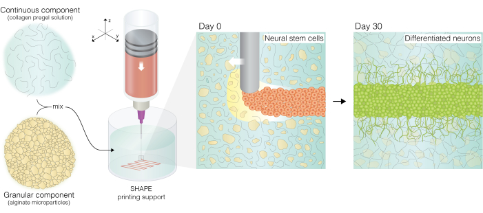
Figure 1: Conceptual illustration of the SHAPE embedded printing approach. A collagen solution is mixed with alginate microparticles to form the SHAPE composite; the SHAPE composite is used as a support material for the embedded 3D printing of hNSCs, which are differentiated into neurons inside the annealed support. The biofunctional properties of the SHAPE composite allow the neurons to extend projections and populate the empty part of the support with their axonal projections. This figure has been modified from Kajtez et al.13. Abbreviations: SHAPE = self-healing annealable particle-extracellular matrix; hNSCs = human neural stem cells. Please click here to view a larger version of this figure.
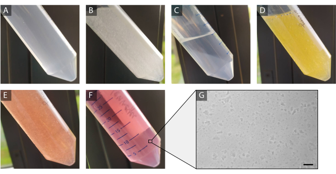
Figure 2: Preparation of alginate microparticles. (A) The alginate solution after gelation overnight. (B) The alginate microparticles generated by homogenization. (C) The particle pellet after centrifugation. (D) The pellet resuspended in DMEM (E) before the pH adjustment and after the pH adjustment. (F) The microparticles after incubation in medium overnight and centrifugation. (G) A brightfield image of the fabricated alginate microparticles. Scale bar = 100 µm. Please click here to view a larger version of this figure.
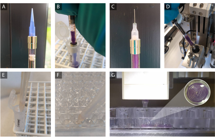
Figure 3: Preparation for the 3D printing process. (A) A slurry plug (~100 µL) is loaded into the syringe followed by (B) the loading of the cellular bioink (here supplemented with colored beads for visualization purposes). (C) The conical plastic tip (21 G) used for ink loading is replaced with a 27 G blunt metal needle tip. (D) The syringe is inserted into the 3D printing head. (E,F) The SHAPE composite is pipetted into a well of a 48-well plate. The tube with the SHAPE composite is kept on ice when not being handled. (G) The printing needle tip is inserted into the SHAPE support, and the printing of the path defined by the computer design is started (here, the printing of a spiral is depicted). Abbreviation: SHAPE = self-healing annealable particle-extracellular matrix. Please click here to view a larger version of this figure.
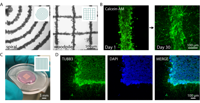
Figure 4: 3D-printed neural constructs inside the SHAPE composite support. (A) Brightfield images of printed hNSCs inside the support hydrogel. Spiral (left) and woodpile (right) constructs designs are displayed. (B) Live-cell imaging of a 3D printed construct the day after printing (left) and after neuronal differentiation (right). (C) A 3D-printed square construct removed from a culturing well with a spatula displays structural integrity. (D) Fluorescence confocal images of the same square construct immunolabelled for a neuronal marker (TUBB3) and with counterstained nuclei (DAPI) confirming the successful differentiation of the hNSCs within the 3D-printed constructs. Scale bars = 500 µm (A, right); 100 µm (B,D). Panels C and D in this figure have been modified from Kajtez et al.13. Please click here to view a larger version of this figure.
The SHAPE composite material approach provides a versatile route for the formulation of annealable and biofunctional support baths for the embedded 3D printing of cellular inks. While this protocol provides an example of the 3D printing of neural constructs, the SHAPE toolbox could easily be adapted to biofabrication with other cell sources for the precise engineering of a range of target tissue types. The printing approach would also allow for the precise patterning of multiple cell types to study their interaction or to engineer tissues with a defined spatial arrangement of the cellular compartments (e.g., neurons and glial cells). In contrast to the traditional granular gels, the SHAPE composite contains an expanded interstitial space (~30% volume fraction for the formulation presented in this protocol). The granular component serves as a rheological modifier that provides the composite with favorable material properties for high-resolution embedded printing. This opens up a route toward a rational design of the cellular microenvironment by altering the formulation of the continuous component while keeping the same granular component. For example, other functional ECM molecules could be introduced to the support bath (e.g., hyaluronic acid, laminins, fibronectin), or different crosslinking mechanisms could be leveraged (e.g., enzymatic, light-based)13. Furthermore, the alginate in the granular component could be replaced with microparticles from different hydrogel materials (e.g., gelatin8, polyethylene glycol14,15, agarose16) or be made in different sizes and shapes in line with the needs of different tissue engineering or disease modeling applications. The ratio between the granular and continuous phase can be tuned according to needs of individual 3D printing projects, but increasing the continuous phase beyond 30% might compromise the printing fidelity and resolution.
During the microgel production steps, it is critical that the stirring of the alginate solution after the addition of acetic acid is effective throughout the volume of the gelling solution. If the stirring speed is too low or the magnetic stirrer is too small, the stirring might not reach the upper layers of the solution, which will turn into a large volume of crosslinked bulk hydrogel, while the lower layers will be sheared. The homogenization of an inconsistently sheared alginate hydrogel will result in the generation of alginate microparticles that are suboptimal for 3D printing applications. Furthermore, the alginate microparticles need to be thoroughly mixed with the collagen solution, since a non-homogenous mixture will result in patches of the support material lacking collagen; these patches would not be annealed and, thus, would lack cell-interactive features. There could also be patches that lack alginate microparticles and would, therefore, not support printing. An inhomogeneously mixed printing support would, therefore, not be able to support high-fidelity printing and would be structurally compromised, as it would not be annealed throughout its volume. Bubbles should also be avoided, not because they could be harmful for the cells, but because air pockets could cause deformations during printing and interfere with the imaging of the constructs. Two common sources of bubbles are vortexing (microbubbles in the cold support that expand during the support annealing at 37 °C) and vigorous pipetting.
The SHAPE composite support in this work was not formulated as a sacrificial material to be removed post printing but rather as a long-term biofunctional support for both stem cell differentiation and neuronal growth and functional maturation. In comparison to granular gels that are not annealed post-printing, the structural stability and transparency of the annealed SHAPE composite material provide a protective environment for delicate neuronal features during the process of fixation and immunolabelling, and as such, this material facilitates morphological characterization via the visualization of antigens. Fluorescence reporters could also be used to track changes in cellular morphology over time, as well as to monitor cellular proliferation and migration within the annealed printing support. Furthermore, calcium imaging approaches could be used to provide information on spontaneous cellular activity (e.g., firing of action potentials in neurons or even synchronous neuronal network activity). However, chemical stimulation of the engineered cellular constructs (e.g., neuronal stimulation using KCl) might be difficult due to the hydrogel layer surrounding the cells, which slows down diffusion and prevents the instantaneous modulation of the cellular microenvironment. Optogenetic stimulation presents a better option for the control of cellular activity, as the SHAPE hydrogels do not obstruct optical access to the cells.
Oxygen-sensitive beads could be incorporated into the bioink or into the support material (via direct printing or dispersion during composite preparation) to allow for live spatial and temporal mapping of the oxygen tension levels inside and around the printed constructs with high sensitivity13. This noninvasive 3D oxygen mapping approach based on phosphorescence lifetime measurements provides a route toward engineering tissue constructs with improved oxygenation, and likely also improved nutrient supply. Poor oxygenation could lead to the formation of necrotic regions within the printed constructs, interfere with stem cell differentiation, and affect neuronal metabolism. Oxygen mapping provides a readout based on which the 3D printing design could be altered to facilitate uniform oxygenation throughout the construct, the fine-tuning of the oxygen levels to match physiological conditions, or the generation of oxygen gradients.
Engineered channels could also be incorporated inside the annealable printing support by printing a sacrificial ink, such as gelatin, that solidifies inside the cold support bath but can easily be evacuated at 37°C4,13. Channels would be required to supply nutrients and oxygen to tissue constructs with high cell density or dimensions that exceed the capabilities of a design-based oxygen tension manipulation approach. Additionally, vascular-like channels could be taken advantage of to create gradients of small molecules that drive the patterning of cellular identity, modulate cellular activity, or guide chemotaxis.
In summary, embedded 3D printing inside the SHAPE composite offers a modular material platform that is easily adaptable and has versatile potential for the functional modeling of mechanically sensitive tissues. The protocol presented here provides a detailed explanation of the necessary steps and basic principles needed to generate the support material and print the cellular ink with high fidelity. The approach takes advantage of affordable materials and accessible equipment while providing room for personalization of the approach to individual researchers' needs and applications.
The authors have nothing to disclose.
The research was primarily funded by the BrainMatTrain European Union Horizon 2020 Programme (No. H2020-MSCA-ITN-2015) under the Marie Skłodowska- Curie Initial Training Network and Grant Agreement No. 676408. C.R. and J.U.L. would like to gratefully acknowledge the Lundbeck Foundation (R250-2017-1425) and the Independent Research Fund Denmark (8048-00050) for their support. We gratefully acknowledge funding for the HORIZON-EIC-2021-PATHFINDEROPEN-01 Project 101047177 OpenMIND.
1 mL Gastight Syringe 1001 TLL | Hamilton | 81320 | |
3DDiscovery 3D bioprinter | RegenHU | ||
Acetic acid | Sigma-Aldrich | A6283 | |
AlbuMAX | ThermoFisher | 11020021 | |
Alexa Fluor 488 secondary antibody | ThermoFisher | A-11001 | Goat anti-Mouse |
Blunt Needle, Sterican (21 G) | Braun | 9180109 | |
Blunt Needle (27 G) | Cellink | NZ5270505001 | |
BioCAD software | SolidWorks | ||
Calcein AM | ThermoFisher | 65-0853-39 | |
Calcium carbonate | Sigma-Aldrich | C5929 | |
Dibutyryl-cAMP sodium salt | Sigma-Aldrich | D0627 | |
Cultrex Rat Collagen I (5 mg/mL) | R&D Systems | 3440-100-01 | |
DAPI | ThermoFisher | 62248 | |
DMEM/F-12, GlutaMAX | ThermoFisher | 10565018 | |
Donkey serum | Sigma-Aldrich | D9663 | |
DPBS | ThermoFisher | 14190094 | |
EGF | R&D Systems | 236-EG | |
FGF | R&D Systems | 3718-FB | |
Formaldehyde solution 4%, buffered, pH 6.9 | Sigma-Aldrich | 100496 | |
GDNF | R&D Systems | 212-GD | |
Geltrex | ThermoFisher | A1569601 | |
Glucose | Sigma-Aldrich | G7021 | |
HEPES Buffer (1 M) | ThermoFisher | 15630080 | |
L-Alanine | Sigma-Aldrich | 5129 | |
L-Asparagine monohydrate | Sigma-Aldrich | A4284 | |
L-Aspartic acid | Sigma-Aldrich | A9256 | |
L-Glutamic acid | Sigma-Aldrich | G1251 | |
L-Proline | Sigma-Aldrich | P0380 | |
Magnetic stirrer RET basic | IKA | 3622000 | |
N-2 Supplement | ThermoFisher | 17502048 | |
Penicillin-Streptomycin | ThermoFisher | 15140122 | |
S25N-10G dispersing tool | IKA | 4447100 | |
Sodium Alginate (80-120 cP) | FUJIFILM Wako | 194-13321 | |
Sodium azide | Sigma-Aldrich | S2002 | |
Sodium bicarbonate | Sigma-Aldrich | S5761 | |
Sodium hydroxide | Sigma-Aldrich | S5881 | |
T18 Digital ULTRA-TURAX homogenizer | IKA | 3720000 | |
Triton X-100 | Sigma-Aldrich | X100 | |
Trypsin/EDTA Solution | ThermoFisher | R001100 | |
TUBB3 antibody | BioLegend | 801213 | Mouse |
Xanthan gum | Sigma-Aldrich | G1253 |