A protocol is presented to evaluate whether small EVs (sEVs) isolated from placental explants cultured under hypoxic conditions (modeling one aspect of preeclampsia) disrupt the blood-brain barrier in nonpregnant adult female mice.
Cerebrovascular complications, including cerebral edema and ischemic and hemorrhagic stroke, constitute the leading cause of maternal mortality associated with preeclampsia. The underlying mechanisms of these cerebrovascular complications remain unclear. However, they are linked to placental dysfunction and blood-brain barrier (BBB) disruption. Nevertheless, the connection between these two distant organs is still being determined. Increasing evidence suggests that the placenta releases signaling molecules, including extracellular vesicles, into maternal circulation. Extracellular vesicles are categorized according to their size, with small extracellular vesicles (sEVs smaller than 200 nm in diameter) considered critical signaling particles in both physiological and pathological conditions. In preeclampsia, there is an increased number of circulating sEVs in maternal circulation, the signaling function of which is not well understood. Placental sEVs released in preeclampsia or from normal pregnancy placentas exposed to hypoxia induce brain endothelial dysfunction and disruption of the BBB. In this protocol, we assess whether sEVs isolated from placental explants cultured under hypoxic conditions (modeling one aspect of preeclampsia) disrupt the BBB in vivo.
Approximately 70% of maternal deaths due to preeclampsia, a hypertensive pregnancy syndrome characterized by impaired placentation processes, maternal systemic endothelial dysfunction, and, in severe cases, multi-organ failure1,2, are associated with acute cerebrovascular complications3,4. Most maternal deaths occur in low and middle-income countries5. However, the underlying mechanisms are still unclear despite the clinical and epidemiological relevance of cerebrovascular complications associated with preeclampsia.
On the other hand, extracellular vesicles (EVs) (diameter ~30-400 nm) are essential mediators of intercellular communication among tissues and organs, including maternal-placental interaction6. In addition to proteins and lipids on the external surface, EVs carry cargo within (proteins, RNA, and lipids). EVs can be categorized into (1) exosomes (diameter ~50-150 nm, also named small EVs (sEVs)), (2) medium/large EVs, and (3) apoptotic bodies, which differ by size, biogenesis, content, and potential signaling function. The composition of EVs is determined by the cells from which they originate and the disease type7. Syncytiotrophoblast-derived EVs express placental alkaline phosphatase (PLAP)8,9, which detects placentae-derived circulating small EVs (PDsEVs) in pregnancy. Also, PLAP helps discern changes in the PDsEVs cargo and their effects in preeclampsia versus normotensive pregnancies10,11,12,13,14,15.
The placenta has been recognized as the necessary component in the pathophysiology of preeclampsia16 or cerebral complications associated with this disease17,18,19. However, how this distant organ might induce alterations in brain circulation is unknown. Since sEVs play pivotal roles in cell-to-cell communication due to their capacity to transfer bioactive components from donor to recipient cells6,20,21, a growing number of studies have associated placental sEVs with the generation of maternal endothelial dysfunction21,22,23,24, including brain endothelial cells25,26in women with preeclampsia. Thus, the compromise of brain endothelial function may lead to disruption of the blood-brain barrier (BBB), a critical component in cerebrovascular complications associated with preeclampsia3,27.
Nevertheless, preclinical findings using rat cerebral vessels exposed to serum of women with preeclampsia28 or human brain endothelial cells exposed to plasma of women with preeclampsia29 reported that circulating factor(s) induce disruption of the BBB. Despite several candidates with the potential to harm the BBB present in the maternal circulation during preeclampsia, such as elevated levels of proinflammatory cytokines (i.e., tumor necrosis factor)18,28 or vascular regulators (i.e., vascular endothelial growth factor (VEGF))29,30,31, or oxidative molecules such as oxidized-lipoproteins (oxo-LDL)32,33, among others34, none of them establishes a direct connection between the placenta and the BBB. Recently, sEVs isolated from hypoxic placentas have shown the capacity to disrupt the BBB in nonpregnant female mice25. Since placental sEVs may carry most of the listed circulating factors with the capacity to disrupt the BBB, sEVs are considered suitable candidates to connect the injured placenta, be the carrier of harmful circulating factors, and disrupt the BBB in preeclampsia.
This protocol allows us to investigate whether sEVs isolated from placental explants cultured under hypoxic conditions can disrupt the BBB in nonpregnant female mice as a proxy for understanding the pathophysiology of cerebral complications during preeclampsia.
The research was carried out following the principles expressed in the Declaration of Helsinki and under the authorization of the respective Ethical Review Boards. All human participants gave their informed consent before sample collection, as reported previously25. Additionally, the Bioethics and Biosafety Committee of the Bío-Bío University approved this project (Fondecyt grant 1200250). The animal work was conducted in accordance with the cardinal principles of the three R's in the use of animals in experimentation35, and according to the recommendations of the guidelines for the Care and Use of Laboratory Animals published by the US National Institute of Health. Animals were kept in appropriate environments at the Vivarium of the University of Bío-Bío. Fresh placentas (n = 4) were obtained within 1 h after elective cesarean section from mothers (ages 28-31 years old) with normal pregnancies at term (38 to 41 weeks of gestation). Cesarean sections were performed at the Herminda Martin Clinical Hospital, Chillan, Chile, as previously reported25. To apply the in vivo model, 4-6-month-old female nonpregnant mice (strain C57BLACK/6) were used. They were divided into three experimental groups: (1) control (without treatment), (2) treated with sEVs from normoxia (sEVs-Nor), and (3) treated with sEVs from hypoxic cultured placentas (sEVs-Hyp), which were used to evaluate the disruption of the BBB in vivo25. All the injected solutions were sterile. Also, the preparation of sEVs was performed in aseptic conditions and under a class II biosafety hood to avoid contamination.
1. Placental culture explants
2. Placental-derived sEVs isolation
3. Mice injection
4. Rapid murine coma and behavior scale (RMCBS)
5. Analysis of Evan's blue extravasation
This protocol evaluates the capacity of sEVs derived from placentas cultured in hypoxia to disrupt the BBB in nonpregnant mice. This method allows one to understand better the potential connection between the placenta and the brain in normal and pathological conditions. In particular, this method may constitute a proxy to analyze placental sEVs participation in the onset of cerebral complications in preeclampsia.
Contrary to mice injected with sEVs-Nor, mice injected with sEVs-Hyp show a progressive decline in the neurological score until 24 h (Table 1), which suggests the sEVs-Hyp capacity to impair brain function.
Also, mice brains of the sEVs-Hyp injected group have higher fresh weight than those isolated from mice injected with sEVs-Nor or control mice (0.51 ± 0.008; 0.46 ± 0.008; 0.47 ± 0.01 g, respectively), which may constitute a gross indicator of brain edema45.
Compatible with this finding, this protocol allows one to identify Evan's blue extravasation as an indicator of disruption of the BBB. In that regard, brains from mice injected with sEVs-Hyp have higher Evan's blue extravasation than brains from the sEVs-Nor group (Figure 5A).
Although the underlying mechanism of disruption of the BBB induced by sEVs-Hyp was not analyzed with this protocol, results also indicate that sEVs-Hyp injected mice showed reduced protein amounts of CLND-5 in the areas in which the BBB was most affected (i.e., posterior areas) (Figure 5B). Therefore, it is feasible that sEVs-hyp impairs the expression of the function of this critical endothelial tight junction protein.
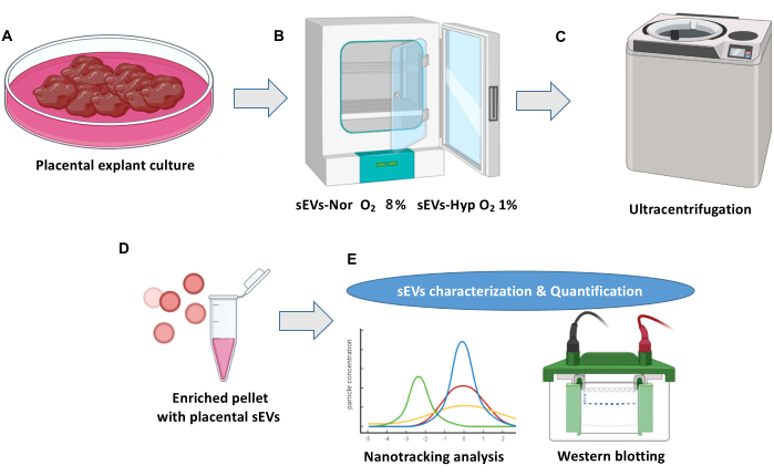
Figure 1: Placental explant culture and extracellular vesicle isolation protocol. (A) Normal placenta explant cultures. (B) Explants are distributed into two conditions for the biogenesis of placental small extracellular vesicles (sEVs). Normoxia (sEVs-Nor, 8% O2) or hypoxia (sEVs-Hyp, 1% O2) for 18 h. (C) Conditioned media is harvested, filtered, and centrifuged to eliminate cell debris. (D) sEVs are isolated by ultra-centrifugations. (E) sEVs are characterized using nano-tracking analysis and western blot. Please click here to view a larger version of this figure.
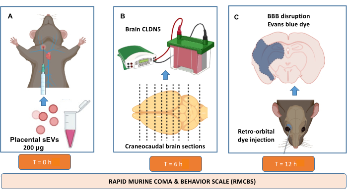
Figure 2: In vivo evaluation of blood-brain barrier disruption protocol. Nonpregnant C57BL6/J mice, 4-6 months old, are used. (A) Via external jugular vein, animals received sEVs (200 μg of total protein) isolated from Normoxic (sEVs-Nor, 8% O2) or hypoxic (sEVs-Hyp, 1% O2) placental cultures. RMCBS is monitored at 0-24 h after injection. (B) 6 h after sEVs injection, brains are extracted and sectioned into nine segments for protein extraction. Claudin 5 (CLDN5) is analyzed in homogenates of those nine sections. (C) Evan's blue extravasation analysis (24 h after sEVs injection) was analyzed after retro-orbital puncture injection in each of the nine segments. Please click here to view a larger version of this figure.
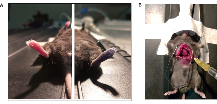
Figure 3: Photo documentaries of Evan' s blue dye and intracardial perfusion protocol. (A) Mouse received Evan's blue via retro-orbital injection. (Left) Animal before and (Right) after injection (15 s) of Evan's blue. (B) Thoracotomy to perform intracardial perfusion of phosphate buffer solution (1x PBS) and paraformaldehyde (4% PFA). The left ventricle is pointed with the tip of the needle. Please click here to view a larger version of this figure.
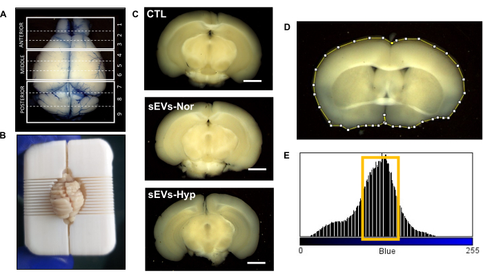
Figure 4: Analysis of Evan's blue extravasation after injection of sEVs. (A) Representative image of the whole brain showing Evan's blue extravasation. The dashed line represents nine sections obtained from the whole brain. (B) Brain dissected using a brain mice slicer. (C) Representative images of brain slices at 24 h after sEVs injection. Control (CTL), placenta in normoxic (sEVs-Nor) or hypoxic conditions (sEVs-Hyp). Scale bar = 0.4 cm. (D) Digital outlining of brain slice using ImageJ. (E) Histogram in the blue channel. Values between 75-110 are associated with Evan's blue extravasation. Please click here to view a larger version of this figure.
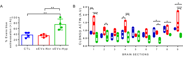
Figure 5: Evan's blue extravasation and claudin-5 levels in mice injected with sEVs isolated from placental explants. (A) Percentage of Evan's blue (EB) extravasation considering the whole brain sections. Control (CTL, blue), placenta in normoxic (sEVs-Nor, red) or hypoxic conditions (sEVs-Hyp, green). (B) Relative levels of claudin 5 (CLDN5) in the nine brain sections were obtained from the three experimental groups. β-actin is used as a loading control. Values are mean ± interquartile range. Each dot represents an individual experimental subject. *p < 0.05, **p < 0.005. ****p < 0.0001. ***p < 0.001; ANOVA test followed by Bonferroni post-test. Please click here to view a larger version of this figure.
Time (h) | Control | sEVs-Normoxia | sEVs-Hypoxia | ANOVA |
0 | 18.75 ± 0.250 | 18.5 ± 0.288 | 18.75 ± 0.250 | ns |
3 | 18.5 ± 0.866 | 17 ± 0.707 | 13.25 ± 1.750*α | 0.006 |
6 | 19.25 ± 0.750 | 17 ± 0.577 | 11.75 ± 1.250*α | 0.002 |
12 | 18.5 ± 0.645 | 16.75 ± 1.109 | 13 ± 0.816*α | 0.001 |
24 | 19.5 ± 0.288 | 17.75 ± 0.250 | 10.25 ± 0.853*α | <0.0001 |
*p < 0.01 versus control. αp < 0.01 versus sEVs-Normoxia |
Table 1: Rapid murine coma and behavior scale (RMCBS) after 24 h post sEVs injection. The score is expressed as mean ± SEM. Animals' scores closest to 20 are standard, while the lower the score, the higher the dysfunction of the CNS.
This study unveils fresh insights into potential harm resulting from sEVs isolated from placental explants cultured in hypoxic conditions on the disruption of the rodent blood-brain barrier. The pathological mechanism involves a reduction in CLND-5 in the posterior brain region25.
Prior investigations have revealed that plasma-sEVs from individuals with preeclampsia induce endothelial dysfunction in various organs using in vitro models46,47. This investigation particularly scrutinized the blood-brain barrier, offering a novel perspective on sEVs isolated from hypoxia-cultured placenta, which disrupts this vital barrier. These discoveries introduce a new realm of research wherein sEVs may serve as a communication channel between the placenta and the brain in both normal and pathological scenarios, like preeclampsia.
The presented protocol is straightforward, but several critical steps warrant mention. This protocol necessitates fresh placentas within 1 h post-delivery. We also advise against extending the culture period beyond 24 h to prevent tissue degradation. This protocol mitigates potential contamination from sEVs produced by mouse placentas. Digital analysis of Evan’s blue extravasation is time-intensive. Furthermore, Evan’s blue staining is less conspicuous after brain sectioning. Therefore, an initial setup for identifying the blue range using a positive control is recommended. A negative control, such as a brain from a mouse not subjected to Evan’s blue injection, can also be employed. Blind analysis of experimental groups is imperative to avert potential observer bias.
Several challenges may arise during this protocol. A significant limitation lies in biological variability originating from both human placentas and injected mice. To ensure the reproducibility of Evan’s blue extravasation experiments, establishing sEV doses isolated from human placentas is suggested. We opted for a dose of 200 µg of total protein; however, this quantity may fluctuate based on vesicle purity, the efficacy of jugular injection, Evan’s blue administration, its clearance from the circulatory system, and, not least, the biological impact of sEVs considering their contents.
The cellular mechanisms by which sEVs from the human placenta can disrupt the blood-brain barrier necessitate additional experimentation. Nevertheless, this method holds relevance, hinting at potential communication between the placenta and the brain, warranting further exploration. Therefore, future investigations are encouraged, concentrating on sEVs and their interactions with the blood-brain barrier, as well as the impact of their cargo on neuronal tissues. Whether blood-brain barrier impairment leads to enduring consequences for the maternal brain warrants further examination.
The authors have nothing to disclose.
The authors would like to thank the researchers belonging to GRIVAS Health for their valuable input. Also, midwives and clinical staff from the Obstetrics and Gynecology Service belong to the Hospital de Chillan, Chile. Founded by Fondecyt Regular 1200250.
Adult mice brain slecer matrice 3D printed | Open access file | Adult mice | Adult mice brain slicer. Printed in PLA filament. |
Anti β-Actin primary antibody | Sigma-Aldrich | Clon AC-74 | Antibody for loading control (Western blot) |
Anti-Claudin5 primary antibody | Santa cruz Biotechnology | sc-374221 | Primary antibody for tight junction protein CLDN5 of mice BBB (Western blot) |
BCA protein kit | Thermo Scientific | 23225 | Kit for measuring protein concentration |
Culture media #200 500 mL | Thermo Fisher Scientific | m200500 | Culture media for placental explants |
D180 CO2 incubator | RWD Life science | D180 | Standard incubator to estabilize explants and culture sEVs-Nor |
Evans blue dye > 75% 10 g | Sigma-Aldrich | E2129.10G | Dye to analize blood brain barrier disruption IN VIVO |
Fetal bovine serum 500 mL | Thermo Fisher Scientific | 16000044 | Additive growth factor for culture media 200 |
Himac Ultracentrifuge CP100NX | Himac eppendorf group | 5720410101 | Ultracentrifuge for condicioned media > 1,20,000 x g |
ImageJ software | NIH | https://imagej.nih.gov/ij/download.html | |
Isoflurane x 100 mL | USP Baxter | 212-094 | Volatile inhalated anaesthesia agent for mice |
Kit CellTiter 96 Non-radioactive | Promega | 0000105232 | In vitro assay for placental explants viability |
Mouse IgG Secondary antibody | Thermo Fisher Scientific | MO 63103 | Secondary antibody for CLDN5 (western blot) |
NanoSight NS300 | Malvern Panalytical | 90278090 | Nanotracking analysis of particles from placental explants condicioned media |
Paraformaldehide E 97% solution 500 mL | Thermo Fisher Scientific | A11313.22 | Fixative solution for brain tissue slices and intracardial perfusion (once diluted) |
PBS 1 X pH 7.4 500 mL | Thermo Fisher Scientific | 10010023 | Wash solution for placenta explants |
Peniciline-streptomicine 100x 20 mL | Thermo Fisher Scientific | 10378016 | Antiobiotics for placental explants culture media |
ProOX C21 Cytocentric O2 and CO2 Subchamber Controller | BioSpherix | SCR_021131 | CO2 regulator to induce Hypoxia in sealed chamber for sEVs-Hyp |
Sodium Thiopental 1 g | Chemie | 7061 | humanitarian euthanasia agent |
Somnosuite low flow anesthesia system | Kent Scientifics | SS-01 | Isoflurane vaporizer for small rodents |
Surgical Warming platform | Kent Scientifics | A41166 | Warming platform for mainteinance anesthesia in mice |
Syringe Filters, Polytetrafluoroethylene (PTFE), Hydrophobic, 0.22 µm, Sterile, 25 mm | Southern labware | 10026 | Filtration of condicioned media harvested from placental explants |
Tabletop High-Speed Micro Centrifuges HITACHI himac CT15E/CT15RE | Hitachi medical systems | 6020 | Serial centrifugations of condicioned media < 1,20, 000 x g |
Trinocular stereomicroscope transmided and reflective light 10x-160x | Center Medical | 2597 | Stereomicroscope to register brain slices |