Visualisation des micro-organismes du sol grâce aux lames de contact et à la microscopie
English
소셜에 공유하기
개요
Source : Laboratoires du Dr Ian poivre et Dr Charles Gerba – Université de l’Arizona
Auteur mettant en évidence : Bradley Schmitz
Sol compose de la mince couche à la surface de la terre, contenant les facteurs biotiques et abiotiques qui contribuent à la vie. La partie non biologique comprend des particules inorganiques variant en taille et en forme qui déterminent la texture du sol. La portion biotique incorpore les résidus végétaux, racines, matières organiques et micro-organismes. Diversité et l’abondance des microbes du sol est large, car un seul gramme de sol contient 107-8 bactéries, actinomycètes 106-8 , 105-6 champignons, 103 levure, 10 protozoaires de4-6 , 10 algues3-4 et 53 nématodes. Les facteurs biotiques et abiotiques forment ensemble des architectures autour des racines des plantes, appelées la rhizosphère, qui offrent des conditions favorables pour les micro-organismes du sol.
Les facteurs biotiques et abiotiques promouvoir la vie dans les sols. Toutefois, elles contribuent aussi stressante dynamique qui limitent les microbes. Les stress biotiques implique une concurrence entre vie à adapter et à survivre dans des conditions environnementales. Par exemple, les microbes peuvent sécréter des substances toxiques ou inhibitrices pour nuire voisine de micro-organismes. Penicillium notatum est un champignon notoire, car elle réduit la compétition pour les nutriments en produisant un antimicrobien, lesquelles les humains récoltent pour créer la pénicilline pharmaceutique. Aux stress abiotiques proviennent des propriétés physiques ou chimiques limitant la survie microbienne, comme la lumière, humidité, température, pH, éléments nutritifs et texture.
Principles
Procedure
Results
Fungi display thick, filamentous hyphae (Figure 2). Actinomycetes display thin, filamentous hyphae. Bacteria display small cocci or rod shapes. They’re often found in clumps, on soil particles, or lining fungal hyphae. Soil particles display irregular, dark shapes (Figure 3).
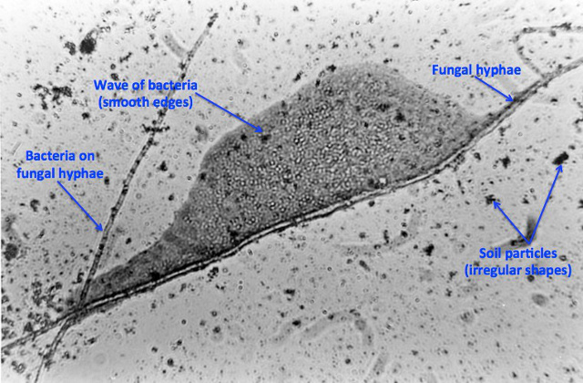
Figure 2. Contact slide image using 100X objective lens.
Photo courtesy W.H. Fuller.
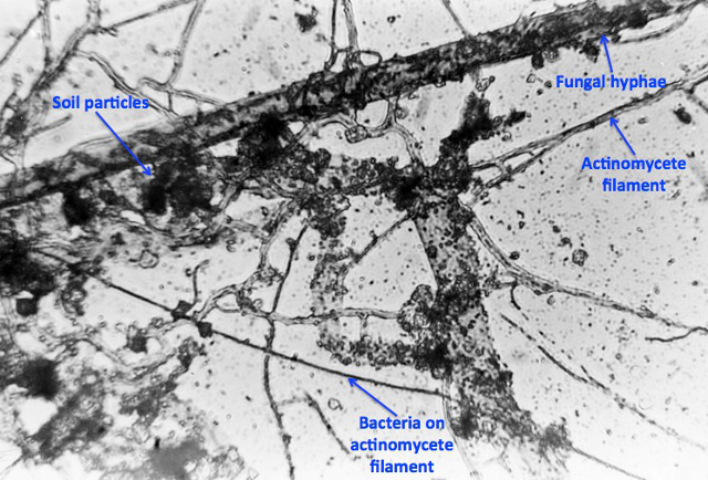
Figure 3. Contact slide image using 100X objective lens.
Photo courtesy W.H. Fuller.
Applications and Summary
The contact slide assay, also referred to as the buried-slide, is a simple technique utilized to qualitatively observe soil biota. This assay qualitatively shows the spatial interactions between fungal hyphae, actinomycete filaments, bacteria, and soil particles. Individuals or industry can employ this assay to gather knowledge on a particular soil’s health in regards to agriculture, gardening, composting, teaching, and studying. However, this technique does not quantify soil micro biota, as it only encompasses a small portrait of a larger heterogeneous environment.
Soil organism relationships can be observed by performing the contact slide assay and viewing the results through 100X oil immersion microscopy (Figures 2 and 3). The simplicity and ease to performing this assay makes it a great starting technique for those who have never been exposed to microbiology and may be viewing microorganisms through a microscope for the first time.
References
- Pepper, I. L., & Gerba, C P. 'Contact Slide Assay.' Environmental Microbiology A Laboratory Manual. 2nd ed. Elsevier 19-25 (2004).
- Pepper, I. L., Gerba, C. P., & Gentry, T. J. 'Earth Environments.' Environmental Microbiology. 3rd ed. Elsevier 59-88 (2014).
- Rossi, G., Ricardo, S., Gesue, G., Stanganelli, M., and Want, T.K. Direct Microscopic and bacteriological investigations of the soil. Soil Science. 41, 52 – 66 (1936).
내레이션 대본
The relationships between the various organisms and inorganic components in soil are vital to understanding soil changes and environmental stresses, but cannot be elucidated without direct visualization.
Soil, an extremely complex system, is a habitat for millions of diverse organisms. The region of soil directly around plant roots in particular, called the rhizosphere, contains a unique array of organisms that are directly influenced by the plant roots.
The abiotic, or non-biological, component of the rhizosphere includes inorganic particles ranging in size and shape that contribute to the soil’s texture. The biotic, or biological, portion includes plant residues, roots, organic matter, and microorganisms.
This video will demonstrate the direct visualization of the biotic and abiotic components of rhizosphere soil, in order to understand factors affecting soil changes and to predict environmental stresses.
Microscopic organisms tend to reside in the water located within soil pores. Bacteria are among the simplest and most plentiful organisms present in soil, and are found in many morphologies including spheres called cocci, rods called bacilli, and filamentous forms.
Fungal species, such as yeast and molds, are the second most abundant organisms in soil. They work to decompose and recycle dead organic matter. Microscopic filamentous fungi visually differ from other microorganisms, as they possess long and branched hyphae that release spores.
Direct observation of the relationships between these organisms is challenging, but can be achieved using a contact slide assay. This method is performed by submerging a glass slide into soil for several days and allowing the organisms and soil particles to adsorb to the slide surface.
The slide is then removed at an angle to prevent smearing of the surface. The microbes are fixed with acetic acid, and stained with Rose Bengal stain to enable visualization via light microscopy.
Now that you understand the principles behind the contact slide assay technique, lets take a look at the process in the laboratory.
First, collect surface garden soil and transfer the soil into the lab. Weigh 150 g of soil into the 2 separate containers. One container should be labeled as the treatment sample, which will be modified with nutrients to encourage rapid proliferation of organisms. Label the other as the control, which will be unchanged.
Calculate the water content in the soil, using the technique shown in this collection’s Determination of Moisture Content in Soil video. Based on this calculation, determine the amount of water in the soil on a dry weight basis. Now calculate the amount of water that needs to be added to give a 15% soil moisture content. This brings the moisture to field capacity, optimal for microorganism growth.
Measure the calculated amount of distilled water using a graduated cylinder. Pour the calculated volume of water into each container. Based on the previously determined dry weight of the soil, calculate the amount of glucose needed to achieve a final soil glucose concentration of 1% by mass, using the dry weight basis. Weigh this amount of glucose and add it to the treatment container only.
Weigh 200 mg of ammonium nitrate, then add it to the treatment container only. The ammonium nitrate serves as the nitrogenous nutrient source for the soil microbes. Mix the soil, glucose, and ammonium nitrate mixture in the container.
Next, label 4 clean microscope slides: two as treatment, and two as control. Insert the two treatment slides into the treatment soil container. Leave a section of each slide exposed above the soil surface, and ensure that there is a gap between the two slides.
Insert the two control slides into the control soil container in the same way. Cover the cups with plastic wrap, and secure with a rubber band. Puncture the plastic wrap several times to allow air transfer, but still prevent excessive evaporation.
Finally, weigh both cups, record their weight, and incubate them in a designated area at room temperature for seven days.
After the seven-day incubation, calculate the soil moisture content by weighing the soil cups. Determine if weight has been lost due to water evaporation, and replace the water if needed.
Remove the plastic wrap from the container, and remove the two slides from the soil by pressing each slide to an inclined position, and withdrawing so that the upper face of the slide is undisturbed.
Gently tap the slides to remove large soil particles. Using a damp paper towel, clean the lower face of the slides. Allow them to dry at room temperature in a fume hood. Once dry, pick up a slide with forceps, and immerse it into acetic acid for 1 to 3 min.
Rinse the top of the slide with a gentle stream of distilled water to remove excess acid. Repeat these steps for all slides. Allow the slides to air dry.
Support the slide on a staining rack over a container to catch excess dye. Using a dropper, gently cover the surface of each slide with phenolic Rose Bengal dye. Allow the slides to stain for 5 to 10 min, taking care to add more dye as needed to keep the slides wet. Gently rinse the slides with water to remove excess stain, and allow the slides to dry at room temperature.
Examine the slides on a light microscope, using an oil immersion objective. The treated soil will have more soil microbes.
The spatial interactions between fungal and bacterial organisms in typical soil samples can easily be visualized. Soil particles display dark irregular shapes.
Fungal organisms display thick filamentous hyphae, while actinomycetes display thin filamentous hyphae.
Bacteria are found as small cocci or rod shapes, typically in clumps, on soil particles or lining fungal hyphae.
The direct isolation of organisms from soil is important to the understanding of soil and environment characteristics.
Entomopathogenic nematodes are microscopic round worms that parasitize insects. While they are not visualized in the contact slide assay, they can be isolated from collected soil samples, as shown in this example.
First, the nematodes were baited in the soil using insects identified from visual examination. Nematodes were isolated from the dead insect bait, by placing the dead insects in a moist and dark environment and allowing the nematodes to migrate out into the surrounding water. The nematodes were then collected from the water, and analyzed.
Filamentous fungi are vital to soil health due to their role in nutrient recycling. The isolation and observation of filamentous fungi from soil was conducted in this example.
Soil samples were diluted with water, and added to separate sterile Rose Bengal streptomycin agar plates. The streptomycin prevented bacterial growth, and enabled fungal growth. Fungal colonies were counted and mounted to a glass slide using adhesive tape. The fungi were then imaged using a light microscope.
Soil microorganisms naturally break down components in soil, such as dead plants and organisms. Biodegradation and colonization of biodegradable plastic films was examined, as shown in this example.
Fungi were isolated from plastic films buried in soil for several months. The fungi were then tested individually for growth on plastic films. Plastic films were then incubated with the selected fungal strain with no growth media, in order to observe direct degradation of the plastic by the fungi.
You’ve just watched JoVE’s introduction to the contact slide assay for qualitative imaging of soil microbes. You should now understand how to prepare the contact slide, and visualize soil microbes. Thanks for watching!


