Détection des micro-organismes environnementaux avec la réaction en chaîne par polymérase et l'électrophorèse sur gel
English
소셜에 공유하기
개요
Source : Laboratoires du Dr Ian poivre et Dr Charles Gerba – Université de l’Arizona
Auteur mettant en évidence : Bradley Schmitz
Réaction en chaîne par polymérase (PCR) est une technique utilisée pour détecter des microorganismes présents dans le sol, l’eau et les milieux atmosphériques. En amplifiant les sections spécifiques de l’ADN, la PCR peut faciliter la détection et l’identification des micro-organismes de la cible vers le bas pour l’espèce, la souche et sérotype/pathovar niveau. La technique peut également être utilisée pour caractériser des communautés entières de micro-organismes dans les échantillons.
La mise en culture de micro-organismes en laboratoire à l’aide de milieux de culture spécialisée est une technique établie de longue date et reste en usage pour la détection des micro-organismes dans les échantillons environnementaux. Beaucoup de microbes dans l’environnement naturel, vivant, maintenir de faibles niveaux d’activité métabolique et/ou le temps de doublement et est ainsi désignés comme viable mais non cultivable organismes (VBNC). L’utilisation de techniques axées sur la culture seules ne peut pas détecter ces microbes et, par conséquent, ne fournit pas une évaluation approfondie des populations microbiennes dans les échantillons. L’utilisation de la PCR permet la détection des microbes cultivables, les organismes VBNC, et ceux qui ne sont plus réceptive ou active, car l’amplification des séquences génétiques ne nécessite généralement pas de pré-enrichissement de micro-organismes présents dans les échantillons environnementaux. Cependant, PCR ne peut pas différencier les États susmentionnés de viabilité et d’activité. Lorsqu’il est combiné avec une ou plusieurs techniques basées sur la culture, la viabilité de certains sous-ensembles de micro-organismes peut-être encore être déterminée.
Principles
Procedure
Results
In Figure 1, the DNA ladder (lane 1) provides a reference for the size and approximate concentration for bands of the PCR products. The negative control (lane 2) does not contain any genetic material, while the positive control (lane 3) is amplified from templates known to contain the target DNA to indicate size and location of target bands. Samples 4, 6, 8, and 9 exhibit similar band pattern as the positive control, therefore indicating that these samples contain the target genetic material. It can be inferred that the organism is present in the environments from which these samples were obtained.
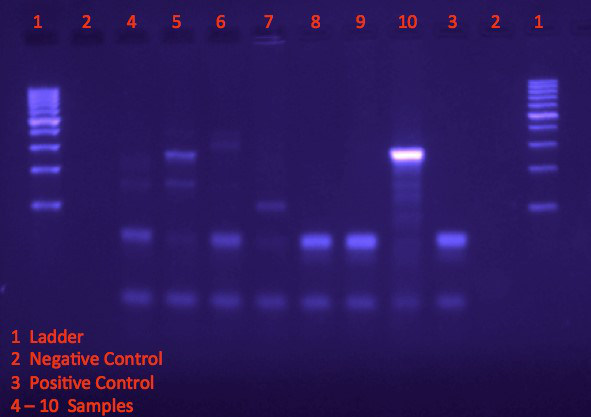
Figure 1. Visualizing bands on agarose gel following electrophoresis.
Applications and Summary
PCR can be employed to quickly determine the presence or absence of pathogens in the environment. For example, primers specific to the brain-eating amoeba, Naegleria fowleri, will amplify DNA and produce strong bands on a gel if the organism is present in a sample. If a single organism is not the main interest, but rather genes associated with toxin production from a variety of organisms, PCR can also be used to determine the presence or absence of these specific genetic materials.
PCR can also be used as a confirmation procedure when analyzing environmental microbes in lab. If a culture method cannot differentiate between certain organisms that are present in an environmental sample, then PCR maybe used to specifically distinguish between the candidate microbes.
Conventional PCR can be modified in several ways for particular experimental purposes. PCR can be used to analyze single-stranded RNA templates by coupling to a reverse transcription step (RT-PCR). Beyond a determination of presence versus absence, quantitative PCR (qPCR) can measure the concentration for specific DNA of interest.
내레이션 대본
The polymerase chain reaction, or PCR, is a fundamental biological technique that is widely applied to detecting and identifying microorganisms present in soil, water, and other environmental samples.
Classically, microorganisms are cultured in labs using specialized growth media. However, many microbes in the natural environment are “non-culturable” – either because they have very low metabolic activity or growth rate, or because they have very stringent growth requirements that may not be replicable in a culture dish. The differences in culturability among microbes also mean that, when microorganisms from an environmental sample are cultured, their relative abundance in culture might not reflect their actual levels in the environment.
The advent of PCR, which can specifically amplify even small amounts of DNA present in a mixed sample, makes it possible to quickly detect and identify specific microbes of interest, even ones that are non-culturable, within the complex assortment of organisms present in an environmental sample.
This video will introduce the principles of PCR. It will then discuss a general protocol for performing PCR on DNA isolated from an environmental sample in order to detect the presence of an organism of interest. Finally, several applications of PCR-based microbe identification will be explored.
The basic premise of PCR is to use repeated cycles of sequential temperature changes to achieve exponential amplification of DNA, usually with a machine known as a thermocycler to automatically cycle through the different temperatures. The DNA synthesis is carried out by DNA polymerase enzymes that are obtained from bacteria living in hot springs, such as Thermus aquaticus or “Taq”. These polymerases are heat stable, allowing them to withstand the high temperatures used during PCR.
The target sequence, known as the amplicon, is amplified from the DNA template using two short stretches of nucleotides known as “primers”. Because of the high specificity of complementary nucleic acid binding, the primers allow for the targeted amplification of very specific sequences of interest. By designing primers that will only amplify a unique sequence, or a unique combination of sequences, from an organism of interest, PCR can be used to differentially detect for the presence of that organism’s DNA among all the genetic materials present in a complex environmental sample.
Each PCR cycle is divided into three phases. The first, known as “denaturation”, is usually set above 92 °C and lasts about 30 s. Denaturation is used to break DNA molecules into single strands, to permit the amplification reaction to proceed.
The second phase, “annealing”, is set 2 to 3 °C below the lower of the melting temperature of the two primers, usually between 50 to 65 °C, and also lasts about 30 s. Melting temperature is the temperature at which 50% of the double-stranded DNA molecules have separated into single strands, and so the annealing step allows the primers to bind to their target sites in the DNA template.
The third phase of a PCR cycle is “elongation” or “extension”, when the DNA polymerase binds to the primer-template duplex and catalyzes synthesis of the new strands. Set at 72 °C for the most commonly used PCR polymerase, Taq, the duration of this phase depends on the length of the amplicon, usually 30 s per 500 basepairs. After each cycle, the amplified DNA is once again denatured and serves as a new template, leading to an exponential increase in the amount of PCR products.
Once the reaction is complete, the PCR products can be resolved by size on a “gel” usually made of the polymer agarose, a process known as electrophoresis. An electric field is applied across the gel, and the negative charges in the backbone of DNA molecules cause them to migrate towards the positive end of the field. Generally speaking, linear DNA molecules that are larger will take longer to travel through the gel matrix.
Now that you understand how PCR works, let’s take a look at how the reaction can be used to identify microorganisms in an environmental sample.
To begin, calculate the volume of each reagent needed based on the number of samples to be processed, plus an additional 10% to account for pipetting errors. A positive control template – which contains the target region – should be included to ensure that the reaction is working; as well as a negative control where no DNA template is included, in order to rule out contamination in any of the reaction components. Keep the Taq polymerase enzyme on ice, and thaw the rest of the reagents and the DNA samples at room temperature at a designated laminar flow hood to prevent contamination.
Once all the reagents have thawed, constitute the reagent “master mix” by adding the calculated volume of each reagent into a low-binding microfuge tube, which minimizes discrepancies in reagent amounts due to adsorption of molecules to the tube surface. Gently vortex and centrifuge each reagent before adding. Once the master mix is prepared, vortex to mix and collect by centrifugation.
Label an 8-tube PCR strip to designate one tube for each sample, including the controls. Dispense the appropriate amount of PCR master mix into each tube of the strip. Then, add each DNA sample to the respective tube.
Place the strip cap securely on the strip tube, and centrifuge briefly in a mini-centrifuge with a strip adaptor. Then, place the tube into the thermocycler, and run the reaction according to the appropriate PCR program.
While the PCR is being run, prepare an agarose gel for the electrophoresis of the PCR products. Weigh out an appropriate amount of agarose powder for a gel with a concentration that can resolve the PCR products based on their expected sizes. Add the agarose into a 125-mL flask, then add the appropriate volume of gel-running buffer into the flask, based on the volume of the gel cast, and swirl to mix. Microwave the agarose solution at high power for 1 min. When complete, verify the agarose has fully dissolved by swirling the flask, and repeat microwaving in 30-s increments if necessary.
Tightly secure the cap onto the flask, and cool the agarose solution to 50 °C by swirling the flask under running cold water. Once cooled, add 1 μL of ethidium bromide to the agarose. Because ethidium bromide is potentially carcinogenic, be sure to wear personal protective equipment such as goggles, a lab coat, and ethidium bromide resistant gloves.
Pour the agarose solution into an electrophoresis gel-casting tray, making sure that no air bubbles are trapped within the agarose. Place a comb with the required number of wells into the solution. Leave the gel at room temperature for 20 to 30 min to solidify. Once the gel is set, carefully remove the comb, making sure not to tear the gel in the process.
Place the solidified gel into the electrophoresis chamber. Add LB buffer into the chamber until the gel is just submerged. Onto a piece of Parafilm, pipette a “spot” of DNA ladder of a suitable range for the expected size of the PCR products. Retrieve the PCR tubes with the completed reactions from the thermocycler. Collect condensates in the PCR tubes by brief centrifugation, and add 8 μL of each sample onto the Parafilm. Add 2 μL of 10x loading dye into each spot of PCR product, so that the final concentration of the dye is 2x.
Load the samples and ladder into the designated wells in the agarose gel, being careful not to poke through the gel. Once loading is complete, put on the lid to the electrophoresis chamber, and connect the electrodes to the power supply. Since DNA is negatively charged and migrates towards the positive electrode, be sure the wells are on the side closer to the negative electrode. Turn on the power supply, and set it to a voltage appropriate for the size of the electrophoresis chamber and the buffer system being used. Set the electrophoresis to “run”. Small bubbles moving up the sides of the chamber will be observed if the electrophoresis is proceeding properly.
Once the dye front has advanced far enough down the gel, turn off the power supply. Carefully transport the gel to a gel imager to visualize the electrophoresed products. With a protective shield, turn on the UV light and visualize the DNA bands on the gel. Analyze the position of the bands to see if it matches the expected pattern that indicates the presence of the species of interest in the environmental sample.
Now that you have seen how PCR is performed, let’s look at various ways it is applied to detect microorganisms of interest in the environment.
One use of PCR-based environmental microbial detection is to identify disease-causing organisms such as the “brain-eating amoeba” Naegleria fowleri, a single-cell organism found in fresh water bodies and unchlorinated pools that can attack the human nervous system, often fatally. The presence of this deadly microbe in either water samples or the cerebrospinal fluid of suspected patients can be tested by performing PCR using primers that target unique DNA sequences in the amoeba’s genome.
Another application for PCR-based microbial identification is to test for the presence of pathogenic bacteria in flies caught in the vicinity of food establishments, as part of public health monitoring and disease outbreak investigations.
Here, investigators looked for the presence of pathogenic bacteria such as Salmonella and Listeria, by first isolating bacteria from both the body surface and the digestive canal of flies, and then using species-specific culture conditions to enrich for these species of interest. After extracting DNA from any bacteria that were cultured, commercially available species-specific detection PCR kits was used to test for their identity.
Finally, different strains of antibiotic-resistant pathogenic bacteria such as Staphylococcus aureus, which present major public health concerns, can be identified and differentiated with PCR.
In this example, researchers isolated and cultured S. aureus from clinical samples, then extracted DNA from the bacterial colonies and performed PCR. The amplification reactions here were “multiplexed”, meaning that multiple primer sets targeting different unique regions of the bacterial genome were combined into the same reaction. Individual primer sets were designed so that PCR products result from DNA of only some strains but not others, so that in combination, unique product band patterns were observed for each strain.
You’ve just watched JoVE’s video on PCR-based microorganism detection. We’ve looked at the principles behind polymerase chain reaction; a protocol for performing PCR on DNA extracted from environmental microorganisms; and finally, several specific applications of this technique to test for organisms of interest in different types of environmental or clinical samples. Thanks for watching!


