Visual attentional control refers to our deliberate state of choosing what to pay attention to.
If, for instance, the goal of an observer is to pick out all of the onions in his soup, then he may not notice the fly that is swirling about.
Even though both were spatially coincident, the item of focus—the onions—stood out because of the individual’s objective. This is an example of object-based attentional control.
Interestingly, the brain—and the visual cortex, in particular—can process the objects separately. But it’s the attended object that gains stronger activation in its associated specialized processing area.
Using functional magnetic resonance imaging, fMRI, and methods originally developed by Nancy Kanwisher and colleagues, this video demonstrates how to locate dedicated brain regions that process particular objects.
We will also investigate how attentional control modulates neural activity in the same regions using voxel-based analysis, and even discuss how mindfulness training can enhance the ability to control attention over time.
In this experiment, participants lie in an fMRI scanner and are shown images of faces and houses in two different phases: passive viewing and superimposed.
During the first phase, they are asked to simply observe images one at a time in a block design, that is, a number of faces are presented followed by a sequence of houses. This type of viewing serves to localize activity within specific regions of interest.
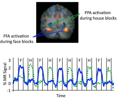
For example, the fusiform face area, the FFA, has been shown to be more active when individuals view faces compared to other common objects, whereas the parahippocampal place area, PPA for short, responds more strongly to houses and places rather than to faces.
Given that these regions respond to specific types of stimuli, the patterns of voxel-based activity—or areas representing some level of activation—are expected to change, depending on the images shown.
Such expectations set-up the second phase, where superimposed images of a face and a house are shown. Over several trials, participants are asked to pay attention to only one of the items at a time, and therefore, must switch their focus between either the house or the face.
In this case, the dependent variable is the amount of activation recorded across image conditions, which can be converted to the magnitude of signal change to observe variation in activation from baseline to face-focused blocks and those centered on the house.
Although both images are presented in a superimposed manner, it is predicted that patterns of activity in the participant’s FFA and PPA will change, based on the specific item they attended to. Such results would highlight object-based attentional control.
After recruiting participants for this study, greet them in the laboratory and verify that they meet the safety requirements as they complete the necessary consent forms. Please refer to another fMRI project in this collection for more details on how to prepare individuals to enter the scanning room and imaging bore.
With the participant now in the scanner, explain the task instructions: They must first passively view a number of images on the screen. During the second phase, text instructions will prompt them to pay attention to either the house or face when they appear superimposed.
Following these directions, begin the scanning protocol by first collecting a high-resolution anatomical scan.
Then, initiate the functional portion with two localizer runs, where participants passively view images in 30-second blocks. For instance, in the first segment, display faces, each for 750 ms, and a fixation cross in between, during an inter-stimulus-interval, or ISI, of 250 ms.
At the end of every block, present the fixation cross for 20 s before alternating the series of images, which should now be houses. Note that this sequence repeats with different images five times, for a total of 10 blocks within one run.
Next, proceed with eight functional runs of the attentional control task. During this phase, instruct participants which object to attend to via text onscreen, and then cycle a superimposed face and house every second, with each run containing 300 overlaid images.
To conclude the study, bring the participant out of the scanner and debrief them.
To preprocess the data, perform motion correction to reduce movement artifacts, temporal filtering to remove signal drifts, and spatial smoothing to increase signal-to-noise ratio.
Subsequently, create a general linear model based on what the expected hemodynamic response should be for each task condition, either faces or houses, in the localizer scan.
Generate a statistical map by fitting the data to this model, where the value at each voxel represents the extent to which it was involved in the task condition.
Based on the regions of interest, identify clusters for each subject with a minimum statistical threshold for each voxel that responded either to faces or houses.
Specifically, focus on the FFA, in the mid-fusiform gyrus, which responds significantly more to faces than to houses, as well as the PPA, which includes all voxels in the parahippocampal gyrus that responds more significantly to houses than to faces.
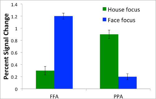
Then, quantify and graph the percentage of signal change for face- and house-focused conditions in the FFA and PPA for each subject.
During the localizer phase, notice that the bilateral FFA was more active when subjects viewed faces compared to houses. Conversely, the PPA was more active when subjects observed houses compared to faces.
Now, from the functional runs, use the same measure—percent signal change—plotted against the brain regions.
When the face was attended to, increased activity was found in the FFA, but not the PPA. Conversely, when the house was focused on, increased activity occurred in the PPA but not the FFA. These findings indicate that neural activity is modulated, depending on what item is being attended to.
Now that you are familiar with how to use functional neuroimaging to study object-based attentional control, let’s look how researchers study other types of attentional processing.
In addition to focusing on static visual images, researchers are also interested in how brain activity is modulated when individuals attend to moving objects—especially relevant for operating a motorized vehicle and avoiding accidents.
For example, if the driver is told to look out for movement—like a dog crossing the street—the motion itself will capture their attention; however, they may not remember other identifying details about the canine. After all, it’s more important to avoid tragedy than to remember fur color.
Another practice, mindfulness, incorporates key elements of attentional switching, by encouraging astute focus away from more stressful thoughts. While engaging in instructor-led meditation, individuals have been shown to enhance their ability to control attention, especially away from adverse views.
However, for individuals with anxiety disorders, including post-traumatic stress, attentional control is more difficult. That is, they are biased towards emotionally negative stimuli, like tragic events in the news, rather than neutral stories.
Such poor attentional control makes them more vulnerable to the effects of threatening images—perpetuating situations that they can’t seem to get off their mind.
You’ve just watched JoVE’s video on how attention modulates neural activity. Now you should have a good understanding of how to design and conduct an attentional control experiment using functional neuroimaging, and finally how to analyze and interpret specific patterns of brain activity related to object-based attention.
Thanks for watching!