光学材料学第1部分:样品制备
English
소셜에 공유하기
개요
资料来源:佐治亚州佐治亚州理工学院材料科学与工程学院费萨尔·阿拉姆吉尔,佐治亚州亚特兰大
固体材料微观结构的成像和成像的结构成分的分析称为材料学。定性信息,例如,材料中是否存在孔隙度,颗粒的大小和形状分布,或者微结构是否存在各向异性,都可以直接观察。然而,我们将在材料学系列的第 2 部分看到,统计方法允许我们定量地测量这些微观结构特征,并将分析从二维横截面转换为材料样品。
本演示将概述为光学显微镜制备固体材料样品所涉及的技术和过程。虽然材料学可以同时进行光学和电子显微镜,但本演示将侧重于专门用于光学显微镜的样品制备。但是,应当指出,为光学材料学准备的样品可用于扫描电子显微镜,并可执行最少的附加步骤(如果有的话)。
Principles
Procedure
Results
From the series of images in Figure 1, particularly from the etched sample (Figure 1e), one can observe that the powder pressing process by which this sample was made rendered the grains to have non-circular, elongated shapes, with non-isotropic grain orientation. There is a significant amount of porosity retained in the material through this processing. Part 2 of the Materialography series will explore the statistics of the grain anisotropy as well the porosity.
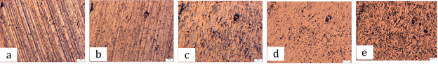
Figure 1: Polishing the sample with a) 600 grit, b) 1200 grit, polishing papers followed by c) 1 µm, d) 0.01 µm alumina suspensions on polishing cloths. Finally, e) etching for 20 seconds a nital solution revealed the porosity.
Applications and Summary
These are the standard methods for preparing cross sections of samples for microscopy. While the procedures detailed here are optimized to provide the best results in optical microscopy, some of the steps are unnecessary for scanning electron microscopy, and are inadequate for transmission electron microscopy. For the latter two, separate sample preparation procedures should be followed.
Materialographic sample preparation described here are the necessary first step towards the analysis of internal microstructure of three-dimensional materials using two dimensional information. For example, one might be interested in knowing how porous a membrane material is since that will affect its gas pearmenability. An analysis of the void structure of the 2D cross section will provide a strong indication of what the porosity is in the actual 3D structure (provided the sampling statistics are high). Another application would be in analyzing, for example the orientation of the polycrystalline grains in oil pipeline alloys. The orientational distribution function (ODF) can be directly related to the axial and transverse mechanical strength of the pipes, and so our sample preparation procedure is an important component of such an analysis.
내레이션 대본
Materialography is a method for microscopic structure imaging and analysis of solid materials. In particular materialography qualitatively studies the porosity in the material, the size and shape distribution of the grains, and the degree of isotropy of the microstructures.
Such detailed analysis requires specific sample preparation of solid materials. This video will illustrate the four major steps performed to prepare a sample four optical materialographic analysis.
Materialography is used to characterize solid materials. With this method, qualitative analysis, as well as quantitative analysis can be performed. In this video we will focus on the qualitative information obtained for a solid. In materialography the sample can either be probed with light, or with an electron beam. Depending on the choice of the probing tool, the sample needs to be prepared in different ways. We demonstrate here the principles of sample preparation for the optical materialography of solid materials of similar hardness to that of steel. This sample preparation is performed in four major steps, cutting, mounting, polishing, and etching. Let us look in detail at each of these steps.
The very first step is sample cutting. For samples with expected isotropic microstructures, meaning evenly distributed microstructures, the orientation of the cut is arbitrary, but for other cases, said as anisotropic samples, the cutting vector should be oriented according to specific directions or planes of the sample. In the second step the cutting sample is mounted on a support. The solid material is fixed to a hot compression thermosetting material like a resin or an epoxy to form a pressed pellet. The third step is sample polishing. It is performed in multiple subsequent steps, from coarse polishing to finer, and finer polishing. The idea is to reveal micro structural features while removing scratches left on the surface of the sample from the previous polishing sub step.
The sample is then ready for the last step that is etching. This is a chemical exposition of the sample to an acid. Some grain boundaries of the solid material have more atomic defects and are therefore more effected by the acid solution. This will have the effect of carving inside the mounted sample. Consequently, this step enhances the contrast between grains that is revealed by optical microscopy. Now that you understand the principles behind sample preparation for optical materialography, let’s see how the main steps of the procedure are performed in the laboratory.
The specimen used in this example is a metal nut. The sample preparation is demonstrated in four main steps as following: First use a linear precision saw to cut the sample normal to the hoop plane. Second, make sure the sample fits the die cavity of the press. Mount the sample in the cavity with the side to be imaged facing down on the mounting press. Then fill the remaining volume of the mounting press cavity with Bakelite.
Find the prescribed heat, pressure, and duration for Bakelite and press the sample accordingly. Note that other thermosetting mounting materials can be used for other types of samples. The third step is polishing of the sample. Start with a coarse 600 grit paper. Employ the rotating polishing wheels for two minutes at a speed of 120 rpm to polish the sample. Then use an optical microscope to check the scratches on the sample surface. Now rotate the sample by 90 degrees from it’s first polishing position and repeat the polishing with a 1,200 grit paper. Make sure to keep the pressure and direction of wheel motion constant.
Check the sample surface with the optical microscope. The previously identified scratches should be removed and new ones will be identified. Rotate again the sample by 90 degrees and polish the sample with finer polishing suspensions of one micrometer alumina particles and again verify with microscope the scratches on the sample surface. Repeat the sequence, this time with 0.05 micrometer alumina particles. At the final polishing step, using the highest magnification of the optical microscope.
There should be no observable scratches on the sample surface. The last step is the sample etching. First prepare a 2% Nital solution by mixing 2% volume concentrated nitric acid in ethanol. Dipped the polished face of the sample in the solution for about 20 seconds. Rinse the sample with ethanol, then observe the etched surface on the microscope. Repeat these etching, rinsing steps until sufficient contrast in the granular structure is observed.
Optical materialography is a very useful technique to characterize solid materials for various applications. For instance, toroidal inductor cores are commonly used in electronic applications to regulate electromagnetic interference. These cores are economically manufactured by compacting iron powder. Porosity and grain size of the core material both impact the electromagnetic properties of the inductor and they can be assessed by optical materialography.
Porus materials, due to their permeability, are used for manufacturing of synthetic membranes. Optical materialography is employed to analyze the void structure of the 2D cross section of the membrane material and in consequence to assess the porosity quality of the membrane.
You’ve just watched Jove’s introduction to sample preparation for optical materialography. You should now understand the four steps of sample preparation, cutting, mounting, polishing, and etching and how these are important for a qualitative analysis of material microstructures.
Thanks for watching.