JoVE 과학 교육
Microbiology
JoVE 비디오를 활용하시려면 도서관을 통한 기관 구독이 필요합니다.  전체 비디오를 보시려면 로그인하거나 무료 트라이얼을 시작하세요.
JoVE 과학 교육 Microbiology
Plaque Assay: A Method to Determine Viral Titer as Plaque Forming Units (PFU)
  • 00:01Concepts
  • 03:09Media Preparation
  • 05:12Preparing Agar Plates
  • 06:21Culturing Host Cells
  • 07:30Phage Serial Dilution and Preparation of Bacteria and Phage Overlay
  • 10:37Data Analysis and Results
  • 11:35Results

Ensayo de placa: Un método para determinar los títulos virales como unidades formadoras de placas (UFP)

English

소셜에 공유하기

개요

Fuente: Tilde Andersson1, Rolf Lood1
1 Departamento de Ciencias Clínicas Lund, División de Medicina de Infecciones, Centro Biomédico, Universidad de Lund, 221 00 Lund, Suecia

Los virus que infectan los organismos procaloicos, llamados bacteriófagos o simplemente fagos, fueron identificados a principiosdel siglo XX por Twort (1) y d’Hérelle (2) de forma independiente. Desde entonces, los fagos han sido ampliamente reconocidos por su valor terapéutico (3) y su influencia en los ecosistemas humanos (4), así como en los ecosistemas globales (5). Las preocupaciones actuales han impulsado un renovado interés en el uso de fagos como alternativa a los antibióticos modernos en el tratamiento de enfermedades infecciosas (6). Esencialmente toda la investigación de fagose se basa en la capacidad de purificar y cuantificar virus, también conocido como un titer viral. Descrito inicialmente en 1952, este fue el propósito del ensayo de placa (7). Décadas y múltiples avances tecnológicos más tarde, el ensayo de placa sigue siendo uno de los métodos más fiables para la determinación del titer viral (8).

Los bacteriófagos subsisten inyectando su material genético en las células huésped, secuestrando las máquinas para la producción de nuevas partículas de fago, y eventualmente causando que el huésped libere numerosos viriones de progenie a través de la lisis celular. Debido a su tamaño minúsmico, los bacteriófagos no se pueden observar utilizando únicamente microscopía de luz; por lo tanto, se requiere microscopía electrónica de barrido (Figura 1). Además, los fagos no se pueden cultivar en placas de agar nutricionales como bacterias, ya que necesitan células huésped para abeste.

Figure 1
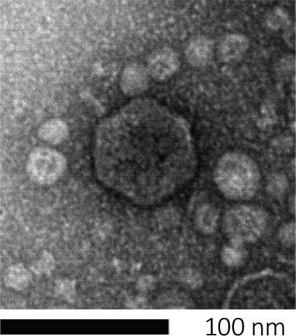
Figura 1: La morfología de un bacteriófago, aquí ejemplificada por un fago E. coli, se puede estudiar mediante microscopía electrónica de barrido. La mayoría de los bacteriófagos pertenecen a Caudovirales (bacteriófagos de cola). Este fago en particular tiene una estructura de cola muy corta y una cabeza icosahedral, colocándola en la familia de Podovirus.

El ensayo de placa (Figura 2) se basa en la incorporación de células huésped, preferentemente en el crecimiento de la fase log, en el medio. Esto crea una capa densa y turbia de bacterias capaces de mantener el crecimiento viral. Un fago aislado puede infectar, replicar y oxidar una célula. Con cada célula lysed, varias de las adyacentes se infectan inmediatamente. Varios ciclos en, una zona clara (una placa) se pueden observar en la placa de otro modo turbia (Figura 2B/Figura 3A),lo que indica la presencia de lo que inicialmente era una sola partícula de bacteriófago. Por lo tanto, el número de unidades formadoras de placas por volumen(es decir, PFU/ml) de una muestra puede determinarse a partir del número de placas generadas.

Figure 2
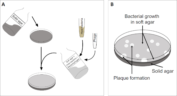
Figura 2: Las pruebas para las unidades formadoras de placas (PFU) son un método común para determinar el número de bacteriófagos en una muestra. (A) La base de una placa estéril de Petri está cubierta con un medio de nutrientes sólido adecuado, seguido de una mezcla de medios blandos, células huésped susceptibles y una dilución de la muestra original de bacteriófagos. Tenga en cuenta que la suspensión del fago podría, en algunos casos, también extenderse uniformemente a través de la superficie del agar blando ya solidificado. (B) El crecimiento de las bacterias anfitrionas forma un césped de células en la capa superior de agar. La replicación de bacteriófagos genera zonas claras, o placas, causadas por la lisis de la célula huésped.

Figure 3
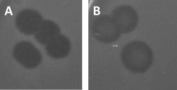
Figura 3: Los resultados de las pruebas de PFU muestran múltiples placas generadas por bacteriófagos. Debido a la lisis de las células huésped susceptibles, las placas pueden ser vistas como zonas de despeje en el césped bacteriano, ya sea con (A) aclaramiento completo, o (B) re-crecimiento parcial causado por la generación de bacterias resistentes (o posiblemente por fagos templados en el lisogénico).

Ciertos fagos templados pueden adoptar lo que se conoce como un ciclo de vida lisogénico, además del crecimiento lítico descrito anteriormente. En la lisógena, el virus asume un estado latente mediante la incorporación de su material genético en el genoma de la célula huésped (9), a menudo confiriendo resistencia a más infecciones por fagos. Esto a veces se revela a través de una ligera opacidad de la placa (Figura 3B). Vale la pena señalar sin embargo, que las placas también pueden aparecer borrosas debido al re-crecimiento de bacterias que han evolucionado resistencia al fago independiente de las infecciones anteriores de fago.

Los virus pueden adjuntarse, o adsorgar, a sólo un rango limitado de bacterias anfitrionas (10). Los rangos de acogida están aún más limitados por estrategias antivirales intracelulares como el sistema CRISPR-Cas (11). La resistencia/sensibilidad hacia fagos específicos mostrados por subgrupos bacterianos se ha utilizado históricamente para clasificar las cepas bacterianas en diferentes tipos de fagos (Figura 4). Aunque la eficacia de este método ha sido ahora superada por nuevas técnicas de secuenciación, la tipificación de fagos todavía puede proporcionar información valiosa sobre las interacciones entre bacterias y fagos, por ejemplo, facilitando el diseño de un cóctel de fago para uso clínico .

Figure 4
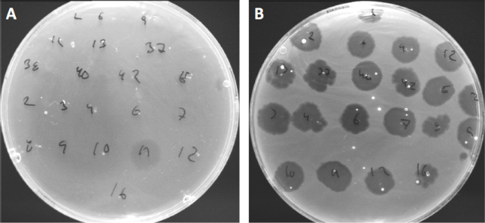
Figura 4: Sensibilidad del fago de diferentes cepas bacterianas. Las placas de agar blandas con cepa Cutibacterium acnes (A) AD27 y (B) AD35, fueron manchadas con 21 bacteriófagos Diferentes de C. acnes. Sólo el fago 11 fue capaz de infectar y matar AD27 mientras que la cepa AD35 mostró sensibilidad hacia todos los fagos. Esta técnica, que se denomina tipificación de fagos, se puede utilizar para dividir especies bacterianas y cepas en diferentes subgrupos basados en la susceptibilidad del fago.

Procedure

1. Configuración Antes de empezar cualquier trabajo que involucre microbios, asegúrese de que el espacio de trabajo esté esterilizado(por ejemplo, limpiado con 70% de etanol). Use siempre una capa de laboratorio y guantes, mantenga el cabello largo atado hacia atrás y asegúrese de que las heridas estén particularmente bien protegidas. Cuando haya terminado, esterilizar todas las superficies y lavar/esterilizar a fondo las manos y las muñecas. <p class="jove_t…

Applications and Summary

Despite multiple technological advances, plaque assays remain the gold standard for determination of viral titer (as PFU) and essential for isolation of pure bacteriophage populations. Susceptible host cells are cultivated in the top coat of a two layered agar-plate, forming a homogenous bed enabling viral replication. The initial event where an isolated bacteriophage in lytic lifecycle infects a cell, replicates within it, and eventually lyses it, is too small to observe. However, the virions released will infect adjacent cells, subsequently giving rise to a clearing zone, or plaque, denoting the presence of a single PFU.

References

  1. Twort, F. An investigation on the nature of ultra-microscopic viruses. Lancet. 186 (4814): 1241-1243. (1915)
  2. d'Hérelle, F. An invisible antagonist microbe of dysentery bacillus. Comptes Rendus Hebdomadaires Des Seances De L Academie Des Sciences. 165: 373-375. (1917)
  3. Cisek AA, Dąbrowska I, Gregorczyk KP, Wyżewski Z. Phage Therapy in Bacterial Infections Treatment: One Hundred Years After the Discovery of Bacteriophages. Current Microbiology. 74 (2):277-283. (2017)
  4. Mirzaei MK, Maurice CF. Ménage à trois in the human gut: interactions between host, bacteria and phages. Nature Reviews Microbiology. 15 (7):397. (2017)
  5. Breitbart M, Bonnain C, Malki K, Sawaya NA. Phage puppet masters of the marine microbial realm. Nature Microbiology. 3 (7):754-766. (2018)
  6. Leung CY, Weitz JS. Modeling the synergistic elimination of bacteria by phage and the innate immune system. Journal of Theoretical Biology. 429:241-252. (2017)
  7. Dulbecco R. Production of Plaques in Monolayer Tissue Cultures by Single Particles of an Animal Virus. Proceedings of the National Academy of Sciences of the United States of America. 38 (8):747-752. (1952)
  8. Juarez D, Long KC, Aguilar P, Kochel TJ, Halsey ES. Assessment of plaque assay methods for alphaviruses. J Virol Methods. 187 (1):185-9. (2013)
  9. Clokie MRJ, Millard AD, Letarov AV, Heaphy S. 2011. Phages in nature. Bacteriophage. 1 (1):31-45. (2011)
  10. Moldovan R, Chapman-McQuiston E, Wu XL. On kinetics of phage adsorption. Biophys J. 93 (1):303-15. (2007)
  11. Garneau JE, Dupuis M-È, Villion M, Romero DA, Barrangou R, Boyaval P, Fremaux C, Horvath P, Magadán AH, Moineau S.. The CRISPR/Cas bacterial immune system cleaves bacteriophage and plasmid DNA. Nature. 468 (7320):67. (2010)

내레이션 대본

Bacteriophages, also called phages, are viruses that specifically infect bacteria and we can confirm their presence and quantify them using a tool called the plaque assay. Bacteriophages infect their susceptible hosts by first attaching to the bacterial cell wall and injecting their genetic material. Then, they hijack the cell’s biosynthetic machinery to replicate their DNA and produce numerous progeny phage particles, which they then release by lysing and killing the host cell.

This lytic activity is the basis of a widely used phage enumerating technique known as the plaque assay or double agar layer assay. Here, a bacteriophage mix is first prepared in a molten nutrient broth containing low concentration agar. All bacteria used in the mix should be alive and actively dividing in the log phase of their growth, which will ensure that a large percentage of the bacteria are viable and able to form a dense lawn around the plaques. Next, this molten bacterial-phage agar mix is spread over a more solid, concentrated agar nutrient medium which is already solidified on a Petri dish. On incubation at room temperature, the low concentration agar-phage-bacteria broth also solidifies to form a soft agar overlay.

Here, the bacterial cells can derive additional nutrients from the bottom layer and should rapidly multiply to produce a confluent lawn of bacteria. However, as phage particles are also present in the soft layer, these will infect and replicate their genetic material within the bacteria, culminating in cell lysis, which releases multiple progeny. These phage progeny then infect the neighboring cells, as the semi-solid state of the bacteria-phage layer restricts their movement to more distantly located host cells. This cycle of infection and lysis continues over multiple rounds, killing a large number of bacteria in a localized area. The effect of the neighboring cells being destroyed, is to produce a single circular clear zone, called a plaque, which can be seen by the naked eye, effectively amplifying the bacteria lytic activity of the phage and enabling their enumeration.

The number of plaques on a Petri dish are referred to as Plaque-Forming Units, or PFUs, and, providing the initial bacteriophage concentration was sufficiently dilute, should directly correspond to the number of infective phage particles in the original sample. This technique can also be used for characterization of plaque morphology, to aid in identification of phage types, or to isolate phage mutants. In this lab, you will learn how to perform the plaque assay for enumerating phages, using the T7 phage of E. coli as an example.

First, identify a suitable medium for the culturing of the host bacterial cells and the bacteriophage. Here lysogeny broth, or LB medium, was used to culture E. coli and the T7 phage. Next, take three clean glass bottles and label them with media, name, and then the first as LB-Broth, the second as LB-Bottom Agar, and the third as LB-Top Agar. Now, weigh out four grams of pre-formulated LB powder in three sets and then transfer one set of weighed dried media into each bottle. Add 200 milliliters of water to the first bottle. Mix the contents using a magnetic stir bar.

Then, using a pH meter and constant stirring, bring the final pH to 7.4 through the addition of sodium hydroxide or hydrochloric acid. Repeat the water addition and pH adjustment for the other two remaining bottles, as well. Now, weigh out three grams of agar powder and add it to the second bottle to make a 1.5 % bottom agar. Finally, weigh 1. 2 grams of agar and add it to the third bottle to make the .6 % LB top agar. The broth condition in bottle one does not need an agar addition. Cap the bottle semi-tightly and then, sterilize the media by autoclaving at 121 degrees Celsius for 20 minutes. Once complete, remove the media bottles from the autoclave and immediately twist the bottle caps to close them fully to prevent contamination. Keep the LB-Broth and LB-Top Agar media on the bench for later use. Place the LB-Bottom Agar to cool in a water bath that is preset to approximately 45 degrees Celsius.

When the LB-Bottom agar reaches approximately 45 degrees Celsius, transfer it to the work bench. Next, sterilize the workspace using 70 % ethenol. Next, add 450 microliters of sterile one molar calcium chloride to the molten bottom agar to make a final concentration of 2.25 millimolar. Gently swirl the bottle to mix. Then, set out seven clean Petri dishes. Label each dish on the bottom with the media name and preparation date. Then, pour 15 milliliters of the bottom agar into each of the seven Petri dishes. Allow the plates to set for a few hours or overnight at room temperature. Once set, the culture plates can be stored at four degrees Celsius for several days if needed, upside down to minimize condensation. Transfer the Petri dishes from the four degrees Celsius refrigerator to a 37 degrees Celsius incubator one hour before the assay.

The day before the assay is to be preformed, the E. coli should be cultured. Here, 10 microliters of E. coli culture was inoculated into 10 milliliters of LB-Broth. Place the bacteria to grow overnight in a shaking incubator set to 37 degrees Celsius at 160 RPM. Then, on the day of the assay, remove the bacterial culture from the incubator. Seed a fresh 10 milliliters of fresh LB broth with 0.5 milliliters of the overnight culture. Place these cells to grow into a shaking incubator set to 37 degrees Celsius at 160 RPM. Next, use a spectrophotometer to check when this culture reaches log phase growth, indicated by an optical density of 0.5 to 0.7. Once the OD reaches this level, stop the incubation by transferring the cell culture to the bench. They are now ready to be used for phage overlay assay.

Phage titers can vary exponentially across different phage types and samples. So in order to count them effectively, they should be diluted to generate a wide range of phage concentrations. On the day of the assay, generate a series of phage dilutions ranging from one tenth to one millionth concentrations, following a 10-fold dilution technique. To obtain statistically significant and accurate data, perform the serial dilution in triplicate.

Next, melt the solidified LB-top agar using a microwave. Then, place it in a water bath that is preset at 45 degrees Celsius for one hour. After one hour, collect the Petri dishes containing the bottom agar layer from the incubator. Label the plates with phage concentration and assay date. Then, set out seven clean test tubes. Label each test tube with the serial phage dilution number and designate one as control.

When the LB-top agar reaches 45 degrees Celsius, transfer it to the working bench. Now, add 450 microliters of one molar calcium chloride to the 200 milliliter agar to make a final concentration of 2.25 millimolar. Gently swirl the bottle to mix. Next, add 35 milliliters of LB-top agar and four milliliters of bacterial suspension to a sterile conical tube. Gently swirl to evenly distribute the cells but avoid shaking to prevent foaming.

Now, aliquot five milliliters of this bacteria- top agar mix to each of the seven test tubes. Then, transfer 100 microliters of the serially diluted bacteriophage samples and control media, which should be simply media with no bacteriophage, to the respectfully labeled test tubes. Swirl the mixture gently to ensure proper mixing. Gently transfer five milliliters of bacteriophage mix onto the respective Petri plate. Evenly spread the mix throughout the whole surface by gently swirling the Petri plate.

Once all of the Petri plates are layered with the mix, allow solidification of the top layer by incubating at room temperature for 15 minutes. After completion of these steps, repeat the process for the second and then the third sets of the Petri dishes using the remaining two sets of phage dilutions. Seal each dish with parafilm and incubate at room temperature for 15 minutes. Place the culture plate upside down at a suitable temperature for 24 hours or until plaques develop. Here, plates were placed in a 37 degrees Celsius incubator for one day, a stimulating growth condition for E. coli and the T7 phage.

Plaques will appear after one to five days of incubation, depending on the bacterial species, incubation conditions, and the choice of medium. Here, plaques were visible after one day of incubation at 37 degrees Celsius. Begin by checking the plates marked control and ensure that no plaques were formed in these plates, as this would indicate viral contamination. To determine the phage titer in the original sample, start with the plates containing the most diluted phage sample first and count the plaques without removing the lids, marking them to indicate which ones have already been counted. Repeat the counting for each plate in every set. Some plates might have too many or too few plaques to be counted. Consider 10 to 150 as an ideal plaque count.

Next, generate a table listing the plaque number values for the different dilutions and replicates. Then, calculate the mean plaque number values for the dilution plates that contained the ideal number of plaque counts. In this example, these were the average number of plaques formed in the 10 to the minus three and 10 to the minus four dilution plates. Now, adjust for phage dilution factor by dividing the obtained mean plaque values by the respective phage dilution factors. Here, the average number of plaques formed to the 10 to the minus three and 10 to the minus four dilution plates, were divided by their respective dilution factors to obtain the number of plaque forming units, or PFUs, in 100 microliters of phage mixture. To convert the value to PFU per milliliter, multiply the generated values by 10, as only 100 microliters of phage dilution mix was used during the bacteriophage overlay preparation step, producing an additional dilution factor of 10. Finally, calculate the average of the values obtained from the different dilution plates. This will give the average number of PFUs per milliliter. The number of PFUs corresponds to the number of infective phage particles in the original sample.

Tags

Cite This
JoVE Science Education Database. JoVE Science Education. Plaque Assay: A Method to Determine Viral Titer as Plaque Forming Units (PFU). JoVE, Cambridge, MA, (2023).