All the procedures followed in this study accorded with the Association for Research in Vision and Ophthalmology Statement for Use of Animals in Ophthalmic and Vision Research and were approved by the Institutional Animal Care and Use Committee of National Taiwan University Hospital.
1. Isolation, Primary Culture Preparation, and Immunostaining of Bovine CECs
2. Rat Corneal Endothelium Cryoinjury Model and Intracameral Injection
3. Harvesting the Rat Corneal Button and Immunostaining
After the isolation of bovine CECs, the cells were cultured in vitro. Figure 1 presents the phase contrast images of the bovine CECs. The hexagonal shape of the cells at confluence indicated that the cells were not contaminated by corneal stromal fibroblast during cell isolation. Figure 2 depicts the immunostaining that was performed using antibodies against ABC, snail, and slug at an indicated time point. Apart from phenotypic changes in the in vitro culture, a corresponding nuclear translocation of ABC and EMT regulators was observed. Figure 3 illustrates the effect of marimastat, a broad-spectrum MMP inhibitor, on the EnMT process of the in vitro cultured bovine CECs. Figure 4 comprises the external eye photographs of rats after cryoinjury followed by intracameral injection. Figure 5 shows the immunostaining of the rat corneal button that was performed using antibodies against ABC after cryoinjury followed by intracameral injection. The nuclear translocation of ABC was observed in the PBS group and was significantly increased in the bFGF group, indicating activation of the Wnt/β-catenin signaling and EnMT process. After intracameral injection of bFGF followed by marimastat injection, the nuclear staining of ABC was diminished, suggesting the EnMT-inhibiting effect of marimastat.
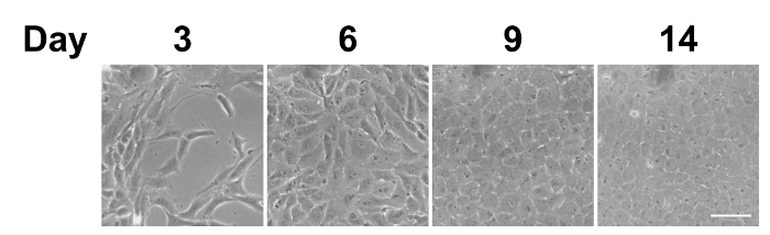
Figure 1: Phase contrast images of in vitro cultured bovine CECs. After being seeded on the culture plate, the bovine CECs initially appeared fibroblast-like on days 3 and 6. They became more hexagonal upon reaching complete confluence on day 9. Scale bar = 50 µm. Representative images of 3 replicates. Please click here to view a larger version of this figure.
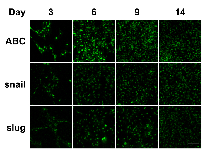
Figure 2: Immunostaining of in vitro cultured bovine CECs. During the in vitro culture of the bovine CECs, the nuclear translocation of ABC, snail, and slug was detected through day 14. ABC: active β-catenin. Scale bar = 100 µm. Representative images of 3 replicates. Please click here to view a larger version of this figure.
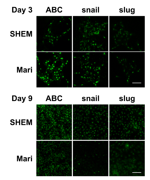
Figure 3: Immunostaining of in vitro cultured bovine CECs with or without marimastat at different cell confluence levels. Immunostaining demonstrated that ABC, snail, and slug were evident in the nucleus of the bovine CECs with or without 10 µM of marimastat on day 3. However, marimastat significantly reduced the nuclear staining of ABC, snail, and slug on day 9 when the bovine CECs became fully confluent. Scale bar = 100 µm. Representative images of 3 replicates. Please click here to view a larger version of this figure.
Figure 4: External eye photographs of rats at indicated time points after cryoinjury. Following cryoinjury for 3 consecutive days, rats were subjected to intracameral injection of 0.02 ml of PBS or 50 ng/ml bFGF on day 3. On day 6, 0.02 ml of 10 µM marimastat was injected intracamerally in the bFGF/Mari group, whereas PBS was injected in the other 2 groups (n = 9 in each group). External eye photographs revealed reduced corneal edema after bFGF injection, whereas marimastat further reduced corneal edema compared with bFGF alone. n = 9 in each group. Scale bar = 1 mm. Please click here to view a larger version of this figure.
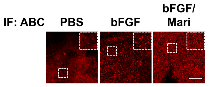
Figure 5: Immunostaining of rat corneal buttons. Immunostaining of the rat corneal buttons that were harvested on day 9 revealed little nuclear staining of ABC in the PBS group. In the bFGF group, there was extensive nuclear staining of ABC, which was significantly reduced in the bFGF/Mari group. Scale bar = 100 µm. Please click here to view a larger version of this figure.
trypsin | ThermoFisher Scientific | 12604-013 | |
Dulbecco’s modified Eagle medium and Ham's F12 medium | ThermoFisher Scientific | 11330 | |
fetal bovine serum | ThermoFisher Scientific | 26140-079 | |
dimethyl sulfoxide | Sigma | D2650 | |
human epidermal growth factor | ThermoFisher Scientific | PHG0311 | |
insulin, transferrin, selenium | ThermoFisher Scientific | 41400-045 | |
cholera toxin | Sigma | C8052-1MG | |
gentamicin | ThermoFisher Scientific | 15750-060 | |
amphotericin B | ThermoFisher Scientific | 15290-026 | |
paraformaldehyde | Electron Microscopy Sciences | 111219 | |
Triton X-100 | Sigma | T8787 | |
bovine serum albumin | Sigma | A7906 | |
marimastat | Sigma | M2699-25MG | |
anti-active beta-catenin antibody | Millpore | 05-665 | |
anti-snail antibody | Santa cruz | sc28199 | |
anti-slug antibody | Santa cruz | sc15391 | |
goat anti-mouse IgG (H+L) secondary antibody | ThermoFisher Scientific | A-11001 | for staining of ABC of bovine CECs |
goat anti-mouse IgG (H+L) secondary antibody | ThermoFisher Scientific | A-11003 | for staining of ABC of rat corneal endothelium |
goat anti-rabbit IgG (H+L) secondary antibody | ThermoFisher Scientific | A-11008 | for staining of snail and slug of bovine CECs |
antibody diluent | Genemed Biotechnologies | 10-0001 | |
4',6-diamidino-2-phenylindole | ThermoFisher Scientific | D1306 | |
mounting medium | Vector Laboratories | H-1000 | |
laser scanning confocal microscope | ZEISS | LSM510 | |
xylazine | Bayer | N/A | |
tiletamine plus zolazepam | Virbac | N/A | veterinary drug |
proparacaine hydrochloride ophthalmic solution | Alcon | N/A | veterinary drug |
0.1% atropine | Wu-Fu Laboratories Co., Ltd | N/A | clinical drug |
0.3% gentamicin sulfate | Sinphar Group | N/A | clinical drug |
basic fibroblast growth factor | ThermoFisher Scientific | PHG0024 | clinical drug |
Corneal endothelial cells (CECs) play a crucial role in maintaining corneal clarity through active pumping. A reduced CEC count may lead to corneal edema and diminished visual acuity. However, human CECs are prone to compromised proliferative potential. Furthermore, stimulation of cell growth is often complicated by gradual endothelial-mesenchymal transition (EnMT). Therefore, understanding the mechanism of EnMT is necessary for facilitating the regeneration of CECs with competent function. In this study, we prepared a primary culture of bovine CECs by peeling the CECs with Descemet’s membrane from the corneal button and demonstrated that bovine CECs exhibited the EnMT process, including phenotypic change, nuclear translocation of β-catenin, and EMT regulators snail and slug, in the in vitro culture. Furthermore, we used a rat corneal endothelium cryoinjury model to demonstrate the EnMT process in vivo. Collectively, the in vitro primary culture of bovine CECs and in vivo rat corneal endothelium cryoinjury models offers useful platforms for investigating the mechanism of EnMT.