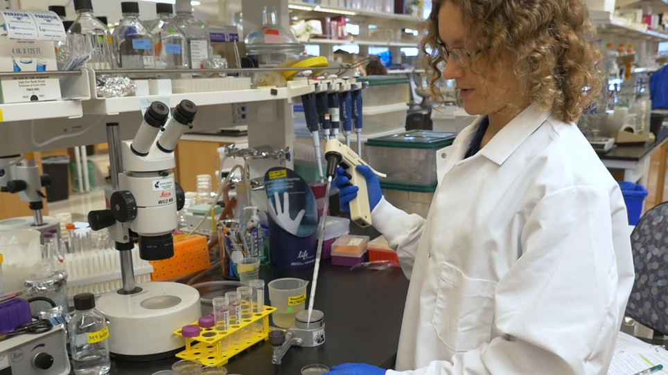
/
/
C. elegans tarmen som en modell for intercellulære Lumen Morphogenesis og I Vivo polarisert membran Biogenesis på encellede nivå: merking av antistoff flekker, RNAi tap av funksjon analyse og bildebehandling
A subscription to JoVE is required to view this content. Sign in or start your free trial.
JoVE Journal
Developmental Biology
The C. elegans Intestine As a Model for Intercellular Lumen Morphogenesis and In Vivo Polarized Membrane Biogenesis at the Single-cell Level: Labeling by Antibody Staining, RNAi Loss-of-function Analysis and Imaging
Chapters
- 00:05Title
- 01:24Introduction to the Accompanying Publication
- 02:47Nematode Fixation
- 04:19Nematode Antibody Staining
- 06:33Applying RNAi to Nematodes Using a Feeding Protocol
- 07:57Analyzing Phenotypes Using Fluorescence Dissecting Microscopy
- 08:41Analyzing Phenotypes Using Confocal Microscopy
- 10:14Results: Immunohistochemistry, RNAi and Microscopic Analysis of Intestinal Apical membrane and Lumen Morphogenesis
- 11:45Conclusion
Den gjennomsiktige C. elegans tarmen kan tjene som en"i vivo vev chamber" for å studere apicobasal membran og lumen biogenesis på én celle og subcellular under flercellet tubulogenesis. Denne protokollen beskriver hvordan kombinere standard merking, tap av funksjon genetisk/RNAi og mikroskopiske tilnærminger å dissekere disse prosesser på molekylært nivå.
Tags
C. ElegansIntestineIntercellular LumenMembrane BiogenesisPolarized MembraneSingle-cell LevelAntibody StainingRNAiLoss-of-function AnalysisImagingMorphogenesisApical MembraneBasolateral MembranePolarityTubulogenesisExcretory CanalTransgenic AnimalsFluorescent Fusion ProteinsLive ImagingImmunohistochemistryMembrane MarkersPromoters


