Here we present a protocol for obtaining entomopathogenic fungi from a forest wood borer and a substitutive way to evaluate their entomopathogenic activities using a Coleopteran model insect. This method is efficient and convenient for exploring entomopathogenic fungal resources from wood-boring insect pests in natural forests.
Forest wood borers (FWB) cause severe tree damage and economic losses worldwide. The release of entomopathogenic fungi (EPF) during the FWB emergence period is considered an acceptable alternative to chemical control. However, EPF resources have been significantly less explored for FWBs, in contrast to agricultural insect pests. This paper presents a protocol for exploring EPF resources from FWBs using wild Monochamus alternatus populations as an example. In this protocol, the assignment of traps baited with M. alternatus attractants to different populations guaranteed the collection of adequate samples with natural infection symptoms, during the emergence periods of the beetle. Following finely dissecting integuments and placing them onto a selective medium, fungal species were isolated from each part of beetle bodies and identified based on both molecular and morphological traits.
Several fungal species were certified as parasitic EPFs via re-infection of healthy M. alternatus with spore suspensions. Their behavioral phenotypes on M. alternatus were observed using scanning electron microscopy and further compared with those on the Coleopteran model insect Tribolium castaneum. For EPFs that present consistent parasitism phenotypes on both beetle species, evaluation of their activities on T. castaneum provided valuable information on lethality for future study on M. alternatus. This protocol helped the discovery of EPF newly reported on M. alternatus populations in China, which could be applied as an efficient approach to explore more EPF resources from other FWBs.
The devastation caused by insect pests has led to great ecological and economic losses in both forest and agricultural ecosystems. Most agricultural pests expose themselves to natural enemies or artificial control agents while damaging host plants. Instead, forest wood borers (FWB) nearly complete their whole developmental cycles inside host tree trunks1, which raises large challenges to explore efficient biocontrol organisms from FWB in the wild field. What is even worse is that FWBs carry a great number of phytopathogens2 or have an intimate relationship with these pathogens as their potential vectors3,4, dramatically amplifying the negative effects of FWB on forest health. Excessive use of chemical insecticides can alleviate FWB severity, but the emergence of insecticidal resistance5,6 limits their environmental application. In certain cases, insect parasitoids, predatory arthropods as well as entomopathogenic microbes were released as biocontrol agents to the distribution areas of FWB7 and were proven to be efficient and economically acceptable alternatives to chemical control8,9,10.
Entomopathogenic fungi (EPF) are regarded to have the advantage in controlling FWB over most other microbial groups. Their spores can be carried by insect hosts and stably fixed on body surfaces via penetration into the cuticle or integument8,11. EPF also present excellent adaptability to environmental stresses and some species colonize well in the tissue of trees as endophytes12,13, facilitating their growth, survival, and transmission. However, compared to that in agricultural industries, the species diversity of EPF used in natural forest ecosystems is remarkably restricted14,15,16. Beauveria bassiana (strain PPRI 5339) was evidenced as the most promising strain to promote an IPM program to Eucalyptus weevils in South Africa17 and the combination of two promising isolates of B. bassiana provided an opportunity for the practical microbial control of red palm weevil, Rhynchophorus ferrugineus, at different life stages in palm tree fields18. In addition to Beauveria and the well-known Metarhizium, other EPF genera of the order Hypocreales, especially species of Lecanicillium (many of which are now classified into the genus Akanthomyces19,20), showed strong pathogenicity and high potential in management of forest pests, such as the Cypress aphid in Chile21.
The pine sawyer beetle Monochamus alternatus is a notorious pine forest pest in China and neighboring countries, which burrows into branches and trunks of pine trees to impede the transportation of nutrients and water22,23,24. Moreover, M. alternatus also promotes the invasion of the plant-parasitic pine wood nematode (Bursaphelenchus xylophilus, PWN) as its main vector beetle. Another congeneric species of the beetle, M. galloprovincialis, has spread PWN in several countries in Europe in recent years25. Previous research reported several genera of natural EPFs from Monochamus spp., such as Beauveria, Metarhizium, and Lecanicillium (Verticillium, an even former name of Lecanicillium), in Spain, Japan, and the Anhui/Zhejiang Provinces of China26,27,28,29. Nevertheless, these collections of EPFs seem to be commonly restricted in a certain location, compared to the wide occurrence of Monochamus beetles in natural fields. As the M. alternatus beetle has a wide geographical distribution in China, it could be regarded as a representative wood borer to explore more potential EPFs across different populations.
In the present protocol, we introduce a specific procedure exploring EPFs from several geographical populations of M. alternatus in southern China. This protocol uses a model Coleopteran beetle as a substitute to perform entomopathogenicity assays, under the condition that the tested fungal species has a consistent behavioral phenotype on both beetle species. This protocol can also provide insights into EPF exploration for other forest wood borers, in which the diversity of their entomopathogenic fungal species is underestimated or less investigated.
1. Isolation of fungi from M. alternatus (Figure 1)
2. Molecular and morphological identification of fungal isolates
3. Induction of the fungal infection symptoms on M. alternatus to observe their behavioral phenotypes
4. Confirm the infection phenotypes of fungi on M. alternatus and model beetle
5. Evaluation of entomopathogenic activity
Isolation and identification of fungal isolates from M. alternatus
With the aid of attractant traps, a large number (approximately 500 beetles in total) of M. alternatus were collected from five geographical regions. Beetle cadavers with typical symptoms of infection by entomopathogenic fungi were picked; then, body integuments of every beetle were dissected into several positions as described in protocol step 1.3. As a result, more than 600 fungal isolates were isolated from different body positions. This allows richer fungal candidates for screening pathogens against pine sawyer beetles. Sequencing of the ITS region preliminarily revealed that these isolates can be categorized into 15 fungal genera and 39 species. In addition, there were significant differences in the fungal community composition among geographical populations of M. alternatus adults. The fungal species, Aspergillus austwickii, Akanthomyces attenuatus (syn. Lecanicillium attenuatum), Penicillium citrinum, Scopulariopsis alboflavescens were the dominant species in Sichuan, Zhejiang, Guangdong, Fujian populations, respectively. Under OM and SEM, the asexual reproductive morphology of these four fungi grown on PDA plates was identified based on the macroscopic characteristics, including the color, texture, and sizes of colonies, and microscopic characteristics, including shape, size of conidia, conidiophores, sporangia and hyphae (Figure 2). These observations can be photographed for measuring the shape, size and arrangement of conidia, hyphae, and conidiophores.
The parasitic fungal infection phenotypes on M. alternatus and T. castaneum
Through the induction method (step 3.3), four region-representative fungal species from different geographical populations were confirmed to function in parasitic or non-parasitic modes. As a result, there was no visible infection phenotypes displayed on M. alternatus by P. citrinum, neither on T. castaneum. Conversely, significant infection symptoms were appeared both on M. alternatus (Figure 3) and T. castaneum body surface after infection by A. austwickii, A. attenuatus (syn. L.attenuatum), and S. alboflavescens. Evidence from SEM also well confirmed the consistency of the morphological characteristics of the parasitic fungi on both beetle surfaces. Furthermore, the process of fungal infection on beetles was clearly observed, in which mycelium carrying conidiophores penetrated from the inside of the beetle to the body surface (Figure 4), indicating strong parasitic abilities of these fungi. The above results indicated that the parasitism of these fungi was consistent for both M. alternatus and T. castaneum. In addition, those pictures and data also suggested the spatial preference and localized niches of three fungi on host beetle bodies.
The entomopathogenic activity of parasitic fungal species
T. castaneum has been regarded as a general model organism of Coleopteran species widely, combined with the results above, it has been used as a substitute to assess entomopathogenic activity in this study. The behavioral phenotypes of the three parasitic fungi against M. alternatus and T. castaneum were similar, so the lethal effect on the model beetle would provide valuable information on the entomopathogenic activity against M. alternatus. In a 9-day pilot assay, respectively inoculated with conidial suspension of parasite fungi (A. austwickii, A. attenuatus (syn. L. attenuatum), S. alboflavescens), three groups performed significantly higher mortality on T. castaneum adults than those treated with Tween-80 (control group), and differences were also found between the survival of three groups (Figure 5). These results suggested parasitic fungal species had varying degrees of pathogenicity to model beetles, revealing that these fungi can also exhibit different levels of entomopathogenic activity to M. alternatus adults. Thus, we provide new candidates for the repository of pathogenic fungi.
Multi-gene phylogenetic analysis
The ITS regions of all fungal isolates were subjected to molecular analysis to gain some preliminary insight into the fungal classification. Then, the sequences of the SSU, LSU, tef-1α, rpb2, and β-tubulin region were performed specifically for parasitic entomopathogens. Based on the five independent loci, the accurate taxonomic status of the three parasitic fungi was classified. As shown in Figure 6, the multi-gene phylogenetic treeclearly indicated the genetic distances of the three fungal species from other species in their respective genera, which also matched the morphological characteristics of these fungi.
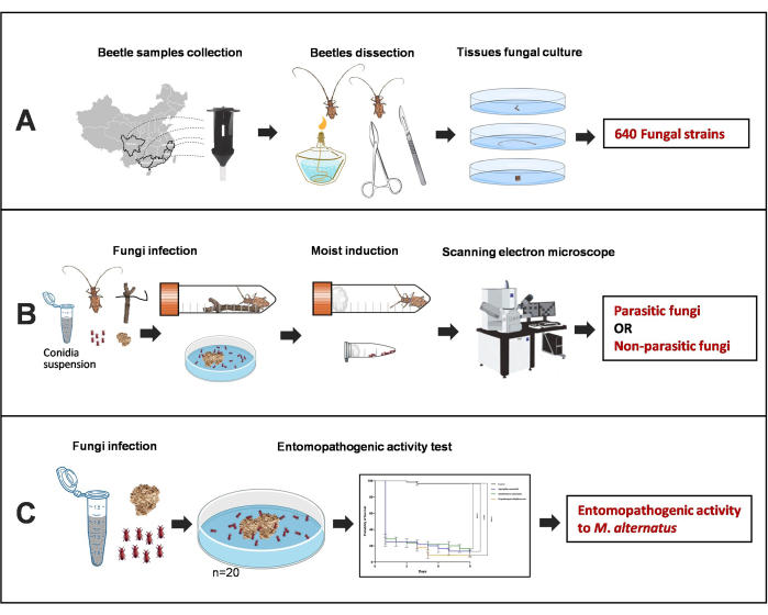
Figure 1: Illustration of screening out entomopathogenic fungi from multiple populations of M. alternatus adults. (A) Fungal isolation from different tissues of M. alternatus with naturally fungal infection. (B) Screening of entomopathogenic fungi. (C) Pathogenicity testing of parasitic entomopathogenic fungi against model beetles. Please click here to view a larger version of this figure.
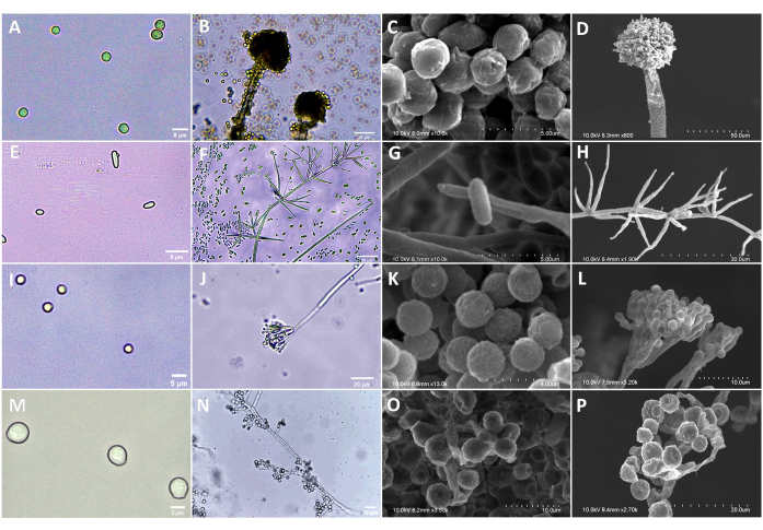
Figure 2: Morphology of A. austwickii, A. attenuatus (syn. L.attenuatum), P. citrinum, S. alboflavescens under OM and SEM. (A) Conidia of A. austwickii (OM). (B) Conidiophores of A. austwickii (OM). (C) Conidia of A. austwickii (SEM). (D) Conidiophores of A. austwickii (SEM). (E) Conidia of A. attenuatus (OM). (F) Conidiophores of A. attenuatus (OM). (G) Conidia of A. attenuatus (SEM). (H) Conidiophores of A. attenuatus (SEM). (I) Conidia of P. citrinum (OM). (J) Conidiophores of P. citrinum (OM). (K) Conidia of P. citrinum (SEM). (L) Conidiophores of P. citrinum (SEM). (M) Conidia of S. alboflavescens (OM). (N) Conidiophores of S. alboflavescens (OM). (O) Conidia of S. alboflavescens (SEM). (P) Conidiophores of S. alboflavescens (SEM). Scale bars are shown in the bottom right corner of each image. Please click here to view a larger version of this figure.
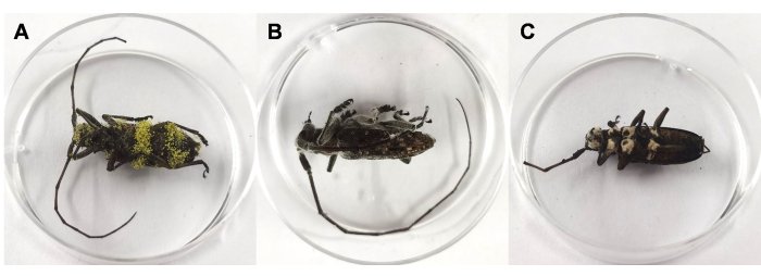
Figure 3: Phenotypes of M. alternatus cadavers infected by A. austwickii, A. attenuatus (syn. L. attenuatum), S. alboflavescens. Beetle cadaver surrounded by mycelium of (A) A. austwickii, (B) A. attenuatus, (C) S. alboflavescens. Please click here to view a larger version of this figure.
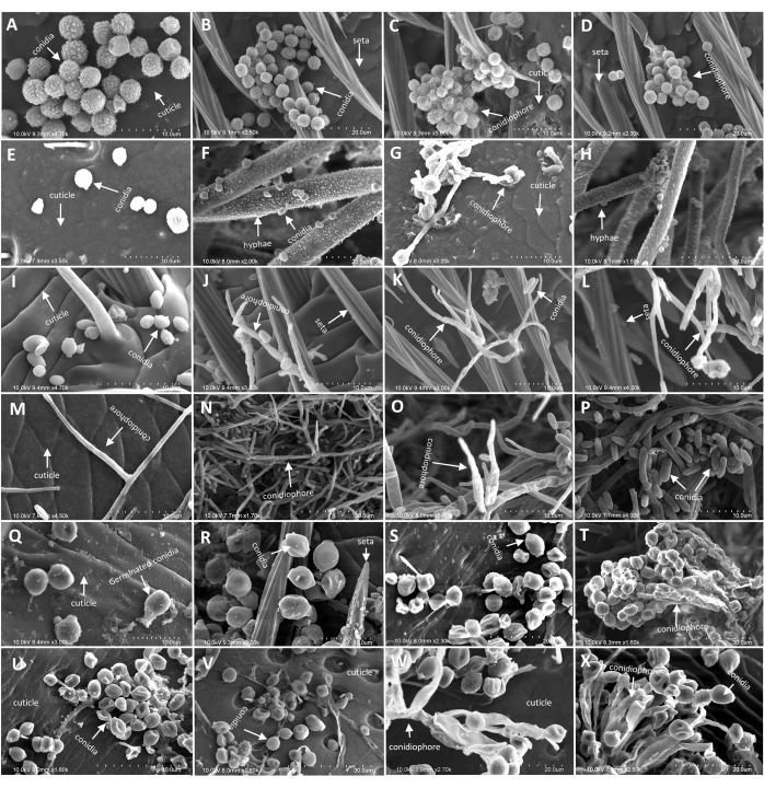
Figure 4: Morphology of M. alternatus and T. castaneum cadaver infected by A. austwickii, A. attenuatus (syn. L.attenuatum), S. alboflavescens under SEM. (A–D) Hyphae and conidiophores of A. austwickii on M. alternatus abdomen. (E–H) Hyphae and conidiophores of A. austwickii on T. castaneum cuticle surface. (I–L) Hyphae and conidiophores of A. attenuatus on M. alternatus antennae. (M–P) Hyphae and conidiophores of A. attenuatus on T. castaneum cuticle surface. (Q–T) Hyphae and conidiophores of S. alboflavescens on M. alternatus head. (U–X) Hyphae and conidiophores of S. alboflavescens on T. castaneum cuticle surface. Scale bars are shown in the bottom right corner of each image. Please click here to view a larger version of this figure.
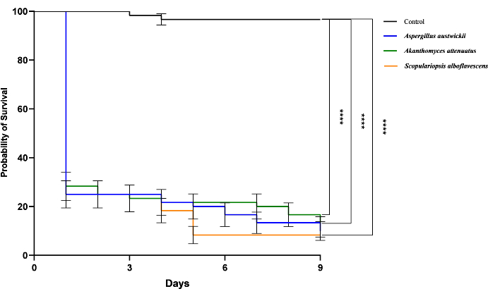
Figure 5: Pathogenicity activities of three parasitic fungi isolated from wild M. alternatus. The survival of T. castaneum beetles infected by three parasitic fungi is shown by Kaplan-Meier curves. Log-rank tests were performed and the significance level were denoted: ns, not significant; ****, P < 0.0001. Please click here to view a larger version of this figure.
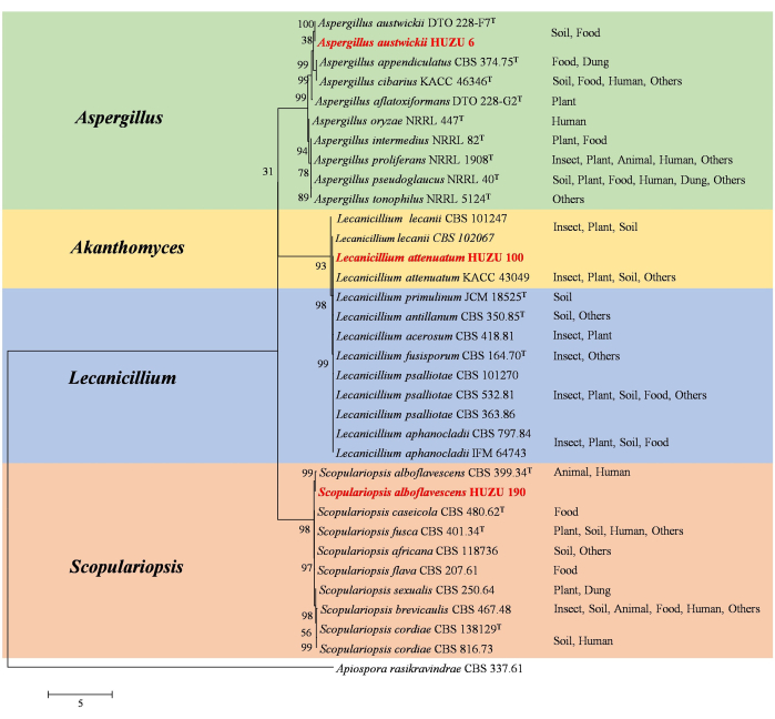
Figure 6: Phylogenetic tree construction of three entomopathogenic fungi inferred from a multi-gene dataset (ITS, LSU, SSU, EF-1α, rpb2, and β-tubulin). The fungal isolates in this study are in red. The nodes indicate supportive values greater than 50%. This figure was modified from Wu et al.43. The habitats where the species were found are indicated behind the species names as Insect, Plant, Soil, Animal, Food, Human, Dung, and others. The T indicates the isolates were from type materials. The GenBank accession numbers of fungal species sequences used for phylogenetic construction see Supplemental Table S1. Please click here to view a larger version of this figure.
Primer name | Region | Sequence (5'-3') | Bibliography | |
ITS1 | ITS | TCCGTAGGTGGACCTGCGG | 34 | |
ITS4 | TCCTCCGCTTATTGATATGC | |||
NS1 | SSU | GTAGTCATATGCTTGTCTC | 34 | |
NS4 | CTTCCGTCAATTCCTTTAAG | |||
LR0R | LSU | ACCCGCTGAACTTAAGC | 37, 38 | |
LR7 | TACTACCACCAAGATCT | |||
EF-983 | tef-1α | GCYCCYGGHCAYGGTGAYTTYAT | 39 | |
EF-2218 | GACTTGACTTCRGTVGTGAC | |||
RPB2-5’F | rpb2 | CCCATRGCTTGTYYRCCCAT | 40 | |
RPB2-5’R | GAYGAYMGWGATCAYTTYGG | |||
TUB1 | β-tubulin | AACATGCGTGAGATTGTAAGT | 41 | |
TUB22 | TCTGGATGTTGTTGGGAATCC |
Table 1: Primer pairs for fungal identification.
Supplemental Table S1: GenBank accession numbers of fungal species sequences used for the phylogenetic construction. Please click here to download this File.
Different geographical populations of FWB may develop varied interactions with the natural entomopathogenic fungi, due to long-term environmental adaptation of EPF species to local climate factors and the specific genotypic population of the host insect44,45. Expansion of the sampling sites to multiple insect occurrence regions helps increase the possibility of acquiring diverse strains or species of EPF from their natural hosts, as described by previous studies on agricultural insect pests46. In this protocol, with the assistance of traps baited with attractants, the pine sawyer beetle M. alternatus is collected at a cross-latitudinal level in five different sites of southern China, which is regarded as an ideal and rich resource reservoir for exploring novel EPF, considering the higher temperature and humidity. This labor-saving method traps beetles alive, which should be individually transported to the laboratory in separate sterile tubes. The natural infection symptoms on beetles require several days to occur. Representative EPF species A. austwickii, A. attenuatus (syn. L.attenuatum), and S. alboflavescens, obtained by the protocol, show significant regional specificity in different M. alternatus populations, which have not been reported on Monochamus beetles by other studies before. In addition, considering the potential distribution areas of the vector beetle M. alternatus in China, more EPF could be expected through deeper field investigation in the future. The result of our study demonstrates the great value of geographical EPF exploration for widely distributed FWB insect species.
This protocol also emphasizes the importance that different body positions of beetle cadavers should be dissected carefully into fine pieces followed by selective growth of mycelium on antibiotics-supplementing plates. As shown in our study with this mode of isolation, more than 600 fungal strains were acquired from different parts of M. alternatus bodies, which could guarantee an adequate quantity of fungal candidates for screening out potential EPF. Although the addition of antibiotics into medium plates seems to cause the loss of certain fungal species during isolation47, this traditional technology would still be the most suitable for various FWB systems to protect EPF growths from being contaminated by bacteria. The fineness of operation in dissection and isolation can compensate for possible species loss owing to the use of antibiotics. Additionally, the isolation of fungi from specific parts of insect bodies promotes the understanding of EPF colonization preferences on the hosts. For instance, A. attenuatus (syn. L.attenuatum) seems to infect M. alternatus antennae more frequently than other body positions, as observed in our previous study43, which would inspire further studies on its behavioral traits.
During strain identification, it is necessary to use a combination of morphological and molecular identification to distinguish closely related fungal species. For morphological identification, both macroscopic and microscopic characteristics should be clearly observed, including the different characteristics of fungal colonies, conidia, conidiophores, and hyphae of each different fungal strain. For molecular identification, the sequencing of the ITS region is not sufficient to reveal the species-level discrimination in the fungal community48. Therefore, it requires the application of different combinations of primer sets targeting more molecular markers (such as SSU, LSU, tef-1α, rpb2, and β-tubulin) to determine the phylogeny of entomopathogenic fungi more accurately.
Observation of fungal infection phenotype on beetles using an optical microscope has the shortcoming that light transmission and resolution affect discrimination of fungal characteristics. Conversely, the use of scanning electron microscopy (SEM) is much more reliable to obtain details on the infection process, which has been applied to record parasitic phenomena on several agricultural nematodes and insect pests49,50. Following artificial infection, the three parasitic entomopathogenic fungi, A. austwickii, A. attenuatus (syn. L.attenuatum), and S. alboflavescens, can be re-isolated from M. alternatus and T. castaneum body surfaces. Their morphological characteristics of conidia, infection pegs, and conidiophores can be determined from the two beetle species via SEM and shown the same with their respective colonies grown on PDA medium. Another insecticidal fungal species, P. citrinum, however, could not be re-isolated from both beetle species and was not observed by SEM from their body surfaces. Thus, the parasitism phenotypes induced by this protocol present excellent consistency between M. alternatus and the model beetle species.
The red flour beetle, T. castaneum is an important postharvest pest51 and also a general model organism of Coleopteran species used for genetics, immunology, and other research52,53,54,55 because it is highly fertile, well-adapted, and easy to manipulate experimentally. The consistency in parasitism phenotypes indicates that T. castaneum could be an appropriate substitute or tool to evaluate entomopathogenic activities of EPF on M. alternatus. This can release the demand for a great number of adult beetles of uniform genetic background, which is difficult for FWB collection in the wild field within a limited period. Following the entomopathogenic bioassay using this model insect, valuable information on EPF lethality can guide field studies on M. alternatus and even other Coleopteran FWB in the future.
To sum up, this present protocol facilitates the EPF resource exploration from forest wood borers, which considers a model beetle as an alternative for entomopathogenic evaluation. It greatly reduces the problems associated with having no access to an adequate quantity of beetles for testing purposes. Hence, this is a practical and efficient protocol for preliminary screening the entomopathogenic candidates on forest wood borers, which will facilitate the provision of natural resources for the future development of biological control strategies.
The authors have nothing to disclose.
This research was supported by the National Key Research and Development Program of China (2021YFC2600100) and the Natural Science Foundation of Zhejiang Province (LY21C040001).
1.5 mL, 2 mL centrifuge tubes | Biosharp | BS-15-M | |
10 µL pipet tips | Sangon Biotech | F601216 | |
10 µL, 20 µL, 100 µL, 200 µL, 1,000 µL pipettes | Rainin | ||
1,000 µL pipet tips | Sangon Biotech | F630102 | |
2 mL cryogenic vials | Corning | 430659 | |
20x PBS buffer | Sangon Biotech | B548117-0500 | |
200 µL pipet tips | Sangon Biotech | F601227 | |
2,000 bp maker | TaKaRar | SD0531 | |
50 mL tubes | Nest | 602052 | |
50% glutaraldehyde solution | Sangon Biotech | G916054 | |
50x TAE buffer | Sangon Biotech | B548101 | |
6x loading buffer | TaKaRar | SD0503 | |
Agarose | Sangon Biotech | A610013 | |
Anhydrous ethanol | Jkchemical | LB10V37 | |
Biochemistry Cultivation Cabinet | Shanghaiyiheng | LRH-250F | |
Chloroform | Juhua | 61553 | |
Commercial beetle traps | FEIMENGDI | BF-8 | www.yinyouji.com |
Gel imager | Bio-Rad | GelDoc XR+ | |
Glycerol | Sangon Biotech | A600232 | |
High speed refrigerated centrifuge | Sigma | D-37520 | |
High-Pressure Steam Sterilization Pot | Mettler Toledo | JA5003 | |
Isopropyl alcohol | General-reagent | G75885B | |
Nucleic acid dye | Sangon Biotech | A616696 | |
Optical Microscope, OM | Leica | DM2000 | |
Parafilm | Parafilm | PM996 | |
PCR meter | Heal Force | Trident960 | |
Penicillin G | Marklin | GB15743 | |
Potato dextrose agar, PDA | Oxoid | CM0139 | |
Potato dextrose broth, PDB | Solarbio | P9240 | |
Primers | Sangon Biotech | / | |
Primers Taq | TaKaRar | RR902A | |
Rapid Fungi Genomic DNA Isolation Kit | Sangon Biotech | B518229 | |
Scanning Electron Microscope, SEM | Hitachi | S-3400N | |
Streptomycin | Marklin | S6153 | |
Tetracycline | Marklin | T829835 | |
Tween-80 | Marklin | T6336 | |
Vacuum freeze dryer | Yamato | DC801 | |
Vortex Shaker | HLD | WH-861 | |
β-Mercaptoethanol | Marklin | M6230 |
.