Here, we describe a protocol for the surgical creation of a volumetric muscle loss (VML) injury in the rat masseter, providing a reproducible and accessible model for the study of craniofacial muscle injuries and their treatment using biomaterials such as the novel hydrogel.
Craniofacial volumetric muscle loss (VML) injuries can occur as a result of severe trauma, surgical excision, inflammation, and congenital or other acquired conditions. Treatment of craniofacial VML involves surgical, functional muscle transfer. However, these procedures are unable to restore normal function, sensation, or expression, and more commonly, these conditions go untreated. Very little research has been conducted on skeletal muscle regeneration in animal models of craniofacial VML. This manuscript describes a rat model for the study of craniofacial VML injury and a protocol for the histological evaluation of biomaterials in the treatment of these injuries. Liquid hydrogel and freeze-dried scaffolds are applied at the time of surgical VML creation, and masseters are excised at terminal time points up to 12 weeks with high retention rates and negligible complications. Hematoxylin and eosin (HE), Masson’s Trichrome, and immunohistochemical analysis are used to evaluate parameters of skeletal muscle regeneration as well as biocompatibility and immunomodulation. While we demonstrate the study of a hyaluronic-acid-based hydrogel, this model provides a means for evaluating subsequent iterations of materials in VML injuries.
Severe trauma, surgical excision, inflammation, and other acquired conditions can result in a degree of tissue loss that overwhelms the endogenous skeletal muscle repair mechanisms. Loss of resident cells and structures that promote the primary regenerative process can result in pathological remodeling and tissue fibrosis, resulting in long-term deficits of function and sensation and are referred to as volumetric muscle loss (VML)1,2,3. The inflammatory response to VML injuries involves a well-documented and complex mechanism involving macrophages, cytokines, and myogenic cells that presents many theoretical targets in regenerative medicine4. While many in vitro studies have utilized these targets in animal models of extremity VML treatment, there is a lack of research on skeletal muscle regeneration in animal models of craniofacial VML5,6,7.
Craniofacial tissue loss can result from conditions as described previously, and deficient craniofacial tissue can also occur in congenital conditions such as clefting, which in some cases involves a true volumetric deficiency of muscle tissue8,9. Because muscles of the craniofacial region are important for function as well as aesthetic appearance, long-term effects of VML may have significant psychological affliction. Several aspects of craniofacial skeletal muscle are different from the somite-derived skeletal muscle found in extremities, including variations in gene expression, embryonic origin, satellite cell phenotype, satellite cell quantity, fiber composition, and architecture10,11,12,13. These variations may result in VML injuries affecting craniofacial muscle differently than somite-derived muscle14,15. To date, tissue engineering approaches shown to increase regeneration in animal models of extremity VML have not translated equivalently to craniofacial VML animal models16. This underscores the need to optimize in vivo approaches to animal craniofacial VML models.
While several in vivo studies of craniofacial VML have been conducted, the studies are small and the creation of a robust craniofacial muscle defect in animal models is challenging8,13. Kim et al. reported the development of a mouse masseter VML model. However, this study only evaluated histology until 28 days following injury and had unclear power to detect differences in histologic outcomes between time points17. Rodriguez et al. reported the development of a sheep craniofacial VML model. However, they reported high variability within experimental groups, suggesting heterogeneity in the severity of the initial surgical injury16. Here, we report the protocol of our rat masseter VML model and demonstrate its utility in evaluating tissue-engineering approaches.
This study was conducted in accordance with the Animal Welfare Act, the Implementing Animal Welfare Regulations, and the Guide for the Care and Use of Laboratory Animals. The UCSF Institutional Animal Care and Use Program approved all animal procedures and postoperative care (IACUC protocol #AN195944-01).
1. VML surgery
2. Masseter harvesting, freezing, and analysis
3. Immunohistochemical analysis
Outcomes for the evaluation of craniofacial VML and tissue regeneration using biomaterials include both quantitative and qualitative outcomes.
Figure 2 depicts an example of qualitative evaluation using the previously described model. The observation of de novo muscle fiber growth within our hydrogel is a qualitative positive outcome (Figure 2A) and suggests a biomaterial can provide sufficient architectural support and growth factor or nutrient delivery. Retention of hydrogel within the wound bed (Figure 2B) is visible on HE stain. Qualitative assessment of other tissues within a biomaterial may be done, such as the identification of blood vessels using HE (Figure 2C) and Masson's trichrome (Figure 2D). The mandibular and buccal branches of the facial nerve are visible on the upper left and upper right corners of the image in Figure 2B, confirming the correct placement of hydrogel in the VML injury bed and providing orientation. A layer of fascia is seen above the hydrogel, confirming the fascial closure was successful and remained intact during the postoperative period. A negative result seen in this stage would include full loss of hydrogel (Figure 2E) or loss of fascial closure and extrusion from the wound bed (Figure 2F), with HE showing evidence of a wound bed or fibrotic scar depending on the elapsed time postoperatively.
Immunohistochemical analysis is critical in the evaluation of biomaterials for the treatment of craniofacial VML in order to identify cell types and molecular interactions at the cellular level. Because VML results in the loss of resident cells and architecture capable of regenerating the skeletal muscle, de novo muscle regeneration at the injured site is an important outcome in treatment4 and the evaluation of efficacy in biomaterials-based therapies for VML. Developmental isoforms of myosin, including embryonic (MYH3) and neonatal myosin (MYH8), are re-expressed in regenerating skeletal muscle fibers and provide a specific marker for regenerating fibers18,19. Figure 3 shows an example of how immunohistochemistry can be used to identify cellular types of interest, in this case, the embryonic myosin heavy chain.
Finally, ImageJ can be utilized to quantify features of histology slides, including immunohistochemistry and specialized stains such as Masson's trichrome, to measure the fibrotic response (Figure 4). Removal of a portion of muscle in VML injuries changes the muscle architecture and composition, leading to increased collagen I deposition and gross compartmental tissue fibrosis2,3. Many studies evaluating biomaterials-based treatments for VML compare fibrosis between their intervention group and control groups4. Using the method described here, the wound beds in animals that underwent VML injury can be identified on Masson's trichrome as fibrotic scars (Figure 4A). Color deconvolution through ImageJ can isolate and quantify the blue channel corresponding to connective tissue (Figure 4B), then set to grayscale for measurement (Figure 4C). The area of the scar can be isolated and measured using Image J (Figure 4D) and calibrated to a scale bar on the image for accurate size and reproducible area. Using this approach, the connective tissue deposition can be measured (Figure 4E).
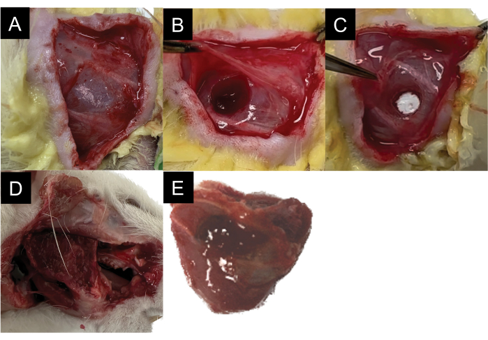
Figure 1: Surgical schematic demonstrating the steps. (A,B) Surgical masseter VML injury. (C) Biomaterial application. (D) Masseter harvesting. (E) Muscle morphology following harvesting. Please click here to view a larger version of this figure.
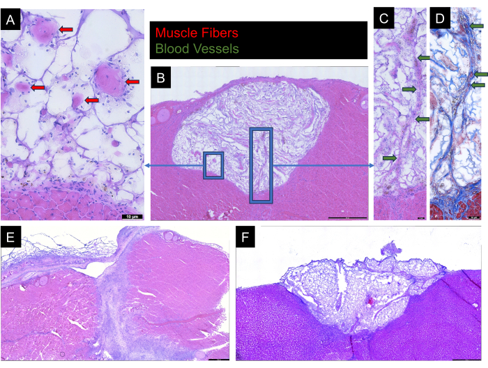
Figure 2: HE and Masson's trichrome staining. (A) HE-stained rat masseter sections from hydrogel-muscle interface with arrows indicating structures morphologically identified as skeletal muscle fibers. (B) HE-stained rat masseter section demonstrating the abundance of skeletal muscle fibers throughout hydrogel. (C) HE-stained and (D) Masson's trichrome-stained tissue with arrows indicating structures morphologically identified as blood vessels. (E,F) Negative results showing full loss of hydrogel with gel adhered to the superficial aspect of the masseter rather than the wound bed and incomplete loss of hydrogel with loss of fascial closure, respectively. Please click here to view a larger version of this figure.
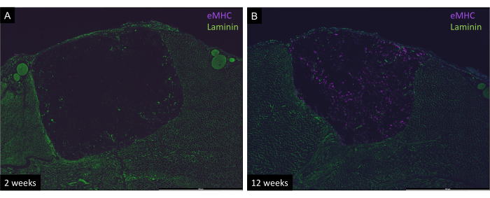
Figure 3: Immunohistologic staining for embryonic myosin heavy chain (eMHC) and laminin on rat masseter cross sections. (A) 2 weeks post-surgery. (B) 12 weeks post-surgery. An increased concentration of eMHC is seen in the 12-week sample relative to the 2-week sample. Images captured at 20x magnification. Please click here to view a larger version of this figure.
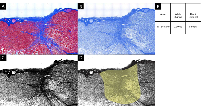
Figure 4: Quantitative fibrosis analysis. (A) Masson's trichrome-stained rat masseter section for quantification of connective tissue deposition as a proxy for wound fibrosis after VML injury at 12 weeks. (B) Image J was used to deconvolute the blue channel corresponding to connective tissue. (C) The image was converted to grayscale for analysis. (D) The area corresponding to the fibrotic scar was isolated, and (E) the cross-sectional area and percentages of the field corresponding to white (background) and black (connective tissue) were calculated. Images captured using light microscopy at 10x magnification. Please click here to view a larger version of this figure.
There are several critical steps in the protocol where special attention is needed to achieve an optimal result. Step 1.4 describes the initial incision and blunt separation of the skin from the superficial masseter fascia. Blunt dissection should be done directly along the skin with scissors pointing away from the underlying muscle and fascia to prevent nicking and unintentionally creating a window through the fascia. The caudal aspect of the superficial masseter should be avoided to prevent unintentional injury to the submandibular vein or external carotid artery. Step 1.5 and 1.6 describe the creation of the fascial window and VML defect, respectively. Care should be taken to ensure the fascial incision is the minimal size required in order to fit the punch biopsy through, as the fascia tends to contract away from the incision, and it can be difficult to locate the edges in step 1.8 if too large an incision is made. Again, blunt dissection beneath the fascia should be done only enough to allow the punch biopsy to fit within the window and angled appropriately to be perpendicular to the mandible. When creating the VML injury, it is important to identify the largest area between the mandibular and buccal branches of the facial nerve. The distal mandibular nerve divides into an upper and lower division, and there is variation in the location where the upper division branches away from the lower division towards the buccal branch of the facial nerve. Because this division courses superiorly, some animals may have the upper division of the mandibular nerve directly in the belly of the masseter where the defect should be created. In these cases, the animal should not be used for analysis, as there will likely be a peripheral nerve injury confounding the data. When creating the punch biopsy, gentle twisting may be required to penetrate the full 5 mm depth. Step 2.4 describes the blotting of the tissue sample after washing it to remove excessive moisture. This is done to prevent ice crystals from forming during the freezing of the tissue and subsequent reduction in freezing artifacts on histology20.
One of the advantages of this method is the ease of access to the superficial masseter muscle and the relative lack of high-risk structures that may be encountered during the surgery. Because the vasculature enters the caudal end of the muscle, significant bleeding can be avoided in the majority of cases. This is important as it not only decreases the risk of morbidity but also increases standardization and decreases the chance that ischemia has a confounding effect on muscle regeneration. Because of the availability of a fascial enclosure, this protocol may be used to evaluate biomaterials in liquid or solid form factors. While an acellular hydrogel was used as an example for this protocol, this method can be replicated to test biomaterials with human stem cell incorporation using immunodeficient rats as has been done in previous studies of extremity VML21.
This study does not formally define the critical size injury in VML, the precise point where endogenous repair mechanisms can no longer regenerate the muscle remains unclear. While it stands to reason that the definition may involve the removal of a certain percentage of the muscle's volume, and indeed 20% is often cited, there is conflicting evidence, and data suggest that other factors, including the location and geometry of injury, age of the animal, among others may also contribute16,21,22,23. Despite this, the observation of fibrous scar and visible contraction of the muscle border observed at 12 weeks following injury (Figure 4A) demonstrates that pathological remodeling has occurred, strongly suggesting that the injuries used here are of critical size with respect to the standard definition.
Because craniofacial muscles in humans, like intrinsic hand muscles, are small compared to proximal limb muscles and carry out more precise movements, they are more prone to having their function impacted by VML. The masseter was selected in this protocol because of adequate bulk for testing and ease of access. However, the geometry and location of injury are important factors in evaluating VML, and conclusions generated using this model may not directly translate to other craniofacial muscles. The masseter is a muscle of mastication, and while it contributes to facial expression in children24, its role in facial expression decreases in adulthood24. Injuries to other muscles of the face, such as the zygomaticus major, may result in greater psychological affliction following VML due to their more direct role in facial expression throughout life relative to the masseter25,26. In addition, this study did not evaluate in vivo force production, a limitation that is an area of need in the field. To date, no reproducible method for craniofacial muscle functional testing with sufficient sensitivity to compare treatment groups has been reported, and this is complicated by the extensive compensatory muscles within the face. Future developments of this protocol will aim to introduce approaches to directly measure force in individual facial muscles.
Alternative methods of histologic analysis of masseter VML in a rodent model are very limited. Kim et al. created a wearable EMG system to assess the functionality of VML-injured masseter muscles in mice. However, their terminal timepoint was 4 weeks, they did not demonstrate the visualization of biomaterial in histology images, did not describe a fascial closure, and focused primarily on validation of their EMG system versus analysis of muscle regeneration and biocompatibility of biomaterials-based treatments17. New treatments are needed for craniofacial volumetric muscle loss, and this masseter VML model is sufficiently reproducible to assess muscle response to VML injury and to evaluate the effects of treatments on regeneration, fibrosis, and contracture, especially in the first 3 months after injury.
The authors have nothing to disclose.
This research is supported by the UCSF Yearlong Research Fellowship Program and the C-Doctor Interdisciplinary Translational Project Program. Thanks to the members of the Pomerantz Lab and Craniofacial Biology Program at the University of California San Francisco for their contributions.
F1.652 Myosin heavy chain (embryonic) monoclonal antibody | DSHB | F1.652 | |
Goat anti-Mouse IgG2b Cross-Adsorbed Secondary Antibody, Alexa Fluor 647 | Invitrogen | A-21242 | |
Goat anti-Rabbit IgG (H+L) Highly Cross-Adsorbed Secondary Antibody, Alexa Fluor 488 | Invitrogen | A-11034 | |
Integra Standard Biopsy Punches, Disposable Standard biopsy punch; 5 mm, Diameter: 0.19 in., 0.5 cm | Integra | 12460411 | |
Mounting Medium with DAPI – Aqueous, Fluoroshield | Abcam | ab104139 | |
Rabbit Anti-Mouse IgG H&L (Alexa Fluor 647) preadsorbed | Abcam | ab150127 | |
Sulfamethoxazole/Trimethoprim Oral Suspension, Cherry Flavored, 473 mL | Med-Vet International | SKU: RXBAC-SUSP |
.