This protocol is designed to demonstrate exposure method of cell cultures to inhaled toxic chemicals. Exposure of differentiated air-liquid interface (ALI) cultures of airway epithelial cells provides a unique model of airway exposure to toxic gases such as chlorine. In this manuscript we describe effect of chlorine exposure on air-liquid interface cultures of epithelial cells and submerged culture of cardiomyocytes. In vitro exposure systems allow important mechanistic studies to evaluate pathways that could then be utilized to develop novel therapeutic agents.
Cell cultures are indispensable to develop and study efficacy of therapeutic agents, prior to their use in animal models. We have the unique ability to model well differentiated human airway epithelium and heart muscle cells. This could be an invaluable tool to study the deleterious effects of toxic inhaled chemicals, such as chlorine, that can normally interact with the cell surfaces, and form various byproducts upon reacting with water, and limiting their effects in submerged cultures. Our model using well differentiated human airway epithelial cell cultures at air-liqiuid interface circumvents this limitation as well as provides an opportunity to evaluate critical mechanisms of toxicity of potential poisonous inhaled chemicals. We describe enhanced loss of membrane integrity, caspase release and death upon toxic inhaled chemical such as chlorine exposure. In this article, we propose methods to model chlorine exposure in mammalian heart and airway epithelial cells in culture and simple tests to evaluate its effect on these cell types.
Exposure to toxic inhaled chemicals (TICs)/gases such as chlorine (Cl2) remains an ongoing health concern in accidental exposures as well as in their potential use as a chemical threat agent. Although the lungs are the primary target, organs such as heart and brain are also affected1-3. In vivo models are generally used for testing toxicity from TICs, but in vitro assays for toxicity assessment are simpler, faster and more cost effective. In vitro models also allow for extensive investigation of agent-cell interactions that may be difficult to evaluate in vivo. Such in vitro exposure systems are rare and moreover, in some conventional models where toxic agents are added to the culture medium in which cells are submerged, the properties of the agents can change due to interactions and binding to components in the medium. In such scenarios cell culture systems such as air-liquid interface (ALI) cultures of primary human airway epithelial cells, proposed here, that can be directly exposed to gaseous agents could be promising.
Epithelial cells lining the airway are the first lines of defense against inhaled toxic chemicals. The human airway epithelium forms a physical barrier between the lumen and the underlying cells in the lung and participates in the response of the lung. It produces a number of cytokines and other pro- and anti-inflammatory agents as well as secretes mucus/airway surface liquid (ASL) covering the epithelium. One of the limitations in conventional submerged in vitro culture systems is also that the ASL and mucus that cover the epithelial surface is removed or diluted. This does not reflect the physiological condition of lung epithelial cells that are exposed to air. Thus, an ideal in vitro system for TIC toxicity testing should replicate this architecture. There is great interest in developing rapid screening methods that predict in vivo toxicity. Epithelial cells grown at the ALI differentiate and have well-differentiated structures and functions compared to cells grown submerged and serve a superior model of the airways.
In this study, we describe the use of air-liquid-interface culture of human airway (tracheobronchial) epithelial cells for testing poisonous inhaled gas toxicity and compare it with a submerged cell culture of cardiomyocyte, hence studying another important target of toxicity.
1. Rat Cardiomyocyte Cultures
2. Differentiated Air-liquid Interface (ALI) Culture of Human Airway Epithelial Basal Cells
3. Chlorine Exposure
4. Transepithelial Electrical Resistance (TER) Measurement
5. Caspase Measurement
6. Western Blot and Immunocytochemistry
Primary rod shaped cardiomyocytes attach on laminin matrices and spread and differentiate into confluent cultures (Figure 1A and its inset). These cells were further characterized on the basis of sarcomeric actin and SERCA2 expression (Figures 1B and 1C). Rat cardiomyocytes are highly susceptible to chlorine toxicity as 15 min exposure to 100 ppm chlorine caused extensive cell rounding and death in submerged cultures and disruption of confluent layers on cells grown on laminin coated membranes (Figure 1D). There was also enhanced apoptotic cell death as indicated by caspase 3/7 release in cardiomyocytes grown on inserts (Figure 1E).
Exposure of differentiated human airway epithelium (Figure 2, inset to panel 1 showing a cross section of cell culture inserts with columnar, ciliated and goblet cells) to chlorine caused sloughing and lifting of cell membranes at both low (100 ppm) and high (300 ppm) concentrations (Figure 2 panel A2 and A3). Damage by low chlorine concentrations was quickly reversed (Figure 2 panel A5), however, cells exposed to higher chlorine concentrations had delayed or no repair ability (Figure 2 panel A6) as shown by visual inspection as well as cell proliferation assessment by Ki-67 staining (Figure 2 panel A9). Trans epithelial electrical resistance, TER measurements and caspase activity further confirmed these results and provide evidence for loss of membrane integrity and apoptotic cell death upon chlorine exposure (Figure 2, panel B and C). Thus our studies describe the development of an in vitro Cl2 exposure system that causes loss of membrane integrity and death of airway epithelium and cardiomyocytes. This effect may not be due to non-physiological pH changes in the cardiomyocytes as the pH of the media after exposure to chlorine was maintained at ~7.4 as measured by using a pH meter in the collected media postexposure.
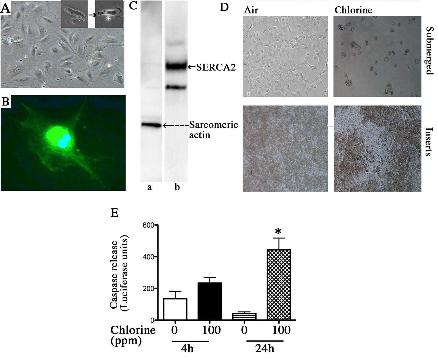
Figure 1. Rat cardiomyocyte isolation and exposure to chlorine. Rat cardiomyocytes were isolated as described in the protocol and plated on laminin-coated plastic dishes (panel A, a representative light microscopic image) or laminin coated inserts. Inset to panel A shows a rod shaped cardiomyocyte spreading and differentiating. The cardiomyocytes were also characterized based on the sarcomeric actin expression (panel B showing a representative image detected by immunofluorescence and lane a panel C showing a representative western blot scan) and abundant SERCA2 expression (panel C lane b). Chlorine exposure (100 ppm 15 min) caused extensive cell death in submerged cultures as well as disruption of the cell monolayer on the inserts (panel D showing the representative photomicrographs). Caspase 3/7 release (panel E) in the supernatant media of cardiomyocytes grown on inserts, at 4 hr and 24 hr post chlorine exposure was also measured as described in the text. Values shown are mean ± SEM and * indicates significant (p < 0.05) difference from 0 ppm control.
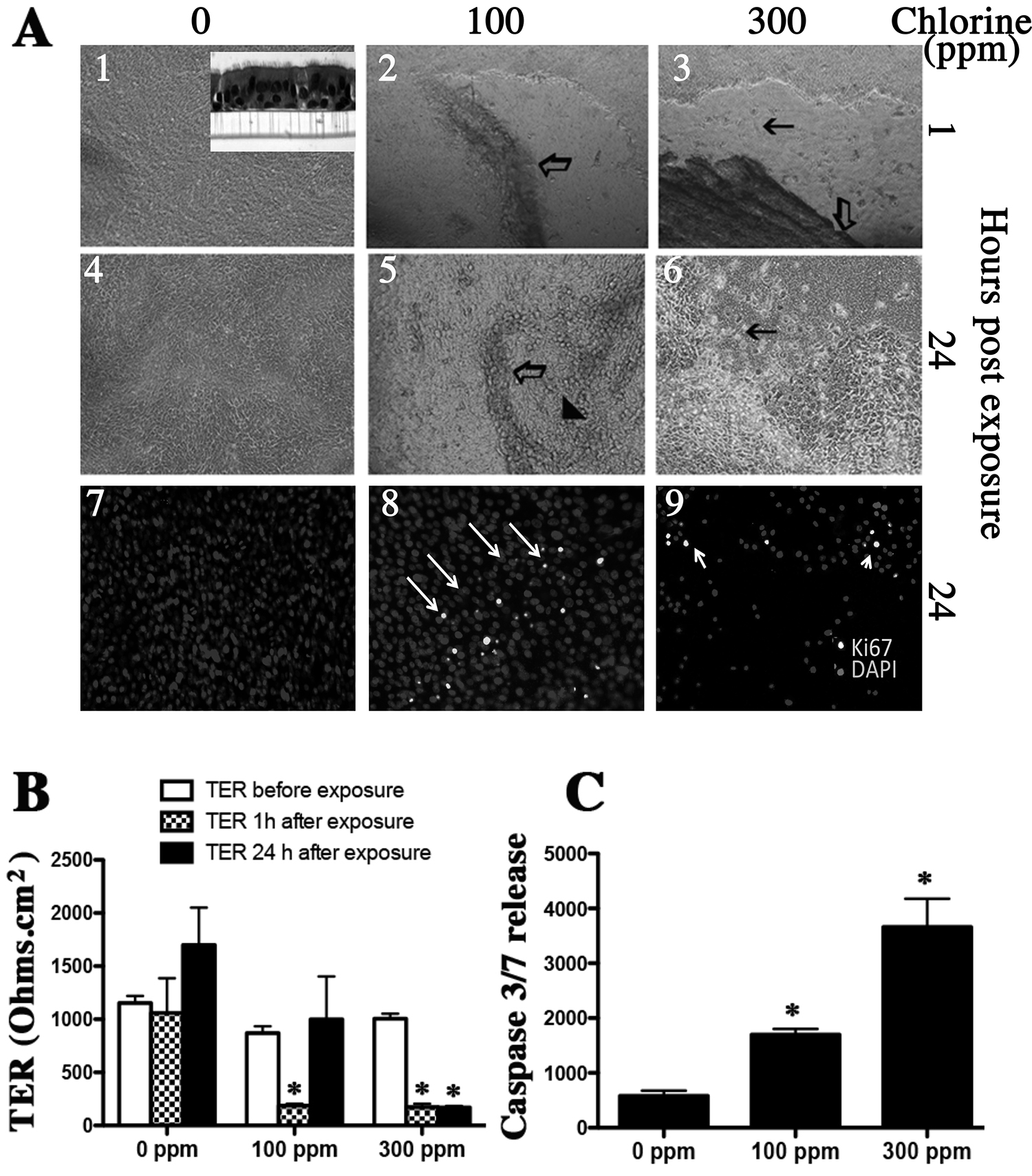
Figure 2. Effect of chlorine exposure on differentiated human airway epithelial air-liquid interface (ALI) cultures. Human airway epithelial basal cells were cultured on collagen coated snapwells. After day 5 the apical media was removed. Differentiated cultures (consisting of basal, ciliated, columnar, and goblet cells as shown in the inset in top left panel of panel A) were exposed to chlorine (100 or 300 ppm) for 30 min. The TER was measured and media was changed and cells incubated for 24 hr. At 24 hr TER was measured again and apical media was collected for caspase release measurement and the cell membranes were fixed for immunohistochemistry. The open arrow in panel A parts 2 and 3 shows sloughed off epithelial layer and black arrows show empty spaces on the insert. The arrowhead in panel A part 5 shows regenerated epithelium. Parts 7, 8, and 9 show cellular proliferation as assessed by Ki-67 immunostaining. Values shown are mean ± SEM and * indicates significant (p < 0.05) difference from 0 ppm control.
The most common type of acute toxic exposures occurs when one breathes a poisonous chemical into the lungs. These chemicals may also be quickly taken up in the bloodstream and may impact other organs such as brain and heart. Inhalation toxicity of various agents using animal models are studied and reported widely, however the mechanisms are less well understood. This is a major hurdle in developing effective therapies. Absence of in vitro exposure systems is a primary reason behind the lack of mechanistic insights. Here we describe cell culture models of heart muscle and airways to study impact of toxic inhaled chemicals such as chlorine exposure. Chlorine reacts rapidly with aqueous surfaces to form hydrochloric and hypochlorous acid3,11,12. Chlorine can also combine with reactive oxygen species (ROS) to produce highly reactive compounds that may lead to oxidation of critical proteins and enzymes of the airway surfaces13. Studies using submerged cultures may only demonstrate effects of by products such as HOCl in case of chlorine rather than the gas itself. Exposure of differentiated ALI cultures of human airway epithelial cells described here would allow study of direct interactions of TICs such as chlorine with cell surfaces in absence of aqueous media similar to what occurs in vivo.
Using primary rat cardiomyocytes, we also describe that chlorine exposure causes rapid cell death in submerged cultures. These cells are unable to grow at ALI as they do not polarize and form tight junctions. Therefore, we also utilized confluent cultures of cardiomyocytes grown on inserts with a thin layer of media. Exposure of these cell monolayers to chlorine demonstrated membrane disruption and enhanced caspase release suggesting apoptosis may play a role.
This study demonstrates that airways (the primary target of inhalation that can be replicated by ALI cultures) as well as organs such as heart that do not grow at ALI may be studied using in vitro exposure systems. These in vitro exposure models can easily be adapted to assess effects of other toxic inhaled chemicals (TICs). Using these models we anticipate to provide detailed mechanistic understanding of the toxicities and develop novel strategies to mitigate the toxic events associated with TIC/chlorine and then evaluate them further in vivo. Although, these exposures are short in duration the exposure system needs to better replicate cell culture conditions. Exposure to gases such as chlorine limits the use of humidity as it may corrode the device. The ALI cultures could be rocked to simulate respiration as previously utilized in our ozone exposure system14,15. We are currently working in these directions.
The authors have nothing to disclose.
This research is supported by the CounterACT Program, National Institutes of Health (NIH), Office of the Director, and the National Institute of Environmental Health Sciences (NIEHS) Grant Number U54 ES015678 (CWW). SA is also supported by Children's hospital Colorado/Colorado School of Mines Collaboration Pilot Award #G0100394 and Children's Hospital Colorado Research Institue Pilot Award #G0100471.
Name | Company | Catalog Number | |
Rats | Harlan Laboratories | Sprague-Dawley | |
Pentobarbital | Sigma-Aldrich | P3761 | |
Chlorine | AirGas, Inc | X02NI99CP163LS1 | |
Caspase 3/7 kit | Promega | G8091 | |
Epithelial voltohmmeter and chopstick electrode | World Precision Instruments | EVOM and STX2 | |
Snapwell inserts | Corning | 07-200-708 | |
70 micron nylon cell strainer | Corning | #352360 | |
Polysulfone biocontainment chambers | BCU, Allentown Cage Equipment | BCU | |
DMEM | Life technologies | 12491-015 | |
Sarcomeric actin antibody | Abcam Cambridge, MA | ab28052 | |
SERCA2 antibody | Affinity Bioreagents, Golden, CO | MA3-9191 | |
Ki-67 antibody | Dako, Carpinteria, CA | M7248 | |
Alexa-488-conjugated secondary antibody | Invitrogen, Grand Island, NY | A11029 | |
BSA | Sigma-Aldrich | A9418 | |
Carnitine | Sigma-Aldrich | C0283 | |
Taurine | Sigma-Aldrich | T8691 | |
Creatinine | Sigma-Aldrich | C6257 | |
Krebs Ringer Buffer | Sigma-Aldrich | K4002 | |
Protease | Sigma-Aldrich | P5147 | |
Collagenase | Sigma-Aldrich | C6885 | |
DNAase | Sigma-Aldrich | DN-25 | |
Lactated Ringer solution | Abott Laboratories | 7953 | |
Donkey serum | Fisher Scientific | 017-000-001 | |
PBS, phosphate buffered saline | Sigma-Aldrich | D1408 | |
4-15% SDS-PAGE gels | Bio-Rad | 456-1083 | |
Nitrocellulose membrane | Bio-Rad | 162-0115 | |
Dergent, Tween | Sigma-Aldrich | P1379 | |
Peroxidase detection kit | Pierce | 3402 | |
DAPI | Sigma-Aldrich | D9542 | |
Mounting media, Fluormount G | eBiosciences | 00-4958-02 | |
Sodium citrate | Sigma-Aldrich | 71497 | |
Collagen | Sigma-Aldrich | C7521 | |
MEM | Sigma-Aldrich | M8028 | |
Laminin | BD biosciences | 354259 | |
Penicillin/Streptomycin | Life Technologies | 15070063 | |
FBS | Gibco | 200-6140AJ |