Focus Ultrasound Based Microbubble Mediated Blood-Brain Barrier Opening: A Technique to Create Localized Transient Openings in Blood-Brain Barrier of Mouse by Sonoporation

Published: April 30, 2023

Abstract

Source: Haumann, R. et al. A High-Throughput Image-Guided Stereotactic Neuronavigation and Focused Ultrasound System for Blood-Brain Barrier Opening in Rodents. J. Vis. Exp. (2020)

This video describes a method of temporarily disrupting the blood-brain barrier (BBB) by creating transient openings using microbubble-mediated focused ultrasound (FUS). These transient local BBB openings can eventually be used to deliver larger-sized drug molecules directly into the brain parenchyma.

Protocol

All procedures involving human participants have been performed in compliance with the institutional, national and international guidelines for human welfare and have been reviewed by the local institutional review board.

1. Focused Ultrasound System

NOTE: The described setup is an in-house built BBB disruption system based on commercially available components and includes a 3D-printed custom-made cone and detachable stereotactic platform. The system is designed modular, which facilitates modifications according to available equipment and specific use. The protocol describes the procedure for the sonoporation of a larger area in the pontine region of the mouse brain. By adjusting the target location, different parts of the brain could be targeted. In this study, a 1 MHz mono-element transducer with a focal length of 75 mm, an aperture of 60 mm, and a focal area of 1.5 x 1.5 x 5 mm (FWHM of peak pressure) was used. The focal plane of the transducer is positioned through the cranium of the animal in the horizontal plane intersecting with the ear bars.

  1. Select an appropriate transducer for BBB opening in rodents.
    NOTE: Based on the properties of the microbubbles and the employed frequency, the acoustic settings, in particular the mechanical index (MI), are subject to change.
  2. Place the transducer in the 3D-printed cone.
  3. Employ an acoustically transparent mylar membrane at the bottom-end of the cone to achieve acoustic coupling of the beam propagation path, and fill the cone with degassed water.
  4. Mount the transducer above the animal on a motorized linear stage as shown in Figure 1 allowing automatic vertical positioning of the transducer.
  5. Design a detachable stereotactic platform based on the requirements of the study, which includes temperature regulated heating, bite and ear bars, anesthesia and multi-modality fiducial markers, as shown in Figure 1 and Figure 2. The mounting of the stereotactic platform consists of a 2D linear stage system, which allows precise automatic positioning (< 0.1 mm) of the animal under the beam.
  6. Connect the transducer to the acoustic emission chain shown in Figure 1 , consisting of a transducer, a function generator and a power amplifier.
  7. Devise an image-processing pipeline to detect the multi-modality fiducial markers that allow precise sonoporation targeting of the brain area of interest and collection of the cavitation data detected by the needle hydrophone.
  8. Calibrate the system and determine the focus point of the transducer in correspondence to vertical positioning of the animal on the stereotactic platform.

2. Animal Preparation

NOTE: The following protocol is specified for mice but can be adapted for rats. For these experiments female athymic nude Foxn1-/- mice (6–8 week old) were used.

  1. Allow the animal to acclimatize for at least one week in the animal facility and weigh the animal regularly.
  2. Administer buprenorphine (0.05 mg/kg) via subcutaneous (s.c.) injection 30 min prior to FUS treatment to start analgesic treatment.
  3. Anesthetize the animal with 3% isoflurane, 2 L/min O2 and verify that the animal is deeply anesthetized. Keep the animals anesthetized during the whole procedure and monitor the breathing frequency and heart rate to adjust the concentration of isoflurane as required.
  4. Apply eye ointment to prevent dry eyes and avoid possible injury.
  5. Remove hair on the top of the head with a razor and depilatory cream and wash afterwards with water to remove any residues to avoid irritation to the skin.
  6. For experiments with BLI tumor models, inject 150 µL of D-luciferin (30 mg/mL) intraperitoneal (i.p.) with a 29G insulin syringe for BLI image-guidance.
  7. Insert a 26–30G tail vein catheter and flush the catheter and vein with a small volume of heparin solution (5 UI/mL). Fill the catheter with heparin solution to avoid blood clotting.
    NOTE: Good catheterization is seen when there is a reflux of blood into the catheter. Avoid air bubbles in the catheter to prevent emboli. To avoid excessive injection pressure, make sure the length of the catheter is as short as possible.
  8. Place the animal on the temperature regulated stereotactic platform to avoid hypothermia.
    NOTE: Hypothermia reduces blood circulation, which can affect the injection/circulation of microbubbles and the pharmacokinetics of the drugs.
  9. Immobilize and fix the head of the animal on the stereotactic platform using ear bars and a bite bar. Fixate the body with a strap and tape the tail of the animal to the platform.

3. In Vivo Image-guided Focused Ultrasound

NOTE: For this protocol a 1 MHz mono-element transducer with a tone-burst pulse with a 10 ms duration, a MI of 0.4 and a pulse repetition frequency of 1.6 Hz with 40 cycles for 240 s was used. The protocol is optimized for microbubbles stabilized by phospholipids containing sulphur hexafluoride (SF6) as an innocuous gas, whereby the mean bubble diameter is 2.5 μm and more than 90% of the bubbles are smaller than 8 μm.

  1. Place the stereotactic platform with the mounted animal in the imaging modality (e.g., BLI or X-ray) and take image(s) of the animal.
  2. Use the multi-modality fiducial markers in combination with the image-processing pipeline to mark the position of the animal according to the focus point of the transducer.
  3. Determine the target area by placing a brain outline over the acquired X-ray image or using BLI images to determine the center of the tumor (Figure 2). The position of specific parts of the brain are specified in the Paxinos Brain Atlas using the skull markings bregma and lambda as reference points. For example the pons is located x=-1.0, y=-0.8 and z=-4.5 from lambda.
  4. Shield the animal’s nostrils and mouth with adhesive tape to prevent ultrasound gel interfering with breathing.
  5. Apply ultrasound gel on top of the animal’s head.
  6. Retract the skin of the animals’ neck, lubricate the needle hydrophone with ultrasound gel and place the needle hydrophone in the direct vicinity of the occipital bone.
  7. Guide the transducer to the correct position using the image-processing pipeline and the focus point.
  8. Apply the preconfigured settings to all attached devices and target the brain region of interest.
    NOTE: Depending on the research question, tumor or brain regions can be sonoporated as a single focal point or as volumetric shape, as shown in Figure 2.
  9. Activate microbubbles as described by the manufacturer. Inject one bolus of 120 µL (5.4 µg) of microbubbles.
  10. Flush the tail vein catheter with saline to check the opening of the catheter.
  11. Inject the microbubbles and start the insonation.
  12. Record microbubble cavitation with the needle hydrophone.
  13. Administer an intravascular contrast agent or drug after sonoporation. The dose, timing and planning are dependent on the purpose of the study and the drug.
    NOTE: Evans blue is a common color agent to assess BBB opening.
  14. Monitor the animal until the predetermined time point or before the humane endpoint.

Representative Results

Figure 1
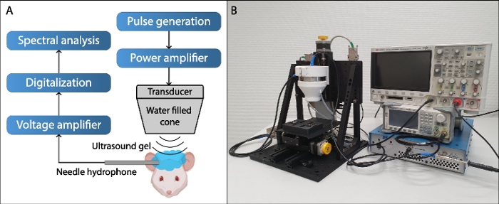
Figure 1: Focused ultrasound setup. (A) Schematic representation of the focused ultrasound setup. (B) Picture of the focused ultrasound setup. The system consists of a top-down mounted transducer on a 1D linear stage over a second 2D stage for automatic 3D positioning. The transducer is built in a water-filled beam-cone, closed at the bottom with an acoustically transparent mylar membrane, which conducts the sound to the cranium of the animal. The transducer is connected to a power amplifier, which is in-turn connected to an arbitrary waveform generator (AWG) for signal generation. For cavitation detection a detachable hydrophone in combination with a low-noise voltage amplifier is used. The hydrophone is placed in the direct vicinity of the occipital bone. The external hydrophone has a 2 mm active surface and is acoustically coupled with ultrasound gel. Both the high-voltage signal of the excitation pulse as well as the recorded cavitation signal are digitalized by a standard 200 MHz oscilloscope and relayed to a control computer (not shown) for on-the-fly processing and real-time control. 

Figure 2
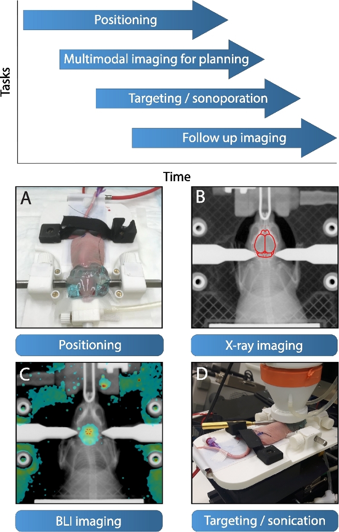
Figure 2: Focused ultrasound workflow. The proposed workflow of the focused ultrasound system starts with (A) the initial positioning of animal on a detachable stereotactic platform, note the application of the acoustic coupling gel (applied post BLI/X-ray). Simultaneously multimodal imaging can be conducted for targeting. (B) At first, X-ray imaging is a possibility, whereas a region of interest can be targeted with the help of an outline of the brain (which in turn is referenced to the mouse brain atlas40, adapted to the size and posture of the skull). (C) Alternatively, a BLI image of a luciferase transfected diffuse midline glioma tumor overlaid on an X-ray maximum intensity projection can be applied for targeting. (D) Subsequently, the stereotactic platform is mounted with the animal in therapy position with both hydrophone and transducer attached. The transducer automatically drives in therapy position and sonicates the chosen trajectory post bolus injection. The system is optimized for high-throughput experiments, whereby multiple platforms allow interleaved work, as shown on top. 

Declarações

The authors have nothing to disclose.

Materials

1 mL luer-lock syringe Becton Dickinson 309628 Plastipak
19 G needle Terumo Agani 8AN1938R1
23 G needle Terumo Agani 8AN2316R1
3M Transpore surgical tape Science applied to life 7000032707 Or similar
Arbitrary waveform generator Siglent SDG1025, 25 mHz, 125 MSa/s
Automated stereotact In-house built Stereotact with all elements were in-house built
Bruker In-Vivo Xtreme Bruker Includes software
Buffered NaCl solution B. Braun Melsungen AG 220/12257974/110
Buprenorfine hydrochloride Indivior UK limitd 0.324 mg
Cage enrichment: paper-pulp smart home Bio services
Carbon filter Bickford NC0111395 Omnicon f/air
Ceramic spoon
Cotton swabs
D-luciferin, potassium salt Gold Biotechnology LUCK-1
Ethanol VUmc pharmacy 70%
Evans Blue Sigma Aldrich E2129
Fresenius NaCl 0.9% Fresenius Kabi Nacl 0.9 %, 1000 mL
Histoacryl Braun Surgical Histoacryl 0.5 mL
Hydrophone Precision Acoustics
Insulin syringe Becton Dickinson 324825/324826 0.5 mL and 0.3 mL
Isoflurane TEVA Pharmachemie BV 8711218013196 250 mL
Ketamine Alfasan 10 %, 10 mL
Mouse food: Teklad global 18% protein rodent diet Envigo 2918-11416M
Neoflon catheter Becton Dickinson 391349 26 GA 0.6 x 19 mm
Oscilloscope Keysight technologies Infiniivision DSOX024A
Plastic tubes Greiner bio-one 210261 50 mL
Power amplifier Electronics & Innovation Ltd 210L Model 210L
Preamplifier DC Coupler Precision Acoustics Serial number: DCPS94
Scissors Sigma Aldrich S3146-1EA Or similar
Sedazine AST Farma 2%
SonoVue microbubbles Bracco 8 µl/mL
Sterile water Fresenius Kabi 1000 mL
Syringe Various syringes can be used
Temgesic Indivior UK limitd 0.3 mg/mL
Transducer Precision Acoustics 1 mhz
Tweezers Sigma Aldrich F4142-1EA Or similar
Ultrasound gel Parker Laboratories Inc. 01-02 Aquasonic 100
Vidisic gel Bausch + Lomb 10 g

Tags

Play Video

Citar este artigo
Focus Ultrasound Based Microbubble Mediated Blood-Brain Barrier Opening: A Technique to Create Localized Transient Openings in Blood-Brain Barrier of Mouse by Sonoporation. J. Vis. Exp. (Pending Publication), e20684, doi: (2023).

View Video