1. Reagent Preparation
- The relaxation and preservation solution (RP Solution) is prepared as previously described with minor modifications1. Briefly, the RP Solution consists of 2.77mM CaK2EGTA, 7.23mM K2EGTA, 20mM imidazole, 0.5mM dithiothreitol, 20mM taurine, 50mM K-MES, 6.56 MgCl2, 5.7mM ATP, 14.3mM phosphocreatine, pH 7.1, adjusted at room temperature (RT). Filter the solution through a 0.45-μm filter to sterilize. Divide into 15 mL portions (Falcon polypropylene tubes) and store at -20 °C See discussion for recipes.
- The Mitochondrial Respirometry Solution (MiR05) is prepared as previously described2 and contains 0.5mM EGTA, 3mM MgCl2.6H2O, 20mM taurine, 10mM KH2PO4, 20mM HEPES, 1g/L BSA, 60mM potassium-lactobionate, 110mM sucrose, pH 7.1, adjusted at 30°C. Filter the solution through a 0.45-μm filter to sterilize. Divide into 50 mL portions (Falcon polypropylene tubes) and store at -20 °C. See discussion for recipes. The oxygen solubility factor for MiR05 at 30°C and 37°C is 0.923.
2. Tissue Preparation
A. Cardiac fibers mechanical preparation
- Procedures were approved by the University of Calgary Animal Care and Use Committee and abide by the Canadian Association for Laboratory Animal Science guidelines for experimentation.
- Cervical dislocation to immobilize the mouse is the preferred method. Alternatively, intraperotineal (IP) injection of ketamine and xylazine (80 and 10mg/kg, respectively) or 0.5mg/kg of sodium pentobarbital may be administered. Note: Sodium pentobarbital is a reversible inhibitor of NADH dehydrogenase (complex I)4.
- Remove the heart and place it in a petri-dish containing the RP solution on ice (Figure 1A). Carefully remove any connective tissue and/or fat using a dissection microscope.
- Cut the heart in half along the septum. Excise 10-25mg wet weight (ww) of tissue by cutting from the endocardium surface of the left ventricle (Figure 1B). Ensure incision is along the fiber orientation to minimize mechanical damage to myocardium.
- Place the dissected, subsample of tissue in a separate petri-dish containing the RP solution on ice. Using a dissection microscope cut the heart into longitudinal strips along fiber orientation with a diameter of 1-1.5mm.
- Shorten the strips with a scalpel to lengths of 2-4mm (Figure 1C).
- Using extreme care and sharp forceps mechanically separate fiber bundles. Final fiber bundles should contain a maximum of 6-8 fibers, connected by small areas of contact, weighing no more than 5mg ww. Cardiac fibers of 1-3mg ww are recommended. Visually, the cardiac tissue should change from original red to a pale pink coloring (Figure 1D).
B. Cardiac fiber bundles chemical preparation
- Place a 12-well plate on ice.
- Rinse the well(s) with RP solution to minimize calcium contamination5.
- Transfer the mechanically prepared fiber bundles to a well containing 3mL ice-cold RP solution with 50ug/ mL saponin. Note: The concentration of saponin does not depend on the amount of cardiac muscle present in the solution.
- Incubate for 20min with mild stirring on ice.
C. Cardiac Fiber Bundle Washing
- Rinse a new well with MiR05 to minimize calcium contamination5.
- Transfer the cardiac bundles to the new well containing 10mL ice-cold MiR05. Incubate for 10min with mild stirring on ice.
- Repeat (1-2) 2-3 times with fresh MiR05. The repetitive washing steps are to ensure the removal or saponin, ATP, ADP and any remaining substrates from the fiber bundles.
D. Wet weight determination
- Directly prior to respiratory analysis, wet weight is obtained by blotting the individual fiber bundles (1-3mg ww) on an absorbent surface (filter paper) using forceps and holding the fiber to the surface for 5s or until all moisture is wicked away.
- Using another absorbent surface, remove excess liquid from forceps.
- Tare the scale and place the fiber bundle on a plastic weigh boat for mass measurement.
- Transfer the cardiac fiber bundle to a new well with ice-cold MiR05. The fiber bundle is ready for oxygen consumption assessment.
3. Respirometric OXPHOS Analysis
A. Respirometric Equipment
- The laboratory utilizes and recommends a two chamber titration-injection oxygraph (Oroboros Oxygraph2-k, Oroboros Instruments). The Oxygraph 2-k offers high resolution respirometry in large part by utilizing instrumental hardware and software (DatLab) that minimizes instrumental background that contribute to oxygen consumption artifacts.
- Calibration of the oxygraph is an essential step to minimize confounding effects of instrumental oxygen consumption. Calibration varies slightly depending on the oxygraph. Refer to oxygraph user manual for specific procedures.
B. Representative Quality Control/Technique Validation Protocol
- Ensure MiR05 has been added to oxygraph chambers housing the polarographic oxygen sensors (POS) and give adequate time for air saturation and equilibration of the MiR05 at 37°C prior to putting the murine cardiac fibers into the oxygraph chambers.
- The cardiac fiber samples are placed in the stirred MiR05 of the oxygraph chambers. Note: Ensure the stir bars are PVDF- or PEEK-coated stirrer bars (6mm diameter).
- Using an oxygen-filled syringe, increase the oxygen concentration of the oxygraph chambers to 250-550uM. It should be noted that oxygen concentration is limiting for permeabilized fiber preparations at even 50% above air saturation6. Allow 5-10min for oxygen concentration stabilization.
- Using a 25μL Hamilton microsyringe, add 10μL of 2M glutamate and 5μL of 800mM malate to obtain a final concentration of 10mM and 2mM, respectively. These titrations allow for the determination of basal complex I-supported respiration (State 2; absence of ADP). Oxygen flux should stabilize within approximately 5min of titration. Proper preparation should provide very reproducible state 2 oxygen consumption.
- Following 2-5min of stable oxygen consumption, add 20μL of 500mM ADP for a final concentration of 5mM (saturating), using a 25μL Hamilton microsyringe, for maximal (state 3) mitochondrial respiration through complex I. Few studies use this high of a concentration of ADP, however, it is important to note that greater than 90% saturation is only reached at concentrations above 5mM7.
- Following 2-5min of stable oxygen consumption, add 5μL of 4mM cytochrome c to obtain a final concentration of 10μM, using a 10μL Hamilton microsyringe, for quality control analysis of outer mitochondrial membrane (OMM) integrity.
- Following 2-5min of stable oxygen consumption, add 1μL of 4mg/ mL oligomycin, using a 10μL Hamilton microsyringe, to inhibit ATP synthase. This titration step will offer validation of inner mitochondria membrane intactness.
- Following the respirometric OXPHOS assessment. The sample is retained for dry weight or mitochondrial marker determination.
- Remove MiR05 from the glass chambers of the oxygraph. Wash the chambers at least three times with distilled water (ddH2O).
- Wash the chambers at least three times with 100% ethanol (EtOH) to remove EtOH-soluble inhibitors such as oligomycin.
- Wash the chambers with 70% EtOH three times with the final 70% EtOH wash lasting 30min to sterilize the oxygraph chambers.
- Prior to addition of MiR05 for new experimental titration protocol ensure the chambers containing the POS are washed at least five times with ddH2O.
- The validation titrations may be performed as an individual protocol (Figure 2) or the titrations may be included into a well-planned titration protocol depending on the experimental and diagnostic aims of the respirometry studies.
4. Representative Results:
Oxygen consumption in properly prepared murine cardiac fibers is evaluated by the quality control protocol as shown in Figure 2. Figures 3-5 provide commonly encountered examples of incorrectly prepared cardiac fibers. The respiratory control ratio (RCR) represents an important index in respirometry. This parameter indicates the coupling between oxygen consumption and oxidative phosphorylation. In permeabilized fiber preparations, the RCR is the rate of respiration in state 3 relative to state 2 or alternatively state 3 over state 4 (induced by oligomycin and/or atractyloside, ATR). Furthermore, RCR can be used as a quality assurance marker and can identify changes in coupling resulting from experimental or pathological interventions5. Well-coupled permeabilized murine cardiac preparations yield an RCR between 3-6 depending on the incubation solution utilized1, 8, 9.
The titration of cytochrome c is used as a validation of proper tissue preparation. Cytochrome c is a protein located in the intermembrane space at the mitochondrial inner membrane10. When the outer membrane of mitochondria is intact, the endogenous cytochrome c remains in the intermembrane space and the titration of exogenous cytochrome c has a negligible effect on respiration (Figure 2). If the outer membrane of mitochondria is damaged, the endogenous cytochrome c can be released from the intermembrane space and will inhibit respiration until exogenous cytochrome c addition (Figure 3). Proper preparations should experience only a slight elevation in oxygen flux following cytochrome c addition in the 5-15% range5. Additionally, this experimental titration allows for the assessment of the pathological or experimental stressor’s influence on mitochondrial outer membrane intactness. If a cytochrome c effect is experienced, ensure care during mechanical preparation of the tissue and/or reduce saponin concentration.
The addition of oligomycin and/or ATR is used to assess alterations in leak respiration; oxygen consumption not contributing to ADP phosphorylation11. Additionally, this titration step may be used as a validation of proper sample preparation. Control or wild-type fibers should be sensitive to this addition and experience a significant reduction in oxygen flux. A low RCR and reduced sensitivity to oligomycin and/or ATR resulting in a relatively elevated oxygen flux indicates damage to the inner mitochondrial membrane during preparation (Figure 4). Damage is likely induced during mechanical separation of the cardiac fibers as the inner mitochondrial membrane is less susceptible to insult by saponin relative to the outer mitochondrial membrane. However, both mechanical and chemical preparation may have to be adjusted accordingly to avoid improperly prepared cardiac fibers5, 12.
The saponin-skinned fiber bundles from mice should not remain in the RP solution for more than 6h or the MiR05 solution for greater than 2h. Lack of response to substrate-inhibitor-uncoupler titrations as seen in Figure 5 may be indicative of prolonged incubation periods. Subsequent efforts should minimize periods between animal sacrifice and oxygen consumption measurements.
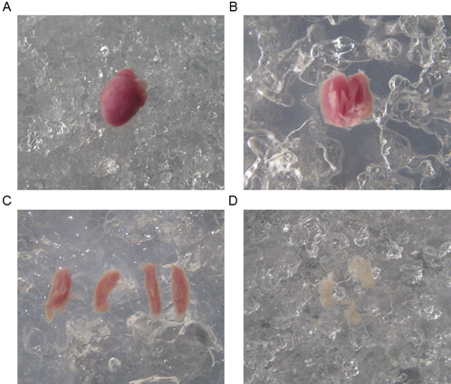
Figure 1. Mechanical preparation of murine cardiac fiber bundles. A. The entire heart immediately following dissection. B. A 10-25mg sample of the anterior left ventricle. C. Cardiac tissue separated into 1mm diameter and 2-4mm length strips. D. Final cardiac fiber bundles ready for chemical permeabilization with saponin.
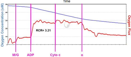
Figure 2. Representative results of proper tissue preparation utilizing polarographic assessment. The state 2 oxygen flux is supported by complex I substrates glutamate and malate (M/G) and is significantly stimulated following the addition of ADP (state 3). No stimulatory effect of exogenous cytochrome c addition indicates the outer mitochondrial membrane is intact. Sensitivity of oxygen consumption to oligomycin suggests the inner mitochondrial membrane integrity is intact. Oxygen concentration in a 2 mL closed chamber is identified by the blue line. Oxygen consumption of the cardiac tissue sample in a 2 mL closed chamber is represented by the red line. Malate and glutamate, M/G; Adenosine Diphosphate, ADP; Cytochrome c, Cyto c; Oligomycin, o.
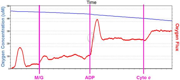
Figure 3. Cytochrome c effect validation test utilizing polarographic assessment. The state 2 oxygen flux is supported by complex I substrates glutamate and malate (M/G) and is significantly stimulated following the addition of ADP (state 3). Stimulatory effect of exogenous cytochrome c addition indicates the outer mitochondrial membrane integrity is compromised. Oxygen concentration in a 2 mL closed chamber is identified by the blue line. Oxygen consumption of the cardiac tissue sample in a 2 mL closed chamber is represented by the red line. Malate and glutamate, M/G; Adenosine Diphosphate, ADP; Cytochrome c, Cyto c.
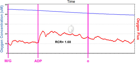
Figure 4. Inner mitochondrial membrane integrity validation test utilizing polarographic assessment. The state 2 oxygen flux is supported by complex I substrates glutamate and malate (M/G) and a relatively weak stimulation of oxygen consumption following the addition of ADP (state 3). Poor sensitivity of oxygen consumption to oligomycin suggests the inner mitochondrial membrane is damaged. Oxygen concentration in a 2 mL closed chamber is identified by the blue line. Oxygen consumption of the cardiac tissue sample in a 2 mL closed chamber is represented by the red line. Malate and glutamate, M/G; Adenosine Diphosphate, ADP Oligomycin, o.
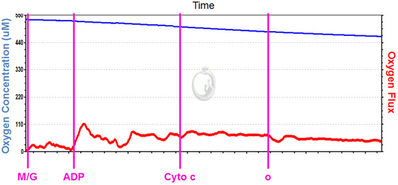
Figure 5. Polarographic assessment following prolonged incubation in MiR05. Oxygen consumption is insensitive to exogenous addition of glutamate and malate (M/G) and oxygen consumption following the addition of ADP (state 3) is reduced. There is no stimulatory effect of exogenous cytochrome c addition and insensitivity of oxygen consumption to oligomycin is experienced. Lack of response to additions to extramitochondrial incubation solution suggests mitochondrial functional stability is compromised. Oxygen concentration in a 2 mL closed chamber is identified by the blue line. Oxygen consumption of the cardiac tissue sample in a 2 mL closed chamber is represented by the red line. Malate and glutamate, M/G; Adenosine Diphosphate, ADP; Cytochrome c, Cyto c.
1. Notes: Reagent Preparation
Preparation of K2EGTA 100mM Stock Solution
| Name of the reagent | Final Concentration (mM) | g/100 mL H2O |
| Ethylene glycol-bis-(2-aminoethylether)-N,N,N‘,N‘-tetraacetic acid (EGTA) | 100 | 3.805 |
| Potassium Hydroxide (KOH) | 200 | 1.15 |
Note: Adjust pH to 7.4 at room temperature.
Preparation of Ca2EGTA 100mM Stock Solution
| Name of the reagent | Final Concentration (mM) | g/100 mL H2O | Comments (optional) |
| Ethylene glycol-bis-(2-aminoethylether)-N,N,N‘,N‘-tetraacetic acid (EGTA) | 100 | 3.805 | Heat to 80°C and stir mildly. |
| Calcium Carbonate (CaCO3) | 100 | 1.001 | The calcium concentration must be precise as calcium regulates the function of various organelles including mitochondria. Ensure complete solubilization of all CaCO3. The final solution must be completely transparent. CaCO3 is initially mixed with EGTA and some water to activate the formation of carbonic acid and CO2 evaporation. This reaction may be accelerated by heating up to 80 °C. |
| Potassium Hydroxide (KOH) | 200 | 1.15 | Neutralize with KOH after evaporation of CO2 is completed. |
Note: Adjust pH to 7.4 at room temperature.
Preparation of Relaxation and Preservation Solution (RP Solution)1
| Reagent | Final Concentration (mM) | Per litre | Comments |
| K2EGTA | 7.23 | 72.3 mL | |
| CaK2EGTA | 2.77 | 27.7 mL | |
| Imidazole | 20 | 1.36g | |
| Dithiothreitol | 0.5 | ||
| Taurine | 20 | 2.52g | |
| Adenosine 5′-triphosphate disodium salt hydrate (ATP) | 5.7 | 3.14g | |
| Phosphocreatine (PCr) | 14.3 | 4.0g | |
| Magnesium chloride (MgCl2) | 6.56 | 0.624g | |
| K-MES | 50 | 14.0g |
Note: Adjust pH to 7.1 at room temperature
Preparation of Mitochondrial Respirometry Solution (MiR05 Solution)2
| Reagent | Final Concentration (mM) | Per litre | Comments (optional) |
| Ethylene glycol-bis-(2-aminoethylether)-N,N,N‘,N‘-tetraacetic acid (EGTA) | 0.5 | 0.190g | Used as a chelator of calcium |
| Magnesium chloride hexahydrate (MgCl2.6H2O) | 3.0 | 0.610g | The quality of fiber preparation cannot be tested without Mg2+. |
| Taurine | 20.0 | 2.502g | Taurine is a membrane stabilizer and antioxidant. 20mM is the intracellular concentration present in the heart. |
| Potassium phosphate monobasic (KH2PO4) | 10.0 | 1.361g | |
| HEPES | 20.0 | 4.77g | |
| Potassium-lactobionate | 60.0 | 120 mL of 0.5 M K-lactobionate stock |
0.5M K-lactobionate Stock: Add 35.83 g lactobionic acid to 100 mL H2O and pH to 7.0 at RT. Adjust volume to 200 mL with ddH2O. Used to replicate the high intracellular K+ concentration. Previously KCl was used, however, the high Cl- inhibits mitochondrial creatine kinase function. |
| Sucrose | 110.0 | 37.65g | Used as a ROS scavenger. |
| Bovine Serum Albumin (BSA) | 1g/L | 1g | Used as a membrane stabilizer, antioxidant, and chelator of calcium and free fatty acids. |
Note: Adjust pH to 7.1 at 30°C
3. Notes: Respirometric OXPHOS Analysis
B. Select Substrates, uncouplers and inhibitors
List of Selected Substrates for Mitochondrial Respirometry Analysis
| Substrate | [Stock] | Preparation | Volume per 2 mL | [Final] | Comments |
| Adenosine 5′-diphosphate monopotassium salt dihydrate (ADP) | 500mM | 246mg/ mL ddH2O. Adjust pH to 7.1 at RT. Store at -80°C in 250 μL aliquots. | 20ul | 5mM | To maintain constant Mg2++ during respirometry experiments add 0.6 mol MgCl2/mol ADP. |
| Ascorbate | 800mM | 0.1584g/ mL ddH2O. Store at -20°C in 200 μL aliquots. Light sensitive | 5 μL | 2mM | Acts as substrate when used in parallel with TMPD. Must correct for oxygen flux for auto-oxidation. |
| Cytochrome C | 4mM | 50mg/ mL ddH2O. Store at -20°C in 250 μL aliquots. | 5 μL | 10uM | |
| Carbonyl cyanide p-(trifluoromethoxy) phenylhydrazone (FCCP) |
0.1mM | 0.254mg/10 mL 100% EtOH. Store in glass vials at -20°C in 500 μL aliquots. | Steps of 1 μL | Acts as an uncoupler. Determines maximal electron transport capacity and any limitation of electron transport by phosphorylation system. | |
| Glutamate | 2M | 0.3742g/ mL ddH2O. Adjust pH to 7.1 at RT. Store at -20°C in 250 μL aliquots. | 10ul | 10mM | Acts as a substrate for NADH dehydrogenase (complex I). |
| Malate | 800mM | 0.1073g/ mL ddH2O. Adjust pH to 7.1 at RT. Store at -20°C in 250 μL aliquots. | 5ul | 2mM | Acts as a substrate for NADH dehydrogenase (complex I). Cannot support respiration alone. |
| Pyruvate | 1M | 11mg/0.1 mL ddH2O. Prepare fresh. | 5 μL | 2.5mM | Acts as a substrate for NADH dehydrogenase (complex I). |
| Succinate | 1000mM | 1.3505g/5 mL ddH2O. Adjust pH to 7.1 at RT. Store at -20°C in 250 μL aliquots. | 20ul | 10mM | Acts as a substrate for succinate dehydrogenase (complex II). |
| N,N,N’,N’-Tetramethyl- pphenylenediamine Dihydrochloride (TMPD) |
200mM | 47.1mg/ mL ddH2O. Add 0.8M ascorbate to final concentration of 10mM to prevent auto-oxidation Store at -20°C in 200 μL aliquots. | 5 μL | 0.5mM | Autoxidation of stock solution evident by appearance of blue coloring. Acts as substrate when used in parallel with TMPD. Must correct for oxygen flux for auto-oxidation. |
List of Selected Inhibitors for Mitochondrial Respirometry Analysis
| Substrate | [Stock] | Preparation | Volume per 2 mL Chamber | [Final] | Comments |
| Antimycin A | 5mM | 27.4mg/10 mL 100% EtOH. Store at -20°C in 250 μL aliquots. | 1 μL | 2.5μM | Inhibitor of coenzyme Q : cytochrome c oxidoreductase (Complex III) |
| Atractyloside | 50mM | 40mg/ mL ddH2O. Store at -20°C in 250 μL aliquots. | 30 μL | 0.75mM | Inhibitor of ATP Synthase. |
| Oligomycin | 4mg/ mL | 4mg/ mL 100% EtOH. Store at -20°C in 200 μL aliquots. | 1 μL | Inhibitor of ATP synthase. | |
| Potassium cyanide | 1M | 65.1mg/ mL ddH2O. Prepare fresh. Adjust pH to 7.1 at RT | 1 μL | 1mM | Inhibitor of cytochrome c oxidase (complex IV). Utilize following TMPD and Ascorbate titration to access autoxidation. |
| Rotenone | 0.1mM | 0.39mg/10 mL 100% EtOH. Store at -20°C in 250 μL aliquots. Light sensitive. | 1 μL | 0.05μM | Inhibitor of NADH dehydrogenase (complex I). Higher concentrations may be required, however, to reduce rotenone retention in chamber begin as outlined. |
