Marine snail Aplysia californica has been widely used as a neurobiology model for the studies on cellular and molecular basis of behavior. Here a methodology is described for exploring the nervous system of Aplysia for the electrophysiological and molecular analyses of single neurons of identified neural circuitry.
A major challenge in neurobiology is to understand the molecular underpinnings of neural circuitry that govern a specific behavior. Once the specific molecular mechanisms are identified, new therapeutic strategies can be developed to treat abnormalities in specific behaviors caused by degenerative diseases or aging of the nervous system. The marine snail Aplysia californica is well suited for the investigations of cellular and molecular basis of behavior because neural circuitry underlying a specific behavior could be easily determined and the individual components of the circuitry could be easily manipulated. These advantages of Aplysia have led to several fundamental discoveries of neurobiology of learning and memory. Here we describe a preparation of the Aplysia nervous system for the electrophysiological and molecular analyses of individual neurons. Briefly, ganglion dissected from the nervous system is exposed to protease to remove the ganglion sheath such that neurons are exposed but retain neuronal activity as in the intact animal. This preparation is used to carry out electrophysiological measurements of single or multiple neurons. Importantly, following the recording using a simple methodology, the neurons could be isolated directly from the ganglia for gene expression analysis. These protocols were used to carry out simultaneous electrophysiological recordings from L7 and R15 neurons, study their response to acetylcholine and quantitating expression of CREB1 gene in isolated single L7, L11, R15, and R2 neurons of Aplysia.
The human brain is extraordinarily complex with almost 100 billion neurons and trillions of synaptic connections. There are almost an equal number of nonneuronal cells that interact with neurons and regulate their function in the brain. Neurons are organized into circuits that regulate specific behaviors. Despite the advances in our understanding of brain functions and neural circuits, little is known about the identity of circuitry components that control a specific behavior. Knowledge of the identities of various components of a circuitry will greatly facilitate our understanding of both cellular and molecular basis of behavior and aid in developing novel therapeutic strategies for neuropsychiatric disorders.
The marine snail Aplysia californica has been a workhorse for determining neuronal circuits underlying specific behaviors1-14. The Aplysia nervous system contains approximately 20,000 neurons that are organized into 9 different ganglia. The neurons of Aplysia are large and can be easily identified based on their size, electrical properties, and position in the ganglia. Aplysia has a rich repertoire of behaviors that can be studied. One of the well-studied behaviors is the gill-withdrawal reflex (GWR). The central components of this reflex are situated in abdominal ganglia. Components of the GWR circuitry have been mapped and contributions of various components determined. Importantly, GWR circuitry undergoes associative and nonassociative learning5,6,15-19. Decades of study on this reflex have also identified several signaling pathways that have a key role in learning and memory20-24.
Several different preparations of Aplysia were used to study cellular and molecular basis of memory storage. These include the intact animal2,3, semi-intact preparation1,7,13,14,16 and reconstitution of major components of neural circuitry25-29. A reduced preparation for exploring Aplysia ganglia for the electrophysiological and molecular analyses of identified neuronal circuits is described here. The following four identified neurons were studied. R15, a bursting neuron, L7 and L11, two different motor neurons and R2, a cholinergic neuron were studied. R2 is the largest neuron described in the invertebrate nervous system. Briefly, this methodology involves protease treatment of ganglia, electrophysiological measurements before and after pharmacological treatments, and isolation of single neurons for quantitative analysis of gene expression. This methodology enables us to combine molecular analyses with simultaneous recording from multiple neurons. This methodology was successfully used to study responses of R15 and L7 neurons to acetylcholine (Ach) by paired intracellular recordings. Following electrophysiological measurements R15 and L7 and other identified neurons such as L11 and R2 were isolated for quantitative polymerase chain reaction (qPCR) analysis of expression of CREB1, a transcription factor important for memory storage.
1. Preparation of Abdominal Ganglia, Electrophysiological Measurements, and Isolation of Single Identified Neurons from Abdominal Ganglion of Aplysia californica
2. RNA Isolation from Single Neurons and Gene Expression Analysis by Quantitative Real Time PCR (qPCR)
The weights of animals that were used in this study ranged from 100-200 g. Following the described protocols, we conducted electrophysiological measurements and molecular analysis of neurons of abdominal ganglia isolated from animals ranging from 2-5 g to 200-300 g.
Standardization of protease treatment is important for successful electrophysiological measurements of neurons in the ganglia. Initially, multiple protease (Dispase) concentrations and durations were used and bursting action potentials from R15 neuron were recorded26,31. These measurements were compared to direct (without protease treatment) recording from the intact ganglia. Concentration of the protease (0.1% protease, see step 1.3) that produced bursting potentials that were comparable to those obtained from exposed ganglion without protease were used in the subsequent experiments.
Using the protocol described, action potentials (AP) in response to Ach exposure to R15, a cholinergic neuron and L7, a motor neuron that does not respond to Ach were recorded. As expected, simultaneous electrophysiological recordings show that Ach induce depolarization in R15 whereas there was no change in firing of L7. Figure 2 shows the application of Acetylcholine (Ach) directly into the cell chamber. 1 M Ach (volume 0.3 μl) was directly applied into the chamber using micropipette in order to obtain a final 1 mM concentration inside the chamber.
Ach was washed off from the bath by perfusion and recorded again from R15 neurons. Figure 3 shows waveforms suggesting that Ach effect is reversible. Analysis of the parameters AP in control, during depolarization and after wash is shown. During depolarization amplitude is decreased and duration of AP increased significantly.
Following electrophysiological measurements, R15 and L7 neurons were isolated and RNAs were prepared for qPCR analyses. A linear T7 RNA Polymerase—driven transcription using MessageAmp II aRNA Amplification Kit from Ambion was used for amplifying RNAs from single neurons. For comparison, identified neurons R2 and L11 and the whole abdominal ganglia from adult (6-9 months old) and young animals (1-2 months old) were also studied. qPCR amplification were carried out in a 7900HT Fast Real-Time PCR System under the following conditions: 95 °C for 10 min, followed by 40 cycles of 95 °C for 15 sec, 60 °C for 1 min. There were four biological replicates in the experiment. Each biological replicate had four technical replicates in the qPCR set up. Ct values from four replicates were averaged and 1/Ct was calculated to find the absolute expression level of CREB1 in each sample (Figure 4). An estimation of the absolute expression levels of CREB1 gene in young and adult Aplysia ganglia, and in four identified single neurons was monitored by the Ct value obtained from qPCR measurements.
The starting mRNA concentration was the same in both young and adult abdominal ganglia. The same forward and reverse CREB1 primer set were used in all qPCRs, both in studies using ganglia and single neurons. The Ct values are very similar between young and adult abdominal ganglia. Similarly, Ct values were measured in the case of single neurons where the starting total mRNA concentrations were low and two rounds of amplification yielded a good amount of RNAs for cDNA synthesis. The Ct values of CREB1 amplification suggested that CREB1 expression in R15 and L7 neurons are comparable whereas R2 and L11 showed significant differences in expression when compared to L7 or R15 (Student's T test, p<0.05) (Figure 4).
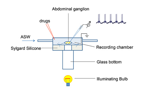
Figure 1. Microscope stage and perfusion system for ganglia electrophysiology. Perfusion system and stage were made using Plexiglas and are transparent. This setup is mounted on to a metal block (not shown) to hold position. The metal block is designed such that samples can be illuminated from the bottom. A sample electrophysiological bursting pattern recorded from R15 neuron is shown.
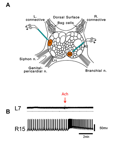
Figure 2. Simultaneous recording from L7 and R15 neuron of abdominal ganglia. (A) Cartoon of abdominal ganglia showing the position of L7 and R15 neurons (modified from Kandel 2001, reprinted with permission from AAAS). Also shown are the positions of R2 and L11 in the abdominal ganglia. (B) Representative intracellular recordings from L7 and R15. Arrow indicates time of Ach application. One could continue these measurements for 8-10 hr.
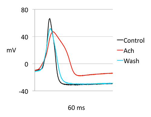
Figure 3. Ach induced changes in action potential (AP) of R15. AP waveforms were analyzed before, during and after Ach exposure. Representative waveforms are shown. ms: millisecond, mV: millivolt.
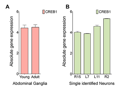
Figure 4. qPCR analysis of expression of CREB1 in abdominal ganglia and single identified neurons. (A) Absolute quantification of CREB1 transcript in the young and adult abdominal ganglia of Aplysia. Total RNA was isolated using standard Trizol protocol as described in the methods. 1 μg of total RNA was used to synthesize cDNA, followed by real time PCR amplification using CREB1 primers. (B) Absolute quantification of CREB1 transcript in identified single neurons of old Aplysia. Total RNA was subjected to two rounds of in vitro amplification before cDNA synthesis. 1 μg of aRNA was used to synthesize cDNA, followed by real time PCR amplification using CREB1 primers.
The neuron R15 is involved in regulating cardiovascular, digestive, respiratory, and reproductive systems30. A regularly rhythmic bursting activity of the AP is a feature of R15. As shown in the results section, paired recording of R15 and L7 show that the ganglia preparation has preserved activity of R15 neurons. R15 and L7 neurons responded appropriately to Ach. This ganglia preparation could be maintained up to 8-10 hr and electrophysiological activity could be continuously monitored. Thus, one could study the electrophysiological effects of brief neurotransmitter exposure (exposed locally to a specific neuron or to the bath) for a long period of time. Furthermore, one could study the effects of exposure to multiple neurotransmitters (simultaneously or in tandem) on the same neuron on firing properties of itself or the follower neuron or both.
An example of gene expression measurements by qPCR is shown in Figure 4. Since the RNA content of a single neuron is low, in vitro amplification is necessary to obtain a sufficient amount of RNA for quantitating expression of multiple genes. Commercial kits are available for the in vitro transcription process. The amplification of the CREB1 transcript was quantified directly by the number of cycles required to obtain a standardized fluorescence signal, the Ct-method31. The amplified RNA from a single neuron is sufficient for the studies to determine gene expression changes in a large number of genes using microarray32 and for the entire transcriptome by RNAseq analyses33.
A limitation of this technique is that this preparation may not preserve all the synaptic connections of the neurons of the abdominal ganglia. Several of the neurons form synapses with neurons in other ganglia and in the ganglia preparation these long-distance connections will be missed. However, the methodology described here could be easily modified to include the entire CNS or could be easily integrated to semi-intact preparation13,14,16 that was described to study cellular and electrophysiological changes in gill and siphon withdrawal reflex.
Recent advances in genomics techniques now allow us to obtain a deep insight into the dynamics of expression of coding and noncoding RNAs34. The reduced preparation that is described here is ideal for studying temporal changes of gene expression in conjunction with electrophysiological measurements. For example, one could apply pharmacological agents and measure their effects on electrical properties of individual neurons or multiple neurons that are part of the same neural circuitry and isolate individual neurons at different times for RNA isolation. The effect of manipulation of a single neuron (such as electrical stimulation, or exposures to pharmacological agents) on the entire identified circuitry could be studied at the level of changes in expression of specific genes in single neurons. Thus this methodology facilitates successful integration of electrophysiological and genomic methodologies to study neural circuitry in Aplysia.
This approach could be easily extended to the study of other invertebrate models such as nudibranch mollusc Tritonia, pond snail Lymnaea, and terrestrial snail Helix whose nervous systems contain several identified neurons35-57. These models are also used extensively for the study of neural control of behaviors such as escape swimming, magnetic field orientation, synapse formation, learning and memory storage. The methodology we described is a simple integration of genomics analysis with physiological measurements of neuronal activity at single cell level. A deeper insight into molecular and physiological basis of neural control of behavior could be obtained from such studies.
The authors have nothing to disclose.
We sincerely thank the Whitehall Foundation for their funding support and startup funds from The Scripps Research Institute for carrying out this work.
Aplysia | National Aplysia Resource Facility, University of Miami | |
NaCl | SIGMA | S 3014-1KG |
KCl | SIGMA | P 9333-500G |
CaCl2•2H2O | SIGMA | C5080- 500G |
MgCl2•6H2O | Fisher Scientific | BP 214-501 |
NaHCO4 | SIGMA | S 6297-250G |
HEPES | SIGMA | H 3375-500G |
Protease | GIBCO | 17105-042 |
Trizol | Ambion | 15596-026 |
Chloroform | MP Biomedicals | 2194002 |
100% Ethanol | ACROS | 64-17-5 |
GlycoBlue | Ambion | AM9515 |
3 M NaOAc, pH 5.5 | Ambion | AM9740 |
Nuclease free water | Ambion | AM9737 |
MessageAmp II aRNA Amplification Kit | Ambion | AM1751 |
qScript cDNA SuperMix | Quanta Biosciences | 95048-100 |
Power SYBR Green PCR Master Mix | Applied Biosystems | 4367659 |
Forceps | Fine Science Tools | 11252-20 |
Scissors | Fine Science Tools | 15000-08 |
Stainless Steel Minutien Pins | Fine Science Tools | 26002-10 or |
26002-20 | ||
Veriti Thermal Cycler | Applied Biosystems | Veriti Thermal Cycler |
5430R Centrifuge | Eppendorf | 5430R Centrifuge |
7900HT Fast Real-Time PCR | Applied Biosystems | 7900HT Fast Real-Time PCR |
Amplifier | BRAMP-01R | NPI Electronics |
Digidata Converter | Instrutech ITC-18 | HEKA ELEKTRONIK |
Micro Manipulator | Patch Star | Scientifica |