This protocol details an assay designed to measure human neutrophil chemotaxis from one droplet of whole blood with robust reproducibility. This approach circumvents the need for neutrophil separation and requires only a few minutes of assay preparation time. The microfluidic chip enables the repeated measure of neutrophil chemotaxis over time in infants or small mammals, where sample volume is limited.
Neutrophils play an essential role in protection against infections and their numbers in the blood are frequently measured in the clinic. Higher neutrophil counts in the blood are usually an indicator of ongoing infections, while low neutrophil counts are a warning sign for higher risks for infections. To accomplish their functions, neutrophils also have to be able to move effectively from the blood where they spend most of their life, into tissues, where infections occur. Consequently, any defects in the ability of neutrophils to migrate can increase the risks for infections, even when neutrophils are present in appropriate numbers in the blood. However, measuring neutrophil migration ability in the clinic is a challenging task, which is time consuming, requires large volume of blood, and expert knowledge. To address these limitations, we designed a robust microfluidic assays for neutrophil migration, which requires a single droplet of unprocessed blood, circumvents the need for neutrophil separation, and is easy to quantify on a simple microscope. In this assay, neutrophils migrate directly from the blood droplet, through small channels, towards the source of chemoattractant. To prevent the granular flow of red blood cells through the same channels, we implemented mechanical filters with right angle turns that selectively block the advance of red blood cells. We validated the assay by comparing neutrophil migration from blood droplets collected from finger prick and venous blood. We also compared these whole blood (WB) sources with neutrophil migration from samples of purified neutrophils and found consistent speed and directionality between the three sources. This microfluidic platform will enable the study of human neutrophil migration in the clinic and the research setting to help advance our understanding of neutrophil functions in health and disease.
Neutrophil trafficking plays a critical role in determining the progress and resolution of many inflammatory conditions, including atherosclerosis1, bacterial infection or sepsis2, and burn injury3. For their major contribution to health and disease conditions, neutrophil count is part of the standard blood analysis often considered in clinical and research laboratories. However, despite being one of the most ubiquitous tests, the value of neutrophil count in the diagnosis of infection and sepsis has been frequently questioned4. For example, one study of neutrophils in burn patients revealed that neutrophil count and neutrophil migration function do not correlate; signifying that neutrophil count alone is not an accurate indicator of immune status3. Although more difficult to measure, neutrophil functional competence has been proposed as more valuable in a broad range of conditions.
Importantly, many of the neutrophil defects are transient and are not triggered by permanent genetic defects, a distinction that has been largely overlooked in the clinic until recently. In the context of burn injuries, neutrophil migration could be monitored during the course of a patient's treatment as an indicator of inflammatory status or infection3. Traditional migration assays currently used in the laboratory (Boyden chamber, Dunn chamber, micropipette assay) cannot be translated into a clinical setting because they require large volumes of blood and cumbersome time-consuming neutrophil isolation techniques (Table 1). These assays also cannot be used to monitor transient changes in neutrophil chemotaxis in small laboratory animals, such as mice, because the volume of blood needed for neutrophil isolation allows for only one sample and often even requires pooling of blood from multiple animals for a single assay. For instance, a study involving multiple conditions and treatments over multiple time-points could potentially require thousands of mice using current chemotaxis assays. This restricts the basic biological research that can be done to understand the complex dynamics of immune function in the context of injury, infection or burn often studied in the murine models5.
To address the need for a neutrophil functional assay that is rapid, robust, while requiring minimal blood volume, we have developed a microfluidic device that measures neutrophil chemotaxis directly from a small droplet of whole blood. It is known that many factors in whole blood, including serum6 and platelets7, affect neutrophil function. It is therefore beneficial that the whole blood microfluidic assay minimizes sample processing to maintain the in vivo microenvironment of the neutrophil when measuring variations in chemotaxis with an in vitro assay8. This approach reduces the time from blood collection to neutrophil migration assays from hours using traditional techniques, to just minutes (Table 1). The whole blood microfluidic platform produces a stable linear chemoattractant gradient for the length of the experiment, has no moving parts, and does not require an external pressure source (i.e. syringe pump). The key feature in the design of the whole blood microfluidic device is the incorporation of a red blood cell (RBC) filtration comb that mechanically filters RBCs from entering the migration channel of the device. The right turns of this filtration comb prevent the need for size exclusion filtration, which would likely be clogged by RBCs and therefore block the chemoattractant gradient from reaching the actively migrating neutrophils in the WB. The incorporation of the whole blood microfluidic device in a 12 or 24-well plate facilitates the screening of multiple mediators of human or murine neutrophil chemotaxis simultaneously.
1. Microfluidic Device Fabrication
2. Microfluidic Assay Preparation
3. Sample Preparation
Human Neutrophils From Capillary Blood
Human Neutrophils From Venous Blood
Neutrophil Separation From Whole Blood – Positive Control
4. Microscopy and Image Analysis for Neutrophil Chemotaxis Measurements
5. Statistical Analysis
The whole blood (WB) neutrophil chemotaxis assay was validated by measuring the accumulation of neutrophils towards a fMLP gradient (Movie S1). Results confirm that RBCs are trapped by the filtration comb while neutrophils (blue) are able to actively migrate out of whole blood (Figure 3A and Movie S1). The stable linear chemoattractant gradient (green) formed by the whole blood microfluidic device was confirmed using FITC-labeled dextran (Figure 3A inset) and measured the fluorescence levels over time. The gradients produced in these devices were stable up to 24 hr for small molecules and up to a week for larger molecular weight. The efficiency of the washing protocol was verified by imaging localized fluorescent signal in the FCC with no signal in the WBLC or surrounding the device. The devices were next employed to assess the differences in neutrophil migration from different blood sources.
Results obtained using the novel WB chemotaxis platform reveal that neutrophils from a finger prick droplet of WB, from venous WB, or isolated from venous blood migrate with consistent velocity (20 ± 2 mm/min, blue line) and with similar total migratory cells (38 ± 10 cells/hr, shaded bars) from all blood sources (Figure 3B). We were also able to quantify the directionality or the ability of the neutrophil to correctly follow the chemotactic gradient. The bifurcation incorporated into the design of the whole blood device allowed us to quantify neutrophils that migrated directionally along chemoattractant gradient toward the FCC compared with neutrophils that migrated randomly under chemokinesis and exited the device. Neutrophils migrating from all three blood sources migrated with a directionality index of 0.9 (9 cells toward FCC for every cell that exited the device).
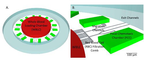
Figure 1. Schematic of donut-shaped device. A) Chemoattractant is primed into the device and a gradient is formed along the migration channel toward the focal chemotaxis chambers (FCCs). Sixteen FCCs surround each WBLC. After washing, the chemoattractant only remains in the FCC, and a linear gradient is formed along the migration channel. The red blood cell (RBC) filtration comb prevents RBCs from clogging the channel and blocking neutrophil active migration. See Movie S1. B) Neutrophils actively migrate out of whole blood and accumulate in the FCC, and their velocity, directionality and numbers can be accurately quantified. Please click here to view a larger version of this figure.
Figure 2. Schematic and overview of cell counting scheme for WB device. Neutrophils actively migrate out of 2 µl whole blood (WB) loaded into whole blood loading chamber (WBLC) past the red blood cell filtration comb and into entrance of the device. Directionality of the cell is measured based on the cell's decision at the bifurcation. Directional neutrophils will follow the chemotactic gradient leading towards the focal chemotaxis chamber and non-directional cells will not follow the gradient and will randomly migrate either towards the exit channel or FCC with an equal distribution. Final cell count is calculated at each time point by counting cells in the FCC. Please click here to view a larger version of this figure.
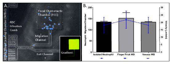
Figure 3. Measuring neutrophil chemotaxis from a droplet of whole blood (WB). A) WB (1 µl) is loaded into the whole blood loading chamber (WBLC). Neutrophils (blue) migrate along the fMLP chemotactic gradient formed in the migration channel towards the FCC. B) Neutrophils from a finger prick droplet of WB, from venous WB, or isolated from venous blood migrate with consistent numbers (bars) and velocity (blue line). Please click here to view a larger version of this figure.
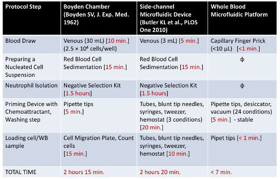
Table 1. Comparison of protocols for neutrophil chemotaxis using the whole blood microfluidic platform vs. a side-channel microfluidic device and the traditional Boyden chamber. The amount of time and reagents to complete each protocol step are compared between different methods to measure neutrophil chemotaxis. The whole blood microfluidic platform reduces the total time required to run the assay by 95% and the required volume of blood >99%.
In this work, we developed a microfluidic platform to measure neutrophil chemotaxis from a droplet of blood (2 µl). The on-chip mechanical filtration of RBCs from actively migrating neutrophils circumvents the need for cumbersome cell separation methods such as density gradients10, positive selection11, or negative selection12, which are prone to introduce artifacts by activating neutrophils. The mechanical filtration of RBCs distinguishes our technology from other microfluidic chips for probing neutrophil migration. For instance, previous works from our lab13 and Beebe et al. 8 utilized selectins on the chip, for separating neutrophils from the whole blood sample, prior to the chemotaxis assay. After separation on selectin surfaces, neutrophil migration occurred in buffer solution. In these conditions, any effect of serum on neutrophil chemotaxis was removed. Moreover, selectins are known to activate the cells by engaging specific receptors and therefore may alter in vitro chemotaxis measurements. The mechanical separation by the RBC filtration comb in our device does not alter the state of neutrophils measured by our device. Our device facilitates the robust measurements of velocity and cell accumulation number as well as cell directionality.
In this study, we have shown that neutrophils from healthy volunteers migrating from capillary whole blood, a drop of venous blood, and separated from venous blood migrate with similar velocity and directionality14. The measurement of neutrophil directionality is important in inflammatory conditions, such as burns3,15 and trauma16, where directionality is known to be impaired. Impaired and over stimulated neutrophils may migrate away from the site of injury and potentially cause injury to healthy tissues. A limitation of the device presented in this paper is that cell enrichment on the device is not possible and therefore cell counts are dependent on the proportion that they are found in the native whole blood. This may present problems in acquiring chemotaxis measurements from a significant number of cells in conditions such as neutropenia, where the neutrophil count is low. Another potential problem in our device is the steric hindrance of migrating cells by RBCs or other cells migrating simultaneously. In our experiments, neutrophils can squeeze past RBCs trapped in the turns of the RBC filtration comb without changing their migration speed. In addition, the chemoattractant concentration and potency is extremely important in inducing cell migration. To troubleshoot the assay with other conditions it may be important to run dose-response curves with varying magnitudes of chemoattractant. Handling and processing more unstable chemoattractants such as lipid mediators is also crucial, so it is important to prime devices immediately prior to WB loading. Finally, it is also critical that the whole blood does not clot before priming the WB device. If it is not possible to run the assay immediately following blood collection blood must be stored on a rocker in heparin. Loading of sample should be done slowly, with care as not to introduce excessive pressure into the WB device.
In the future, our device can be used to measure perturbations in neutrophil function in burn or trauma patients without removing the neutrophil from the patient's native serum, which likely plays a key role in neutrophil dysfunction17. This device can also be utilized in the future to screen potential pro-resolution therapeutics to restore normal neutrophil function in these patients. Finally, this device may be further engineered to measure chemotaxis from other migratory cells such as monocytes, macrophages, and t-cells.
In conclusion, we have developed a novel method for measuring neutrophil chemotaxis directly from a droplet of whole blood. The incorporation of the whole blood microfluidic device in a 12 or 24-well plate facilitates the screening of multiple conditions simultaneously. This device will facilitate the development of mediators of inflammation resolution to treat burn and other inflammatory conditions. In the future, this device will be utilized to measure perturbations in neutrophil chemotactic function over time in patient samples and murine models where sample volume is limited.
The authors have nothing to disclose.
Support from the National Institutes of Health (grants GM092804, DE019938) and Shriners Burns Hospital.
Name of Material/ Equipment | Company | Catalog Number | Comments/Description |
Device Fabrication | |||
SU8 | Microchem | Y131273 | |
Polydimethylsiloxane (PDMS) | Ellsworth Adhesives | Sylgard 184 1.1 lb. Kit | |
Standard glass slides | Fisher Scientific | 125495 | 1 X 3 inches |
Glass-bottom plate | MatTek | P12G-1.5-14-F | |
Harris Uni-Core, Tip Diameter 5.0mm | Ted Pella, Inc. | 15081 | |
Harris Uni-Core, Tip Diameter 1.5 mm | Ted Pella, Inc. | 15072 | |
Microfluidic Assay Preparation and Analysis | |||
Gel-loading pipet tip | Fisher Scientific | 02-707-139 | |
Syringe | Fisher Scientfic | 309602 | |
Blunt tip needle, 30g ½ in. | Brico Medical Supply | BN3005 | |
Vacutainer, Heparin | Becton Dickinson | ||
HBSS | Sigma-Aldrich | ||
Human serum albumin | Sigma-Aldrich | A5843-5G | 0.2% final concentration in HBSS |
Fibronectin | Sigma-Aldrich | F0895-1MG | |
fMLP | Sigma-Aldrich | F3506-10MG | |
SurgiLance safety lancet, 2.2mm depth, 22 gauge | SLN240 | ||
Hoescht stain | Life Technologies | H3570 | |
Positive Control | |||
HetaSep | STEMCELL Technologies Inc. | 7906 | |
EasySep Human Neutrophil Enrichment Kit | STEMCELL Technologies Inc. | 19257 | |
Equipment | |||
Plasma Asher | March Instruments | P-250 | |
Lindberg/Blue M Oven | Thermo Scientific | 13-258-30C | |
Stainless Steel Precision Tweezers | Techni Tool | 758TW458 | |
Bel-Art Scienceware Chemical-Resistant Vacuum Desiccator | Fisher Scientific | 08-594-15A | |
Dataplate Digital Hot Plate | Alpha Multiservices | PMC 720 | |
Nikon TiE inverted microscope | Nikon – Micro Video Instruments Inc. | MEA53100 | |
CFI Plan Fluor DL 10x na 15.2wd Objective | Nikon – Micro Video Instruments Inc. | MRH20101 | |
Lumen 200 with 2 Meter Light Guide for Nikon | Nikon – Micro Video Instruments Inc. | 500-L200NI2 | |
DAPI/Hoechst/AMCA Narrow Band 32mm Exciters – 25mm Emitters | Chroma – Micro Video Instruments Inc. | 31013v2 | |
Retiga R 2000 cooled CCD Camera 1600×1200 pixels | Qimaging – Micro Video Instruments Inc. | RET-2000R-F-M-12-C |