Tissue Engineering of Tumor Stromal Microenvironment with Application to Cancer Cell Invasion
Summary
Tissue engineered fibroblast-derived native matrix is an emerging tool to generate a stromal substrate which supports epithelial cell proliferation and differentiation. Here a protocol applying this methodology to assess the impact of different stromal cell types on tumor cell biology is presented.
Abstract
3D organotypic cultures of epithelial cells on a matrix embedded with mesenchymal cells are widely used to study epithelial cell differentiation and invasion. Rat tail type I collagen and/or matrix derived from Engelbreth-Holm-Swarm mouse sarcoma cells have been traditionally employed as the substrates to model the matrix or stromal microenvironment into which mesenchymal cells (usually fibroblasts) are populated. Although experiments using such matrices are very informative, it can be argued that due to an overriding presence of a single protein (such as in type I Collagen) or a high content of basement membrane components and growth factors (such as in matrix derived from mouse sarcoma cells), these substrates do not best reflect the contribution to matrix composition made by the stromal cells themselves. To study native matrices produced by primary dermal fibroblasts isolated from patients with a tumor prone, genetic blistering disorder (recessive dystrophic epidermolysis bullosa), we have adapted an existing native matrix protocol to study tumor cell invasion. Fibroblasts are induced to produce their own matrix over a prolonged period in culture. This native matrix is then detached from the culture dish and epithelial cells are seeded onto it before the entire coculture is raised to the air-liquid interface. Cellular differentiation and/or invasion can then be assessed over time. This technique provides the ability to assess epithelial-mesenchymal cell interactions in a 3D setting without the need for a synthetic or foreign matrix with the only disadvantage being the prolonged period of time required to produce the native matrix. Here we describe the application of this technique to assess the ability of a single molecule expressed by fibroblasts, type VII collagen, to inhibit tumor cell invasion.
Introduction
The use of biomaterial in 3D tissue culture has enabled researchers to study cell behavior in the laboratory under physiological conditions more akin to an in vivo environment than that of one recapitulated with 2D adhesion and a plastic substrate. In particular, great strides forward have been made in modeling stratified epithelia with the adoption of 3D culture methods at the air-liquid interface1-4. Such techniques faithfully mimic keratinocyte differentiation and tumor cell invasion enabling greater flexibility and fidelity for researchers studying these processes. The choice of biomaterial substrate to mimic the stromal environment has primarily involved the use of type I collagen, Engelbreth-Holm-Swarm mouse sarcoma matrix and de-epidermized dermis. For example, cancer-associated fibroblasts have been shown to contribute toward cancer invasion5, initiation, and progression via stromal-epithelial interactions6,7 when grown in such substrates.
The gold standard for mimicking the stromal environment in skin, the largest and most widely studied stratified epithelia using such techniques, is regarded to be de-epidermized human dermis (DED). Preparation of DED involves the removal of the epidermis via trypsinization or physical disassociation from human cadaver skin3,4. However, access to such skin can be very difficult for laboratories not associated with clinical institutions, and diseased dermis is near impossible to obtain. As an alternative, laboratories frequently use a combination of type I collagen (isolated from rat tails) and/or Engelbreth-Holm-Swarm mouse sarcoma matrix.
After the discovery in 1927 by Nageotte8 that collagen can easily be isolated using acetic acid and salt precipitation, its application to tissue culture was subsequently pioneered by Huzella and colleagues9. Collagen coating proved to be superior to glass for cell culture of 29 strains and tissue explants as interrogated by Ehrmann and Gey9. Currently, the major type of collagen used in tissue culture is isolated from rat-tail tendons, and is usually bought from commercial sources. However, the drawback for faithful substrate recapitulation is that the rat tail collagen is not identical to human collagen, or the human dermis, where type I and III collagens are present as major constituents, and isolated rat tail collagen is invariably fragmented.
Engelbreth-Holm-Swarm mouse sarcoma matrix is a gelatinous protein mixture secreted by cultured Engelbreth-Holm-Swarm mouse sarcoma cells10. The major constituents are laminin, type IV collagen, heparin sulfate proteoglycan, entactin and nidogen and the exact ratios of these proteins will vary from batch to batch. Aside from structural proteins, this matrix also contains significant levels of growth factors such as transforming growth factor β, epidermal growth factor, insulin-like growth factor 1, bovine fibroblast growth factor, and platelet-derived growth factor which would alter cellular behavior11,12. Pointing towards the sheer complexity of Engelbreth-Holm-Swarm mouse sarcoma matrix, a total of 1,851 proteins were identified in a recent proteomic study13. In light of the rich and complex nature of this matrix, caution has been advised when interpreting and comparing different experiments with the use of it11.
Our laboratories have a keen interest in genetic skin diseases, particularly those with a predisposition to developing cutaneous squamous cell carcinoma (cSCC)14. In the case of recessive dystrophic epidermolysis bullosa (RDEB), a severe blistering disease with germline mutations in COL7A1 gene15-17, we have determined that the dermal microenvironment in these patients is tumor promoting18. During the course of this study we were unable to assess the tumor promoting properties of dermal fibroblasts embedded within collagen I/ Engelbreth-Holm-Swarm mouse sarcoma matrix and investigated ways of assessing cells’ own, native matrix. To achieve this, we modified a previous technique from the laboratory of Lucie Germain working on human skin equivalents19,20. Germain’s technique was able to reconstruct human skin with well-organized basement membrane using primary human keratinocyte and fibroblast cultures in the absence of a synthetic or cadaveric scaffold.
In this paper the steps used to recapitulate the cutaneous tumor stromal microenvironment (native matrix) derived directly from primary stromal fibroblasts in vitro are described18. Native matrices produced by long-term culture of fibroblasts were used as a dermal equivalent to assay for cSCC cell invasion. We present data using native matrix derived from either the extracellular matrix secreted by RDEB fibroblasts (deficient in type VII collagen (C7)) or from RDEB fibroblasts retrovirally transduced with a type VII collagen expressing construct and demonstrate the profound effect of a single collagen on tumor cell invasion.
Protocol
This study was conducted according to the Declaration of Helsinki Principles and was approved by the appropriate Ethics Committees.
1. Preparation of Media and Reagents
- Preparation of 200x of L-ascorbic acid 2-phosphate stock
- Dissolve 29 mg of L-ascorbic acid 2-phosphate per 5 ml of Dulbecco’s Modified Eagle’s Medium (DMEM) solution and filter through 0.22 μm membrane filter. Store as 0.25 ml sterile aliquots in -20 °C.
- Add 0.25 ml aliquot of 200x L-ascorbic acid 2-phosphate stock to every 50 ml of fibroblast media (DMEM with 1% L-glutamine and 10% fetal bovine serum) on the day required, for a final concentration of 0.1 mM of L-ascorbic acid 2-phosphate.
- Preparation of keratinocyte growth media
- Isolation and culture of primary SCC keratinocytes have been described previously21. Preparation of keratinocyte growth media is described therein as well.
- Briefly, prepare the media as follows:
300 ml of DMEM and 100 ml of Ham’s F-12 supplemented with 10% FBS
0.4 mg/ml hydrocortisone
5 mg/ml insulin
10 ng/ml EGF
5 mg/ml transferrin
8.4 ng/ml cholera toxin
13 ng/ml liothyronine
1x penicillin-streptomycin solution
2. In Vitro Construction of Fibroblast-derived Native Matrix in L-Ascorbic Acid 2-Phosphate Supplemented Media
- Seed 200,000 fibroblasts per well in 6-well plates (20,000 cells/cm2) in fibroblast media supplemented with of L-ascorbic acid 2-phosphate. Refeed every 2-3 days with 2-5 ml of media. (Note: Refeeding frequency and volume may be adjusted to suit the individual needs of different cells. See “Discussions”).
- A thick layer of cells embedded in extracellular matrix will form at the end of 6 weeks, visible to the naked eye (Figure 1). To release this layer from the tissue culture plate, gently scrape the circumference of matrix with a 1 ml micropipette tip and then push the edges of the matrix towards center of the well. This layer of cells and matrix (native matrix) should be easily detachable from the plate surface, and now be floating in the media. Spread the native matrix out in the media to avoid it folding up onto itself.
- Let the native matrix float and remodel for 5 days, changing media every 2-3 days. Due to tensile strength and intrinsic remodeling within matrix, the matrix will contract drastically and become reduced to a smaller, but thicker native matrix, and is ready to be utilized for invasion assay.
3. Invasion Assay with Tumor SCC Keratinocytes
- Pick up the native matrix gently with blunt forceps and transfer to Nylon net. Once on the Nylon net, spread the matrix gently to lie as flat as possible using a 1 ml micropipette tip and blunt forceps.
- Prepare the sterile clonal cylinders by smearing a small amount of sterile Vaseline on one end.
- Place the clonal cylinders on the native matrix, with the Vaseline side down. This is to ensure a tight seal between the native matrix and the clonal rings.
- Add cSCC cells to the clonal cylinders (250,000 cells in 100 µl of keratinocyte growth media).
- Remove the clonal cylinders after 6 hr when the cSCC cells have settled down on the native matrix.
- Lift the Nylon net with the native matrix and cSCC cells to air-liquid interface onto bent stainless steel wire mesh support.
- Add keratinocyte growth media supplemented with ascorbic acid until the media level touches the bottom of the native matrix.
- Change media every 2-3 days and harvest at 7 and 14 days post-seeding of cSCC cells.
4. Harvesting of 3D Cultures and Preparation for Histology
- Fix in 4% paraformaldehyde overnight at room temperature.
- Bisect the samples and embed in wax for formalin-fixed paraffin embedded blocks, with the cut surface facing outwards.
- Alternatively, embed the bisected samples in OCT and snap-freeze immediately in liquid nitrogen for fresh frozen tissue blocks.
- Cut 4 μm sections on microtome and dewax (if necessary). Stain using standard haematoxylin and eosin. To visualize the cSCC cells, mouse monoclonal antibody against keratin 14 (LL001, in-house) can be used in immunohistology staining.
Representative Results
This technique opens up the possibility of examining and comparing the invasive behavior of tumor cells (in this case cSCC) under different 3D stromal environments. Using this technique, not only C7-deficient native matrices that recapitulated the RDEB dermal environment can be generated, but also additional matrices that have been genetically engineered to over-express C718. As seen in Figure 2, invasion of RDEB cSCC keratinocytes was significantly retarded in C7-overexpressing native matrices compared to C7-deficient RDEB control. This invasion is visualized via standard histological methods, where the gels were fixed, embedded in paraffin blocks and then sectioned and immunostained. Suggested antibodies to use for staining are anti-keratin 14 (LL001) for cSCC cells and anti-vimentin (V9) for fibroblasts.

Figure 1. Workflow of generation of fibroblast-derived native matrix and the tumor invasion assay. Click here to view larger image.
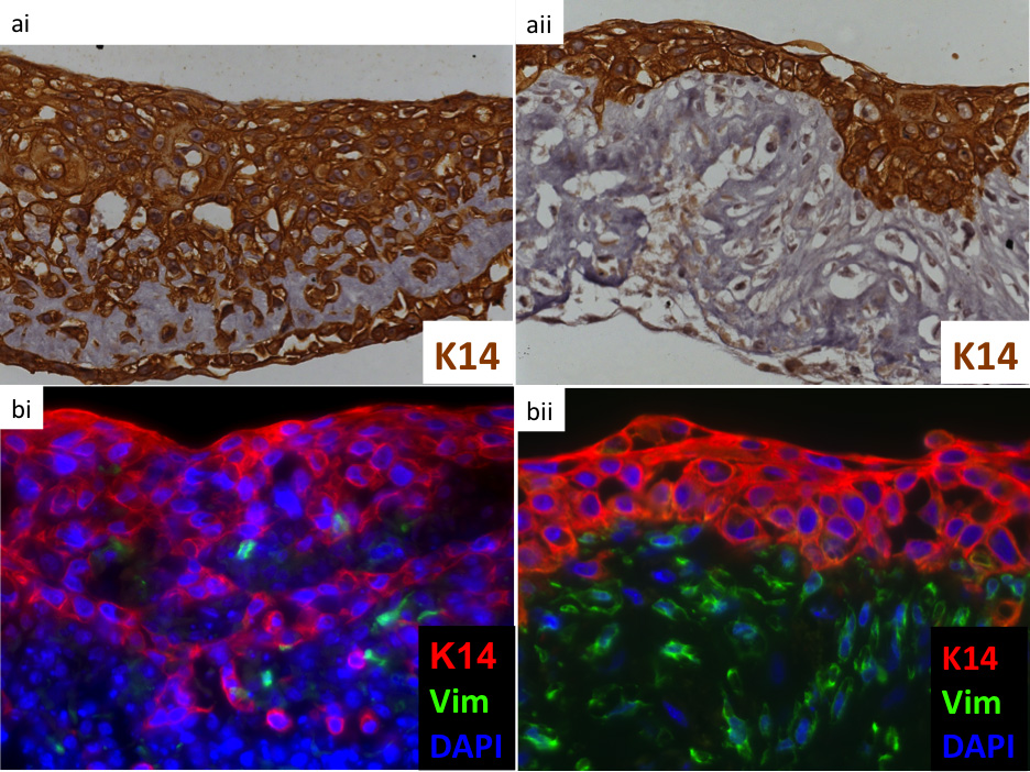
Figure 2. Keratin 14 (LL001) staining of the cSCC invasion into the native matrix derived from control RDEB fibroblast deficient in C7 (ai & bi), or in RDEB fibroblasts over-expressing full-length C7 expression (aii and bii), clearly demonstrates that the re-expression of C7 in extracellular matrix can retard cSCC keratinocyte invasion. Nuclei were stained with DAPI (in blue) and fibroblasts with vimentin (in green) in bi and bii.
Discussion
Due to the nature of this experiment, the total time required for completion can be up to two months. Throughout this time, utmost care and sterile tissue culture practices must be employed to prevent microbial contamination.
Aside from its role as a cofactor in the synthesis of hydroxyproline and hydroxylysine of collagens, ascorbic acid stimulates collagen specific mRNA expression in fibroblasts22. The ascorbic acid of choice here is the more stable L-ascorbic acid 2-phosphate23. Refeeding is advised three times a week but this will invariably be dependent on the cells being studied. The initial fibroblast cultures will consume more nutrients as the weeks pass, and more frequent refeeding with larger media volumes may be advisable. It is important to be gentle when changing media and care should be taken to minimize disturbances to the cell layer during native matrix production.
The matrix should become visible usually after 2 weeks, especially around the circumference of the well. Infrequently, the matrix will free itself without any mechanical disruption. This is a reflection of the intrinsic remodeling of the matrix and tensile strength, which pulls the matrix inwards, but may also be a sign that the circumference of the native matrix was disturbed during media changes.
When releasing and freeing the matrix cell layer from the plate, one must be careful not to poke holes through the matrix. Small blunt cell scrapers may also be used in lieu of 1 ml micropipette tips. Placement of the released native matrix onto a Nylon mesh first (being sure to lay the native matrix flat) makes for much easier handling and subsequent lifting to the air-liquid interface.
This protocol is the first to describe the use of native fibroblast matrix as a dermal substrate to model the microenvironment in tumor cell assays in order to obtain more accurate and physiologically relevant data.
Declarações
The authors have nothing to disclose.
Acknowledgements
A.P.S is supported by DebRA International and the British Skin Foundation. Y.Z.N is supported by A*STAR – University of Dundee Partnership PhD Programme.
Materials
| L-Ascorbic acid 2-phosphate | Sigma | A8960 | |
| DMEM with l-glutamine, | Life Technologies | 11995-073 | |
| 4,500 mg/L d-glucose, 110 mg/L sodium pyruvate | |||
| 100x Penicillin-Streptomycin | Life Technologies | 15070 | |
| Vaseline | VWR | PROL28908.290 | |
| Clonal cylinders | Sigma | Z370789 | |
| Nylon Net Filter Disc Hydrophilic 100um 25um diameter 100/pk | Millipore | NY1H02500 | |
| Bent stainless steel wire mesh support | Made in house | Dimensions were made so that the mesh would fit into 6-well plates |
Referências
- Bell, E., Ehrlich, H. P., Buttle, D. J., Nakatsuji, T. Living tissue formed in vitro and accepted as skin-equivalent tissue of full thickness. Science. 211 (4486), 1052-1054 (1981).
- Asselineau, D., Bernhard, B., Bailly, C., Darmon, M. Epidermal morphogenesis and induction of the 67 kD keratin polypeptide by culture of human keratinocytes at the liquid-air interface. Exp. Cell Res. 159 (2), 536-539 (1985).
- Pruniéras, M. M., Régnier, M. M., Woodley, D. D. Methods for Cultivation of Keratinocytes with an Air-Liquid Interface. J. Invest. Dermatol. 81 (1 Suppl), 28-33 (1983).
- Prunieras, M., Régnier, M. New procedure for culturing human epidermal cells on allogenic or xenogenic skin: preparation of recombined grafts. Ann. Chir. Plast. 24 (4), 357-362 (1979).
- Gaggioli, C. Fibroblast-led collective invasion of carcinoma cells with differing roles for RhoGTPases in leading and following cells. Nature. 9 (12), 1392-1400 (2007).
- Bhowmick, N. A., Neilson, E. G., Moses, H. L. Stromal fibroblasts in cancer initiation and progression. Nature. 432 (7015), 332-337 (2004).
- Erez, N., Truitt, M., Olson, P., Hanahan, D. Cancer-Associated Fibroblasts Are Activated in Incipient Neoplasia to Orchestrate Tumor-Promoting Inflammation in an NF-κB-Dependent. Manner. Cancer Cell. 17 (2), 135-147 (2010).
- Harkness, R. D., Marko, A. M., Muir, H. M., Neuberger, A. The metabolism of collagen and other proteins of the skin of rabbits. Biochem. J. 56 (4), 558-569 (1954).
- Ehrmann, R. L., Gey, G. O. The growth of cells on a transparent gel of reconstituted rat-tail collagen. J. Natl. Cancer Inst. 16 (6), 1375-1403 (1956).
- Kleinman, H. K. Basement membrane complexes with biological activity. Bioquímica. 25 (2), 312-318 (1986).
- Vukicevic, S., Kleinman, H. K., Luyten, F. P., Roberts, A. B., Roche, N. S., Reddi, A. H. Identification of multiple active growth factors in basement membrane Matrigel suggests caution in interpretation of cellular activity related to extracellular matrix components. Exp. Cell Res. 202 (1), 1-8 (1992).
- . BD Biosciences – Discover Labware. SPC-356230 Rev 5.0 at Rev 5.0. , .
- Hughes, C. S., Postovit, L. M., Lajoie, G. A. Matrigel: a complex protein mixture required for optimal growth of cell culture. Proteomics. 10 (9), 1886-1890 (2010).
- Ng, Y. Z., Dayal, J. H., South, A. P. Genetic Predisposition to Cutaneous Squamous Cell Carcinoma. , .
- Christiano, A. M. A., Greenspan, D. S. D., Lee, S. S., Uitto, J. J. Cloning of human type VII collagen. Complete primary sequence of the alpha 1(VII) chain and identification of intragenic polymorphisms. J. Biol. Chem. 269 (32), 20256-20262 (1994).
- Hovnanian, A. A. Genetic linkage of recessive dystrophic epidermolysis bullosa to the type VII collagen gene. J. Clin. Invest. 90 (3), 1032-1036 (1992).
- Ryynänen, M. M., Ryynänen, J. J., Sollberg, S. S., Iozzo, R. V. R., Knowlton, R. G. R., Uitto, J. J. Genetic linkage of type VII collagen (COL7A1) to dominant dystrophic epidermolysis bullosa in families with abnormal anchoring fibrils. J. Clin. Invest. 89 (3), 974-980 (1992).
- Ng, Y. Z. Fibroblast-derived dermal matrix drives development of aggressive cutaneous squamous cell carcinoma in patients with recessive dystrophic epidermolysis bullosa. Cancer Res. 72 (14), 3522-3534 (2012).
- Larouche, D., Paquet, C., Fradette, J., Carrier, P., Auger, F. A., Germain, L. Chapter 15. Methods Mol. Biol. 482, 233-256 (2009).
- Pouliot, R. R. Reconstructed human skin produced in vitro and grafted on athymic mice). Transplantation. 73 (11), 1751-1757 (2002).
- Purdie, K. J., Pourreyron, C., South, A. P. Isolation and culture of squamous cell carcinoma lines. Methods Biol. 731, 151-159 (2011).
- Pinnell, S. R. Regulation of collagen biosynthesis by ascorbic acid: a review. Yale J. Biol. Med. 58 (6), 553-559 (1985).
- Hata, R., Senoo, H. L-ascorbic acid 2-phosphate stimulates collagen accumulation, cell proliferation, and formation of a three-dimensional tissuelike substance by skin fibroblasts. J. Cell. Physiol. 138 (1), 8-16 (1988).
