The adult structures of Drosophila are derived from sac-like structures called imaginal discs. Analysis of these discs provides insight into many developmental processes including tissue determination, compartment boundary establishment, cell proliferation, cell fate specification, and planar cell polarity. This protocol is used to prepare imaginal discs for light/fluorescent microscopy.
A significant portion of post-embryonic development in the fruit fly, Drosophila melanogaster, takes place within a set of sac-like structures called imaginal discs. These discs give rise to a high percentage of adult structures that are found within the adult fly. Here we describe a protocol that has been optimized to recover these discs and prepare them for analysis with antibodies, transcriptional reporters and protein traps. This procedure is best suited for thin tissues like imaginal discs, but can be easily modified for use with thicker tissues such as the larval brain and adult ovary. The written protocol and accompanying video will guide the reader/viewer through the dissection of third instar larvae, fixation of tissue, and treatment of imaginal discs with antibodies. The protocol can be used to dissect imaginal discs from younger first and second instar larvae as well. The advantage of this protocol is that it is relatively short and it has been optimized for the high quality preservation of the dissected tissue. Another advantage is that the fixation procedure that is employed works well with the overwhelming number of antibodies that recognize Drosophila proteins. In our experience, there is a very small number of sensitive antibodies that do not work well with this procedure. In these situations, the remedy appears to be to use an alternate fixation cocktail while continuing to follow the guidelines that we have set forth for the dissection steps and antibody incubations.
For more than a century the fruit fly, Drosophila melanogaster, has been a premier system to study development, behavior and physiology. Development in the fly can be divided into two broad stages: embryonic and post-embryonic with much of the latter taking place within monolayer epithelia called imaginal discs 1-3. Drawings of imaginal discs were first published in 1864 by August Weismann as part of his broad monograph on insect development 1. These discs begin their development during embryogenesis, are patterned during the larval stages, survive the massive histolysis of the early pupal stages, and ultimately give rise to a high percentage of adult structures that are found within the adult fly 1-14. During larval development each disc makes several critical decisions regarding fate, shape and size. Within the first and second larval instars, the discs are tasked with adopting a primary fate, establishing compartment boundaries, adopting the correct shape and generating the requisite number of cells 15-16. During the third larval instar and early pre-pupal stage, the imaginal discs continue to divide and are patterned as cells adopt their terminal fates 16.
During the early history of Drosophila developmental biology, imaginal discs were studied nearly exclusively in the context of normal development and in the limited cases in which a loss or gain-of-function mutant was viable. The use of X-rays to induce mitotic recombination allowed for lethal mutations to be analyzed in cell clones within the larval and adult tissues. This method has been improved by the introduction of transgenic methods to analyze loss and gain-of-function mutations in both larval and adult tissues. The number of antibodies, transcriptional reporters and protein traps for describing the molecular landscape of wild type and mutant tissues is also constantly growing. Using these molecular markers to analyze loss and gain-of-function mutant cell clones has made it increasingly feasible to gain a real-time understanding of how mutant cells deviate from their wild type cousins during development. To properly take advantage of these tools and reagents it is critical to have high quality preparations of imaginal discs that can be viewed, photographed and analyzed. The goal of this manuscript is to provide an optimized protocol for the isolation and preparation of the eye-antennal disc complex (Figure 1A). It can also be successfully used to isolate a wide variety of additional discs including those that give rise to the wings, halteres, T1-T3 legs and the genitals (Figure 1B-E). This procedure, with minor modifications, has been used to isolate imaginal discs from Drosophila for nearly eighty years.
As described above, since most genes are expressed during multiple stages of development and in a multitude of tissues, it is often impossible to study the effects that null mutants have on the entire eye as the animal dies well before the third instar larval stage. Four methods have made the study of more developed tissues such as the retina significantly more tractable. The first is the Flippase (FLP)/Flippase Recombination Target (FRT) method of generating mutant cell clones within an otherwise wild type tissue 17-19. In this instance the mutant tissue is identified by the absence of a visual marker such as Green Fluorescent Protein (GFP) and can be compared to the surrounding wild type tissue in which GFP is present (Figure 2D). The second is the “flp-out” method in which a transgene is expressed in a population of cells 20. In this instance the cell clones are identified by the presence of GFP and compared to the surrounding wild type tissue that lacks the GFP reporter (Figure 2E). The third is the Mosaic Analysis with a Repressible Cell Marker (MARCM) technique, which combines elements of the FLP/FRT mutant clone and flp-out expression systems 21. With this method a transgene can be expressed within a population of cells that are simultaneously mutant for an individual genetic locus. Like flp-out clones, MARCM clones are identified by the presence of GFP and compared to the surrounding wild type tissue that lacks the GFP marker (Figure 2F). And lastly, the genes and RNAi constructs can be expressed within imaginal tissues under the control of specific promoter-GAL4 constructs. These four methods have increased the interest in studying imaginal discs since mutant or over-expression clones or patterns can be directly compared to adjacent wild type tissue. The method described in this procedure has been developed so that researchers who study the post-embryonic development of adult tissues in Drosophila, particularly those derived from the eye-antennal disc, will be able to obtain high quality tissue for analysis. Although individual researchers have made slight modifications, the core of this procedure (which we describe here) has remained largely unaltered. Since obtaining high quality tissue is critical to the study of the imaginal discs we hope this written protocol and accompanying video will serve as a valuable teaching resource.
1. Preparation of Larvae
2. Coarse Dissection of Larvae
3. Fixation and Staining of Tissue with Antibodies
4. Fine Dissection of Eye-Antennal Disc Complexes and Mounting onto Slides
The method that is described above reliably produces high quality material for analysis with in situ probes, transcriptional reporters, protein traps and antibodies. In Figure 1 we display eye-antenna, genital, wing, haltere and leg discs that are routinely recovered with this method. These discs have been treated with a phalloidin-conjugated fluorophore, which binds to F-actin and therefore outlines each cell. If the tissue has been fixed properly then the morphogenetic furrow of the eye disc, the edges of the concentric tissue folds in the genital, antennal and leg discs, and the dorsal-ventral axis of the wing and haltere discs will all appear as sharp edges. If a tissue is properly fixed then antibodies, fluorescent proteins and in situ probes will also reveal sharp patterns. Several examples are shown in Figure 2. The top three panels display discs that have been stained with different antibodies while the lower three panels show discs in which GFP is used to mark populations of cells.
One of the most striking features of the eye-antennal disc is the morphogenetic furrow (Figure 1A), which can be seen as an indentation within tissue running along the dorsal-ventral axis 1, 22. Prior to the third larval instar all cells within the developing eye are unpatterned, undifferentiated, and morphologically indistinguishable from one another. At the start of the third larval instar the morphogenetic furrow initiates at the posterior margin of the eye field and progresses anteriorly towards the eye/antennal border 22. As the furrow progresses across the eye field the sea of disordered cells is transformed into an ordered array of periodically spaced unit eyes or ommatidia (Figure 1A) 22-23. Ahead of the furrow a gene regulatory network that includes the Pax6 homolog Eyeless (Ey) channels cells towards an eye fate (Figure 2A) 15. The initiation and progression of the furrow itself is dependent upon the activities of the Hedgehog (Hh) and Decapentaplegic (Dpp) signaling pathways 24-29. Indeed a dpp-lacZ reporter faithfully reflects the expression of the dpp locus within the furrow (Figure 2B) 30-31. As cells exit the furrow and begin to adopt their terminal fates, they express cell specific markers such as embryonic lethal abnormal vision (elav), which encodes a pan-neuronal RNA binding protein (Figure 2C) 32-34.
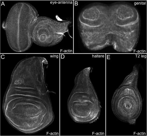
Figure 1. The imaginal discs of Drosophila melanogaster. (A–E) Confocal images of wild type eye-antenna, genital, T2 leg, wing and haltere imaginal discs. (A) As the morphogenetic furrow progresses across the eye field, a sea of unpatterned and undifferentiated cells is transformed into columns of unit eyes that are also called ommatidia. All discs are treated with phalloidin-conjugated fluorophores, which bind to and reveal F-actin distribution. Anterior is to the right and dorsal is up.
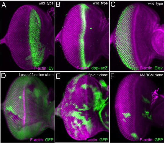
Figure 2. Clonal and expression analysis of the eye imaginal disc. (A–F) Confocal images of eye imaginal discs. (A) The Pax6 protein Eyeless (Ey) is distributed broadly ahead of the morphogenetic furrow. (B) A dpp-lacZ transcriptional reporter responds to Hh signaling and is expressed within the morphogenetic furrow. (C) The pan-neuronal RNA binding protein Elav is distributed in all developing photoreceptors behind the morphogenetic furrow. (D) An eye disc containing loss-of-function clones generated by the FLP/FRT system. The clones are identified by the lack of GFP (E). An eye disc containing over-expression clones generated with the flp-out system. Clones are positively marked with GFP. (F) An eye disc containing MARCM clones. Like the flp-out system, the MARCM clones can be identified by the presence of GFP. All detected proteins and genotypes are listed within the figure. Anterior is to the right and dorsal is up.
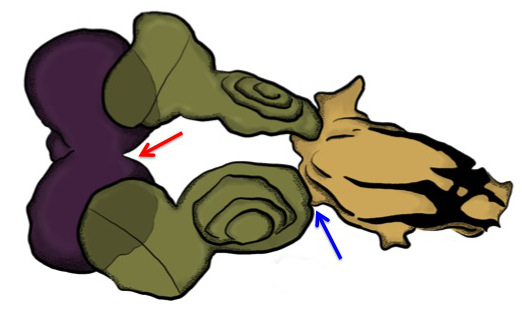
Figure 3. Eye-antenna-brain complex. A schematic drawing of the first day coarse dissection products. The only tissues that should be fixed are the mouth hooks, eye-antenna discs and brain (often times the ventral ganglion will remain attached as well — not shown). Purple = brain, green = eye-antenna discs, brown = mouth hooks. Anterior is to the right. If dissecting leg, wing, haltere and genital discs, the outer cuticle of the larva should still be attached to these tissues. Do not remove the overlying cuticle as you might lose the imaginal discs during subsequent transfers through various antibody, block and washing solutions. The excess tissue can be removed during the fine dissection steps (4.1–4.2).
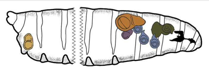
Figure 4. Position of imaginal discs within the Drosophila larva. A schematic drawing of the relative position of the eye-antenna, leg, wing, haltere and genital discs within a third instar larva. The eye-antennal disc is colored in green, the leg discs are in blue, the halter disc is in purple, the wing disc is in brown/orange and the genital disc is in light brown. Anterior is to the right.
Name of Solution |
8% Paraformaldehyde |
0.2 M Sodium Phosphate Monobasic |
0.2 M Sodium Phosphate Disbasic |
1 N Sodium Hydroxide |
10% Triton |
Distilled Water |
0.1 M Sodium Phosphate Buffer (Dissection Buffer) |
0.1 M Sodium Phosphate Buffer + 0.1% Triton (Wash Buffer) |
Lysine Buffer |
2% Paraformaldehyde-Lysine-Periodate Fixative (PLP) |
10% Normal Goat Serum |
Table 1: List of Required Solutions.
8% Paraformaldehyde stock solution |
To a 50 ml Erlenmeyer flask add the following: |
2.0 g paraformaldehyde |
23.0 ml of distilled water |
4.0 drops of 1 N sodium hydroxide (from a glass pasteur pipet) |
Mix and heat on a stir plate until solution reaches a gentle boil |
Allow to gently boil until paraformaldehyde is completely dissolved |
Place on ice until cold (make fresh prior to each dissection) |
0.1 M Phosphate Buffer (Dissection (P) Buffer) |
To a 50 ml conical tube add the following: |
18.0 ml 0.2 M Sodium Phosphate dibasic |
7.0 ml 0.2 M Sodium Phosphate monobasic |
25.0 ml distilled water |
Store at 4 °C (1 week shelf life) |
0.1 M Phosphate + Detergent Buffer (Wash (W) Buffer) |
To a 50 ml conical tube add the following: |
18.0 ml 0.2 M Sodium Phosphate dibasic |
7.0 ml 0.2 M Sodium Phosphate monobasic |
25.0 ml distilled water |
0.5 ml 10% Triton |
Store at RT (1 week shelf life) |
Lysine (L) Buffer |
To a 50 ml conical tube add the following: |
0.4 g Lysine |
1.2 ml 0.2 M Sodium Phosphate dibasic |
8.0 ml Dissection (P) Buffer |
10.0 ml distilled water |
Shake solution until lysine is completely dissolved |
Place on ice until cold (make fresh prior to each dissection) |
2% Paraformaldehyde – Lysine – Periodate (PLP) fixative |
0.1 g Sodium Periodate |
15.0 ml of Lysine (L) buffer |
5.0 ml of 8% Paraformaldehyde |
Shake solution well until sodium periodate is completely dissolved |
Place on ice until cold (make fresh prior to each dissection) |
Table 2: Recipes for Required Solutions.
Although this procedure has largely focused on the isolation and subsequent treatment of eye-antennal discs, it is amenable to being used to isolate and analyze the wing, haltere, leg and genital discs (Figure 4). The only required modification of the protocol for isolating these discs (as opposed to the eye-antennal disc) is the method of coarse dissection (section 2 of the protocol). The first thoracic leg (T1) pair is found at the anterior of the larva and can be recovered by following the protocol for eye-antennal disc isolation. However the second thoracic leg (T2) discs are attached to the cuticle. To isolate these discs a pair of forceps should be used to hold the mouth hooks (as described above) while another pair of forceps should be used to clasp the ventral cuticle of the animal (make sure to clasp the larva about 1/3 from the mouth hooks. The larva can simply be fillet by tearing the ventral cuticle away from the rest of the larva. The T2 legs will remain attached to the cuticle. The third thoracic (T3) leg is attached to the wing and haltere discs as part of a complex, which itself is also attached to the cuticle. You can choose to separate the disc complex and the T2 legs from the cuticle at this stage or keep the cuticle-disc complex together during the subsequent fixation/antibody incubation steps and isolate the discs during the fine dissection portion of the procedure (section 4). For recovering genital discs, it is best to grasp the larvae at midsection with one pair of forceps and then peel the ventral cuticle away with the other pair of forceps starting at the midsection and ending at the posterior end of the larva. It is recommended that forceps be used to clear away all extraneous tissue and then transfer just the genital disc to the fixative solution using a P-200 pipetman and a yellow tip whose end has been cut with a razor blade.
This procedure is best suited for tissues of limited thickness such as imaginal discs and works best if the antibodies being used are of high titer and specificity. However, it can be adapted to work well with thicker tissues such as the adult ovary, testis, and brain as well as with low titer antibodies or those that give higher than desired “background” staining. When working with thicker tissues, it is suggested that optimal results can be obtained by simply increasing the concentration of paraformaldehyde and/or incubating in the PLP fixative for longer periods of time. Since proper fixation of dissected tissues is essential for success with this protocol it is also suggested that the efficiency of increasing the paraformaldehyde concentration and/or increasing the fixation length be assessed with phalloidin (Figure 1). In particular, close attention should be paid to the “sharpness” of cell outlines and other physical landmarks within the tissue (i.e., the morphogenetic furrow). Another way to ensure that your tissue (particularly thicker samples) is fixed properly is to prepare the 8% paraformaldehyde and PLP solutions fresh prior to each dissection. And finally, the overall quality of the dissection can be increased if tissue is dissected for short periods of time and tissue is transferred into fixative as soon as possible. It is suggested that dissecting for no longer than 15–20 min. Dissecting for shorter durations increases the quality of tissue preservation.
This protocol works well with a wide range of antibodies, but it is true that some antibodies do not work well with the PLP fixative. One notorious example is the antibody that recognizes the Rough (Ro) transcription factor. Rough is expressed within and is required for the specification of a subset of developing photoreceptors 35. The anti-Ro antibody works best, not with PLP, but rather with a PIPES – EGTA – MgSO4 (PEM) buffer 36. Similarly, other antibodies may work best on tissues that have been incubated in still other fixatives. It is suggested that, unless stated otherwise, the PLP fixative should be tried first. If unsuccessful then the process of finding an alternate fixative should begin.
One common issue to confront is the working concentration of the antibody in question. Often times you may be the first researcher to use a particular antibody to detect proteins in your tissue of interest. The working concentration can deviate from what is reported for other tissues. It is suggested that the recommended concentration be tried first. Increase the concentration of the antibody if a signal cannot be seen. On the other hand, if the recommended concentration gives high background staining then diluting the antibody should be tried. Working concentrations among antibodies can vary. For example, the anti-Eyes Absent (Eya) antibody is diluted 1:5 while the anti-Elav antibody works well even at a 1:500 dilution. Some antibodies can even be diluted as far down as 1:3,000. Additionally, this procedure, as written, works best with high specificity antibodies. However, as many have experienced, some antibodies can bind non-specifically to the tissue and this can create a sub-optimal image. This situation can be often corrected by incubating the diluted antibody with fixed embryos or larval carcasses prior to being added to the fixed imaginal discs. Depending upon the level of non-specific background staining, the required length of “pre-absorption” can vary and will have to be worked out on a trial basis. It is also possible to increase the signal/noise ratio by re-using antibodies several times or by conducting several rounds of pre-absorption.
When deciding on which discs should be used in publications and/or presentations, it is best to choose discs that have been oriented with the apical side oriented upwards. It is also best to use discs that are not folded. This is particularly true of the eye-antennal disc. The ventral side of disc tends to fold and unfortunately, there is little one can do to prevent this from happening. One solution is to dissect younger discs as these tend to fold far less. Another option is to just dissect large numbers of discs until you get one that is completely flat. The overall shape of the eye-antennal disc can be affected by the rate at which the larva is torn apart. If one pulls the larva apart too quickly the hole through which the brain-disc complex passes is small and the eye-antennal disc comes out folded and/or stretched. It is best to pull slowly until the larva begins to tear since the hole will be larger. Then you can continue to pull the brain-eye-antennal disc complex slowly. Bear in mind that you can never pull too slowly. Also note, that the rate at which you tear the tissue is not a factor in the isolation of other tissues.
The life cycle of Drosophila consists of three larval phases. Important developmental events take place during each one of these phases, thus it may be important to isolate tissues not just from third larval instars (which is the main focus of this procedure) but also from both first and second larval instars as well. With one minor exception, this procedure can be used, as written, to isolate second instar discs. During the fine dissection portion of the procedure (section 4) it is recommended that sharp tungsten wire should be used to separate the eye-antennal disc from extraneous tissue like the mouth hooks, brain and salivary glands. The tungsten wire can also be used to separate the T2/T3 leg, wing and haltere discs from each other and the overlying cuticle. It is recommend that one end of the tungsten wire (the end that will be used to separate tissues) be sharpened by heating in a boiling sodium nitrate bath. The other end can be inserted into a pin vise; this will allow you to hold the tungsten wire more easily. Unfortunately, it is considerably more difficult to isolate intact first instar discs, therefore it is recommended that you place the entire eye-antennal disc/brain/mouth hook complex on the slide and cover with a coverslip. The amount of dissected tissue can be minimized by using a sharp tungsten wire to remove the salivary glands and overlying cuticle prior to mounting the disc/brain/mouth hook complex onto the slide. It is relatively easy to identify the first instar eye-antennal disc when it is still attached to the brain and mouth hooks (see Figure 6A,D of Kumar and Moses, 2001) 37. The other imaginal discs will have to be separated from the cuticle and placed on a slide for viewing.
The authors have nothing to disclose.
We would like to thank Donald Ready and Kevin Moses for teaching JPK the original imaginal disc dissection procedure. We also thank Bonnie Weasner for the genital disc in Figure 1B and the eye disc in Figure 2A, Brandon Weasner for Figure 3, the Bloomington Drosophila Stock Center for fly stains and the Developmental Studies Hybridoma Bank for antibodies. CMS has been supported by a stipend from the National Institutes of Health (NIH) GCMS Training Grant (T32-GM007757), the Frank W. Putnam Research Fellowship, and the Robert Briggs Research Fellowship. JPK is supported by a grant from the National Eye Institute (R01 EY014863)
Name of Material/Equipment | Company | Catalog Number | Comments |
Sylgard 184 Silicone Elastomer Kit | Dow Corning | 184 SIL ELAST KIT 0.5KG | Used to create base for dissection plate |
Pryex Glass Petri Dish 150x20mm | Dow Corning | 3160-152CO | Use the cover for dissection plate |
#5 Dissecting Forceps | Ted Pella | 525 | Forceps must be kept very sharp |
9 well watch glass | Vairous Vendors | N/A | Used for fixation of imaginal disc complexes |
50ml Erlenmeyer Flask | Various Vendors | N/A | |
Small Stir Bar | Various Vendors | N/A | Small enough to fit into Erlenmeyer Flask |
50ml Conical Tubes | Various Vendors | N/A | |
1.5ml Microfuge Tubes | Various Vendors | N/A | Clear or Dark depending upon application |
Microfuge Rack | Various Vendors | N/A | |
Benchtop Rotator | Various Vendors | N/A | 100ul volume should not splatter at low setting |
Paraformaldehyde | Macron Chemicals | 2-26555-1 | Serves as fixative |
Sodium Phosphate Monobasic | Sigma Chemical Co | S-3139 | Used to make dissection and wash buffers |
Sodium Phosphate Dibasic | Sigma Chemical Co | 71636 | Used to make dissection and wash buffers |
Lysine | Acros Organics | 125221000 | Used in the fixative solution |
Sodium Periodate | Sigma Chemical Co | S-1878 | Used in the fixative solution |
Triton X-100 | EMD Chemicals | MTX1568-1 | Used to perforate imaginal discs |
Sodium Hydroxide | EM Science | SX0593-3 | Used to dissolve paraformaldehyde |
100% Normal Goat Serum` | Jackson Laboratories | 005-000-121 | Serves as a blocking solution |
Primary Antibodies | Various Vendors | N/A | Dilute in 10% goat serum as directed by manufacturer |
Seondary Antibodies | Various Vendors | N/A | Dilute in 10% goat serum as directed by manufacturer |
Vectashield | Molecular Probes | H-1000 | Prevents bleaching of samples |
Microscope Slides | Fischer Scientific | 48312-003 | |
Glass Cover Slips 18x18mm | Fischer Scientific | 12-542A | |
Kimwipe Tissue | Various Vendors | NA | Prevents Glass slides from adhering to silicone base |
Panit Brush 000 | Various Vendors | NA | Use to gently lower coverslip on to samples |