We outline a methodology for the processing of whole blood to obtain a variety of components for further analysis. We have optimized a streamlined protocol that enables rapid, high-throughput simultaneous processing of whole blood samples in a non-clinical setting.
Collection and processing of whole blood samples in a non-clinical setting offers a unique opportunity to evaluate community-dwelling individuals both with and without preexisting conditions. Rapid processing of these samples is essential to avoid degradation of key cellular components. Included here are methods for simultaneous peripheral blood mononuclear cell (PBMC), DNA, RNA and serum isolation from a single blood draw performed in the homes of consenting participants across a metropolitan area, with processing initiated within 2 hr of collection. We have used these techniques to process over 1,600 blood specimens yielding consistent, high quality material, which has subsequently been used in successful DNA methylation, genotyping, gene expression and flow cytometry analyses. Some of the methods employed are standard; however, when combined in the described manner, they enable efficient processing of samples from participants of population- and/or community-based studies who would not normally be evaluated in a clinical setting. Therefore, this protocol has the potential to obtain samples (and subsequently data) that are more representative of the general population.
Multiple studies have characterized differences in gene expression, DNA methylation and cell subset in blood among individuals with and without mental (or other) illnesses1-4. These studies, however, have been obtained from clinical settings in which disease-associated differences may be magnified due to the generally more severe nature of the illnesses for which patients are seeking treatment. Due to advances in “omics” approaches, the past decade has seen an explosion of interest in obtaining biologic samples from community and/or epidemiologic settings5-7, in order to provide population-based estimates of disease prevalence and a broader picture of the environmental determinants of these mental and/or physical illnesses.
A key challenge in this regard is the requirement for rapid processing of the collected specimens. Degradation of mononuclear cells, key immune system components that are frequently used to assess the health of an individual, begins immediately upon blood draw with a significant decrease in recovery after 2 hr of collection8-10. To address this challenge, we present an optimized protocol in which multiple components of human whole blood are simultaneously isolated from samples obtained in homes of subjects living in a large metropolitan area. The protocol is based upon our compilation and modification of current techniques, including storage of all “extra” fractions in the event future techniques allow for further isolation/analyses. While alternative methods or kits may be employed in place of the individual methods described here, those outlined have proven to be a reliable and efficient means for processing samples in a high-throughput manner. High-quality fractions (PBMCs, DNA, serum, and RNA) of fresh blood can be produced within 2 hr of collection and all assay-ready specimens can be available within 2 days (Figure 1).
This protocol was developed to enable the efficient processing of samples collected from community-dwelling, adult residents of the city of Detroit for testing in the Detroit Neighborhood Health Study (DNHS; DA022720, RC1MH088283, DA022720-05-S1), a population-based study of the social and biological determinants of post-traumatic stress disorder (PTSD) and other mental illnesses. The prevalence of PTSD in Detroit is more than twice the national average11,12. Identifying biological determinants of PTSD in this population may help to develop appropriate pharmacologic and/or cognitive-behavioral interventions to aid those suffering from the disorder, both in this urban population, and in other high-risk populations (e.g., returning military veterans). Our laboratory, previously located at Wayne State University in Detroit, Michigan, was selected for processing based on our expertise in handling fresh tissue samples derived from a variety of sources, the necessity to begin processing the samples within 2 hr of collection, and our proximity to the collection sites. With this unique opportunity at hand, our goal was to optimize the processing for greatest yield of DNA, RNA, serum and peripheral blood mononuclear cells (PBMCs) from each specimen (a total of N=1,639 samples over 5 waves of specimen collection). The procedures outlined here can be performed simultaneously in a non-clinical setting, thus producing starting material (see Table 1 for average yields) for a multitude of downstream applications including microarray, epigenetic, real-time RT-PCR, and flow cytometry analyses.
Figure 1. Overall work flow. The overall process depicted here includes the logistics of obtaining the blood specimens from identifying consenting participants to the blood draw itself. High-quality, fractions (peripheral blood mononuclear cells; PBMCs, DNA, serum, and RNA) of fresh whole blood can be produced within 2 hr of collection and all assay-ready specimens can be available within 2 days. Moreover, the fractions prepared through this method are suitable for long-term storage if samples are not to be tested immediately. The entire timeline outlined here could be completed in a single day (~5 hr total). However, such a day would be extremely labor intensive especially for a single technician with substantial experience with the techniques. Thus, we recommend dividing the procedures on Day 1 between at least two technicians and completing the RNA processing on Day 2. Please click here to view a larger version of this figure.
The Detroit Neighborhood Health Study was reviewed and approved by the University of Michigan's Institutional Review Board. All participants provided informed consent prior to their participation in the study.
1. Overview
2. Day 1 Setup
3. Day 1: Processing Whole Blood Sample for DNA, PBMCs and Leukocyte RNA (2 hr)
4. Day 2 Setup
5. Day 2: Long Term Sample Storage and Leukocyte Filter Processing (3 hr)
It is essential that the overall procedure produce high quality material for analysis in a multitude of downstream applications including gene expression via microarray and RT-PCR analysis, detection of epigenetic modifications, and cell subset variations. Table 1 indicates the average yield and quality of materials from each of the processes. Figure 3 provides an example of the quality output of the leukocyte RNA isolation and filter processing methods. The image on the upper left of Figure 3 is the gel image resulting from the capillary electrophoresis. Each lane should produce two distinct bands with minimal shadowing which would indicate degradation. The chromatograms below the gel provide an additional look at the level and type of degradation that can be determined based upon the location and size of the peaks. The RNA Integrity Number (RIN) is another quality measure that ranges from 1 (low; degraded) to 10 (high; pure, good quality RNA).
Such a high-throughput methodology lends itself to an occasional error, but there are several checkpoints throughout the various stages of processing to ensure quality control. Figure 2 displays the proper separation following centrifugation of the Ficoll containing vacutainer. There are several reasons a vacutainer may not display this separation including an error in centrifugation speed, low collection volume, or lack of a fasting participant as indicated in Table 2.
Subsets (n=100) of the specimens isolated using this procedure have been analyzed with success by HumanMethylation27 (HM27) DNA Analysis BeadChips including validation of those results by pyrosequencing11. Genotyping, targeted bisulfite pyrosequencing, and real-time RT-PCR has been successfully performed in the Wildman and Uddin laboratories20,21,22,23. Serum, isolated in parallel with the DNA isolation for both the Beadchip and genotyping analyses, was successfully analyzed for IL-6 and C-reactive protein activity24. T-cell subsets from the isolated PBMCs have been successfully analyzed by flow cytometry25-28. Additionally, RNA from the modified leukocyte procedure has been subjected to Genome Wide gene expression profiling 29.
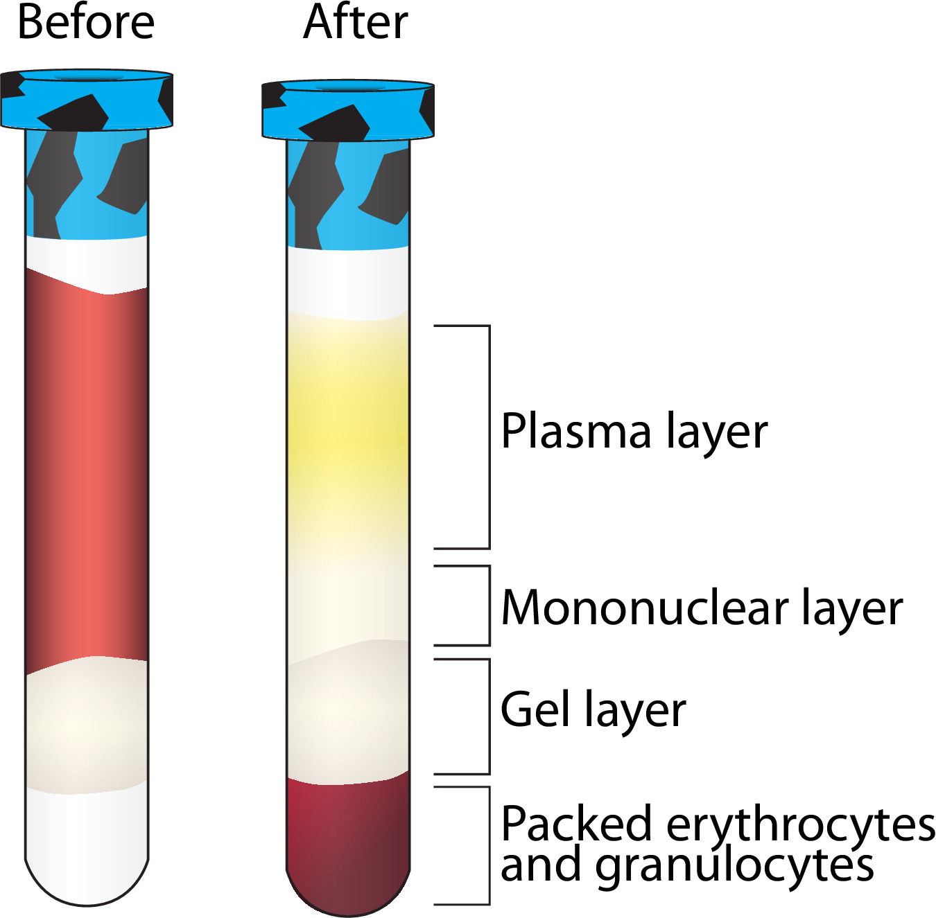
Figure 2. Layer separation following centrifugation of Ficoll containing vacutainer. A visualization of the separation of multiple blood components following centrifugation. The mononuclear layer is further purified while the other layers are stored at -80 °C. Please click here to view a larger version of this figure.
Figure 3. Expected results from leukocyte RNA processing. Bioanalyzer results displaying two distinct bands representing 18S and 28S ribosomal RNA with RNA integrity numbers (RINs) above 8 isolated with the described leukocyte RNA isolation and filter processing methods (see protocol 3.6 and 5.3). Please click here to view a larger version of this figure.
Total Average Yield (N≈500) | Average Quality | |
Serum | 2.30 ml | N/A |
DNA from whole blood | 39.97 µg | Absorbance at 260/280 = 1.74 |
PBMC | 22.25 million viable cells | At least 95% viability of overall isolation |
RNA from Leukocytes | 44.09 µg | Absorbance at 260/280 = 2.01 RNA Integrity Number (RIN) = 6.48 |
Table 1. Expected yields. Average quantity and quality of samples processed using the described methods.
Problem | Potential Solution |
No separation after Ficoll containing vacutainer centrifugation | Vacutainer is not filled to capacity — the minimum collection volume for adequate processing is 6 ml. |
Check centrifuge settings, be sure the speed is in g-force. If incorrect, set to g-force and respin. | |
Low PBMC count | Volume of blood draw too low. |
Aspirated too close to the pellet in step 3.7.8 | |
Non-fasting participant. | |
No separation after serum vacutainer centrifugation | Check centrifuge settings, be sure the speed is in g-force. If incorrect, set to g-force and respin. |
Unexpected concentration using the Nanodrop 1000 | Make sure the measurement is for the appropriate sample type (e.g., DNA or RNA). |
Table 2. Troubleshooting. Common problems encountered with the described methods and potential solutions.
Supplementary Table 1. Tracking sheet. An example of tracking the vacutainers from blood collection to laboratory delivery. Please click here to view this table.
Supplementary Table 2. Specimen log. An example of the document used to document the processing of the vacutainers upon delivery to the lab. Please click here to view this table.
Supplementary Table 3. Cryovial Numbering System. An example of the numbering system used for each participant. Please click here to view this table.
Supplementary Information 1. Preprocessing details. Details the duties of the Study Coordinator, Courier and Phlebotomist. Please click here to view supplementary information 1.
Supplementary Information 2. Recipes. A list of recipes for the necessary reagents. Please click here to view supplementary information 2.
We have described a streamlined protocol that has been successfully applied to process more than 1,600 whole blood samples in the Detroit Neighborhood Health Study. Although many of these techniques are available in the existing literature, our step-by-step compilation, including precisely timed alterations between each step, reflects an optimized, efficient protocol that successfully produces a variety of biologic specimens with a wide range of downstream applications, including DNA methylation, mRNA expression and immunological analysis. These specimens have already been tested in a variety of experiments, results of which have been published in peer-reviewed literature and/or presented at national meetings11,20,21,23,24,29. This protocol should thus be of interest to other investigators seeking to collect biological specimens in population-based studies similar to the DNHS.
When processing samples in such a high-throughput manner, it is of utmost importance to maintain accurate records at all stages. We recommend developing a database to store all of the cryovial information at the outset. This database should include all aspects of the sample within each cryovial including the volume, concentration, quality, barcode, and storage location (storage box number and location within the box). We recommend preparing pre-labeled cryovials and storage boxes which contain a barcode. Further, we have found it convenient to have a “master” document containing the barcodes for each tube, specific to each participant (Table S3). This enables rapid input of the cryovial data into the database without excessive handling of the samples, introducing unnecessary freeze/thaw cycles to the samples and eliminates the potential for human error in entering in the barcodes manually. It is also essential that the labels adhere at extreme (e.g., vapor phase of liquid nitrogen -178 to -150 ºC) temperatures. The documentation can be time consuming, and with the necessity of efficient processing upon delivery, we have found that having at least two technicians divide the processes produces the greatest efficiency.
A limitation of this method is the proximity of the laboratory to the collection site. For PBMC isolation in particular, the samples must be processed within 2 hr of collection to avoid significant increases in red blood cell contamination and decreases in viable mononuclear cells. As such, the laboratory should be no more than 30 min from a collection site to account for any traffic related issues that may arise. In addition, any laboratory proposing to undertake the protocol outlined here will require two technicians on hand to ensure optimal sample processing. Furthermore, each laboratory must have access to the requisite equipment for each step outlined above. Thus, laboratories with fewer staff and/or limited equipment would likely be unable to undertake this protocol.
An advantage of this protocol is the ability to collect specimens directly in the homes of consenting individuals. This allows the study to reach individuals with mental or other health issues who would not typically seek medical help, perhaps because of a lack of insurance or transportation. It also the enables comparison of affected and unaffected individuals living within the same community who have experienced similar triggers, but differ in terms of their mental health symptoms. Obtaining specimens in this manner requires precise timing and coordination of phlebotomists in the field. Because our laboratory was located within the community we were studying, a phlebotomist could typically collect samples from two-three homes and deliver the samples to the laboratory within the 2 hr window of the first collection. Efficiency is key to our sample processing, and as such, we had multiple phlebotomists in the field, increasing the need for precise coordination. The laboratory received multiple blood deliveries with up to eight participants per delivery spaced 2 hr apart. The phlebotomists met at a designated location and combined their collections with only one phlebotomist delivering the specimens to the laboratory as a single batch. The methods described here can easily be adapted to study many other phenotypes and the biologic specimens collected can be used in a multitude of downstream assays.
The authors have nothing to disclose.
We would like to thank Henriette Mair-Meijers for invaluable attention to detail and hours devoted to processing the blood collections. We are grateful for the graphic design expertise of Natalie Jameson Kiesling. We also appreciate the approval of the manufacturers (Qiagen (Valencia, CA), BD Biosciences (San Jose, CA), Life Technologies (Grand Island, NY)) mentioned herein to publish the use of their products as described. Funding for this work was generously provided by the National Institutes of Health award numbers DA022720, RC1MH088283, and DA022720-05-S1.
QIAamp DNA Blood Mini Kit | Qiagen | 51104 | Day 1: DNA isolation |
Phosphate-buffered saline (PBS) | Sigma | P5493-1L | Day 1: PBMC isolation |
5 ¾” Pasteur pipets | Fisher | 13-678-6A | Day 1: PBMC isolation |
Fetal Bovine Serum (FBS), heat inactivated | Life Technologies | 10082147 | Day 1: PBMC isolation |
Dimethyl Sulfoxide (DMSO) | Sigma | D8418-500ml | Day 1: PBMC isolation |
RPMI Medium 1640, liquid | Invitrogen | 11875119 | Day 1: PBMC isolation |
0.4% trypan blue stain | Invitrogen | T10282 | Day 1: PBMC isolation |
Countess Cell Counting Chamber | Invitrogen | C10283 | Day 1: PBMC isolation |
Countess Automated Cell Counter or cell counting device such as a microscope and hemocytometer | Invitrogen | C10281 | Day 1: PBMC isolation |
LeukoLOCK Fractionation & Stabilization Kit | Ambion | 1933 | Day 1: Leukocyte RNA isolation |
25G x 5/8 in. needles | Becton Dickinson | 305122 | Day 1: Leukocyte RNA isolation |
Syringes (5ml) | Becton Dickinson | 309646 | Days 1 and 2: Leukocyte RNA isolation |
Denaturing Lysis Solution | Ambion | 8540G | Day 2: Leukocyte RNA isolation |
5M NaCl | Life Technologies | 24740011 | Day 2: Leukocyte RNA isolation |
TRI Reagent | Ambion | 9738 | Day 2: Leukocyte RNA isolation |
Bromo-3-chloro-propane (BCP) | Sigma | B-9673 | Day 2: Leukocyte RNA isolation |
spin cartridges | Ambion | 10051G | Day 2: Leukocyte RNA isolation |
0.1mM EDTA | Ambion | 9912 | Day 2: Leukocyte RNA isolation |
DNA-free Kit | Ambion | AM1960 | Day 2: DNase treament |
RNA 6000 Ladder | Agilent | 5067-1529 | Day 2: Bioanalyzer analysis |
RNA 6000 Nano Series II Kit | Agilent | 5067-1511 | Day 2: Bioanalyzer analysis |
RNaseZAP | Ambion | AM8782 | Day 2: Bioanalyzer analysis |
Ethanol >99% | Sigma | E7023-500ml | |
Isopropanol >99% | Sigma | I9516-500ml | |
Nuclease-free ultra pure water | Invitrogen | 9938 | |
Pipette tips (nuclease-free) | Eppendorf | 22491253 | |
Pipetter (serological) | Eppendorf | 2223020-4 | |
Pipetters (for volumes under 1ml) | Eppendorf | 3120000-054 | |
Pipettes (serological) | Fisher | 13-678-27E | |
Controlled rate freezing containers | Nalgene | 5100-0001 | |
Cryoboxes (to hold 2ml and 5ml cryovials and 1.5ml microcentrifuge tubes) | Fisher | 03-395-464 | |
Test tube rack | Thermo Scientific | 14-804-134 | |
15ml polypropylene tubes | Fisher | 14-959-49D | |
1.5ml and 0.65 microcentrifuge tubes | Fisher | 07-200-534 and 07-200-185 | |
2ml and 5ml cryovials | Fisher | 10-500-26 and 10-269-88F | |
8ml CPT vacutainer | BD Biosciences | 362761 | 2 tubes |
6ml K2 EDTA vacutainer | BD Biosciences | 367863 | 2 tubes |
8.5ml SST vacutainer | BD Biosciences | 367988 | 1 tube |
Vortexer | Fisher | 2215365 | |
Dry bath incubator with heating block for microcentrifuge tubes | Fisher | 11-715-1250 | |
Filtration/vacuum system for use within the cell culture hood | Fisher | 01-257-87 | |
Fixed-angle rotor for microcentrifuge tubes with aerosol-tight lid | Eppendorf | 22637002 | |
Refrigerated centrifuge with a swing-bucket rotor and aerosol-tight caps for 16 x 125mm vacutainers and 15ml polypropylene tubes | Eppendorf | 22628157 | 2, one does not need to be refrigerated |
Nanodrop 2000 (recommended for accuracy of small volumes) or other spectrophotometric device | Fisher | 13-400-411 | |
Agilent Bioanalyzer | Agilent Technologies | G2940CA | |
Liquid nitrogen tank | Thermo Scientific | 11-676-56 | |
-80 ˚C freezer | Thermo Scientific | 992RAK | |
Sharps container | Fisher | 22-037-970 | |
Biological waste container | Thermo Scientific | 1223P52 | |
Biosafety Level 2 certified cell culture hood | Thermo Scientific | 13-261-315 |