Thromboembolic stroke models are vital tools for optimizing the recanalization therapy. Here we report a murine thrombotic stroke model based on transient cerebral hypoxic-ischemic (tHI) insult, which triggers thrombosis and infarction, and responds favorably to tissue plasminogen activator (tPA)-mediated fibrinolysis in a therapeutic window similar to those in stroke patients.
Stroke research has endured many setbacks in translating neuroprotective therapies into clinical practice. In contrast, the real-world therapy (tPA thrombolysis) rarely produces benefits in mechanical occlusion-based experimental models, which dominate preclinical stroke research. This split between the bench and bedside suggests the need to employ tPA-responsive models in preclinical stroke research. To this end, a simple and tPA-reactive thrombotic stroke model is invented and described here. This model consists of transient occlusion of the unilateral common carotid artery and delivery of 7.5% oxygen through a face mask in adult mice for 30 min, while maintaining the animal rectal temperature at 37.5 ± 0.5 °C. Although reversible ligation of the unilateral carotid artery or hypoxia each suppressed cerebral blood flow only transiently, the combination of both insults caused lasting reperfusion deficits, fibrin and platelet deposition, and large infarct in the middle cerebral artery-supplied territory. Importantly, tail-vein injection of recombinant tPA at 0.5, 1, or 4 hr post-tHI (10 mg/kg) provided time-dependent reduction of the mortality rate and infarct size. This new stroke model is simple and can be standardized across laboratories to compare experimental results. Further, it induces thrombosis without craniectomy or introducing pre-formed emboli. Given these unique merits, the tHI model is a useful addition to the repertoire of preclinical stroke research.
Thrombolysis and recanalization is the most effective therapy of acute ischemic stroke in clinical practice1. Yet, the majority of preclinical neuroprotection research was performed in a transient mechanic obstruction model (intraluminal suture middle cerebral artery occlusion) that produces rapid recovery of cerebral blood flow upon removal of the vascular occlusion and shows little to no benefits by tPA thrombolysis. It has been suggested that the dubious choice of stroke models contributed, at least in part, to the difficulty in translating neuroprotective therapy to patients2,3. Hence, there is an increasing call for employing tPA-responsive thromboembolic stroke models in preclinical research, but such models also have technical problems (see Discussion)4-7. Here we describe a new thrombotic stroke model based on unilateral transient hypoxic-ischemic (tHI) insult and its responses to intravenous tPA therapy8.
The tHI stroke model was developed based on the Levine procedure (permanent ligation of the unilateral common carotid artery followed by exposure to transient hypoxia in a chamber) that was invented for experiments with adult rats in 19609. The original Levine procedure faded into obscurity because it only produced variable brain damage, but the same insult caused consistent neuropathology in rodent pups when it was re-introduced by Robert Vannucci and his colleagues as a model of neonatal hypoxic-ischemic encephalopathy (HIE) in 198110. In recent years, some investigators re-adapted the Levine-Vannucci model to adult mice by adjusting the temperature in the hypoxic chamber11. It is plausible that the inconsistent brain lesions in the original Levine procedure may arise from fluctuating body temperatures of adult rodents in the hypoxic chamber. To test this hypothesis, we modified the Levine procedure by administering hypoxic gas through a facemask, while maintaining the rodent core temperature at 37 °C on the surgical table12. As expected, stringent body temperature control greatly increased the reproducibility of HI-induced brain pathology. The HI insult also triggers coagulation, autophagy, and gray- and white-matter injury13. Other investigators have also used the HI model to investigate post-stroke inflammatory responses14.
A unique feature of the HI stroke model is that it closely follows the Virchow’s triad of thrombus formation, including the stasis of blood flow, endothelial injury (e.g. due to HI-induced oxidative stress), and hypercoagulability (HI-induced platelet activation) (Figure 1A)15. As such, the HI model may capture some pathophysiological mechanisms relevant to real-world ischemic stroke. With this idea in mind, we further refined the HI model with reversible ligation of the unilateral common carotid artery (therefore to create a transient HI insult), and tested its responses to tPA thrombolysis with or without Edaravone. Edaravone is a free radical scavenger already approved in Japan to treat ischemic stroke within 24 hr of onset9. Our experiments showed that as brief as 30 min transient HI triggers thrombotic infarction, and that combined tPA-Edaravone treatment confers synergistic benefits8. Here we describe detailed surgical procedures and methodological considerations of the tHI model, which can be used to optimize reperfusion treatments of acute ischemic stroke.
This protocol is approved by the Institutional Animal Care and Use Committee (IACUC) of Emory University and follows the National Institutes of Health Guideline for Care and Use of Laboratory Animals.
1. Setup
2. Transient Cerebral Hypoxia-ischemia (Figure 1B)
3. Laser Speckle Contrast Imaging
Note: Although this is not an essential procedure of the tHI model, a two-dimensional laser speckle contrast imaging system16 can be used to characterize the changes of cerebral blood flow (CBF) during or after transient hypoxia-ischemia. To document the alterations of CBF under tHI, record immediately after the step 2.6. Alternatively, to compare CBF recovery after tHI insult, these procedures can be performed following the step 2.10.
4. tPA Administration
5. Brain Damage Detection with Several Different Options
Note: To collect brain samples, euthanize the mice at 1, 4 or 24 hr after tHI.
Two-dimensional laser speckle contrast imaging (LSCI)16 was used to compare the alterations of cerebral blood flow (CBF) by 30 min transient unilateral carotid occlusion (tCCAO), 30 min exposure to hypoxia (7.5% oxygen), and 30 min unilateral carotid ligation under hypoxia (tHI). This experiment revealed that tCCAO under normoxia suppressed the CBF on the carotid ligated hemisphere to ~50% of the baseline value, which quickly recovered to above 85% after release of the carotid occlusion (R in Figure 2A). Exposure to systemic hypoxia alone reduced the CBF to approximately 75% of the baseline value, which transiently rebound to ~130% after returning to the normoxic atmosphere (Figure 2B). In contrast carotid ligation under hypoxia (tHI) quickly reduced CBF in the ipsilateral hemisphere to less than 20% of the baseline value around 10 min, which rarely recovered to above 30% at 20 min after release of the carotid ligation and returning to normoxia. CBF on the contralateral hemisphere (L), however, fluctuated between 20 and 50% during hypoxia, and quickly returned to above 80% after the tHI insult (Figure 2C).
At 24 hr after tCCAo (30 min) or tCCAO plus 30 min hypoxia (tHI), in vivo TTC stain was used to detect infarction17. This analysis showed no obvious injury by the tCCAO insult, but sizable infarction in the middle cerebral artery-supplied territory following tHI insult (Figure 3A). Anti-fibrin(ogen) immunostaining was used to compare the tCCAO- and tHI-injured brains at 1 hour recovery and showed widespread deposition of fibrin(ogen), an indicator of thrombosis, in the tHI-injured, but not tCCAO-challenged mouse brains (Figure 3B, C). Tail-vein injection of Evans blue dye was also used to compare vascular perfusion of tCCAO- and tHI-injured brains at 4 hr recovery. This analysis showed diminished cerebral perfusion and intense extravasation of the Evans blue dye in tHI-injured, but not the tCCAO-challenged mouse brains (Figure 3D, E).
Finally, the outcomes of tHI insult in mice receiving tail-vein injection of vehicle (at 0.5 hr recovery) or recombinant human tPA (Activase, 10 mg/kg, at 0.5, 1, or 4 hours post-tHI) were compared using in vivo TTC stain at 24 hr recovery (Figure 4A). In vehicle-treated mice, the mortality rate at 24 hr post-tHI was 23.8% and only one in 21 tHI-injured mice was beyond the mean and 2 SD (the outlier). The 24 hours mortality rate in mice receiving tPA treatment at 0.5 hr recovery dropped to 8.3%, but this effect was lost when tPA was administrated at 1 or 4 hours after the tHI insult (Figure 4B). Figure 4C plotted the infarct size of all survived mice in the four treatment groups. Of note, both 0.5 and 1 hr tPA-administration significantly reduced infarct size, when compared to vehicle treatment. The 0.5 hr tPA-treatment group also showed a significantly reduced infarct size than the 4 hr tPA-treatment group. Figure 4D showed representative TTC-stain results after each treatment.
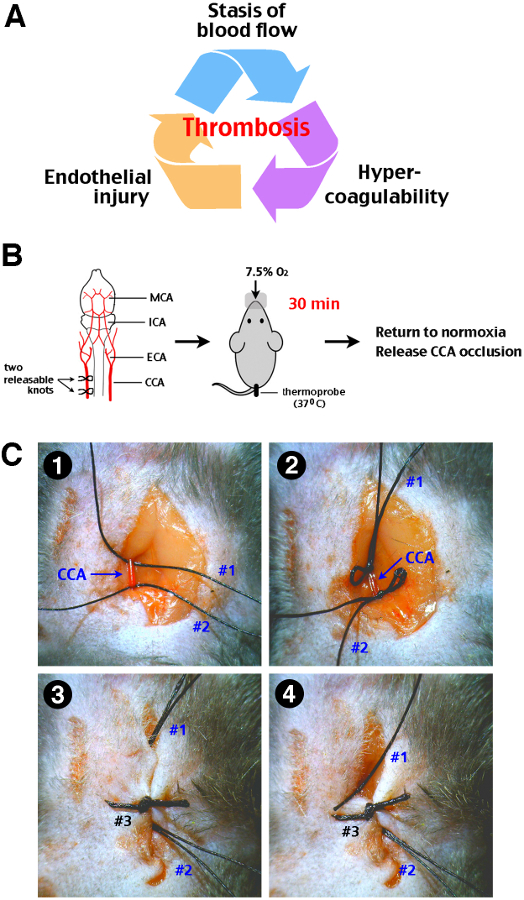
Figure 1: Procedure of transient cerebral hypoxia-ischemia (tHI) insult in adult mice. (A) The Virchow’s triad that propels thrombosis includes stasis of blood flow, endothelial injury, and hypercoagulability of the blood. (B) A schematic diagram of the tHI stroke procedure. Two releasable knots were tied onto the right common carotid artery (CCA), and followed by delivery of 7.5% oxygen via a nose cone for 30 min, while the mouse rectal temperature was maintained at 37-38 °C. After the transient systemic hypoxia, the CCA ligation was released by pulling out one end of the releasable suture knots. MCA, middle cerebral artery; ICA, internal carotid artery; ECA, external carotid artery; CCA, common carotid artery. (C) Surgical procedures for transient right CCA occlusion. 1. Two precut suture (#1 and #2) were placed under an isolated right CAA. 2. Two releasable knots were made. 3. The incision line was closed up by suture #3. Make sure that the ends of suture #1 and #2 were approachable outside the incision line. 4. Carefully pull suture #1 and #2 from outside to release the CCA. When performed gently, this procedure will not cause laceration of the CCA.
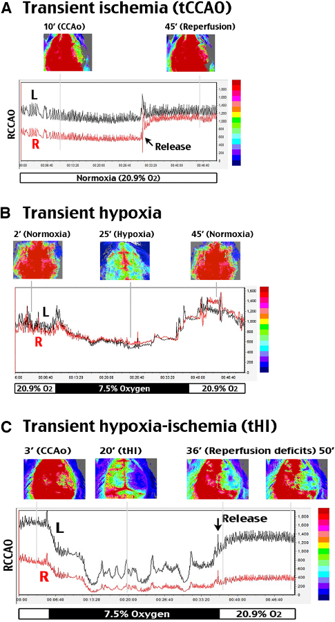
Figure 2: Analysis of cerebral blood flow alterations during and after tHI insult. A two-dimensional laser speckle contrast imaging (LSCI) system was used to evaluate cerebral blood flow (CBF). R (right) indicates the carotid-ligated hemisphere; L (left) is the contralateral hemisphere. (A) tCCAO under normoxia suppressed CBF to ~50% of the baseline value on the carotid-ligated hemisphere (R) for at least 30 min, which recovered to above 85% within 3 min upon release of the carotid ligation. (B) In hypoxia (7.5% oxygen, 30 min) without carotid artery ligation, CBF declined to 76% of the baseline value and transiently rebounded to around 130% after returning to normoxia. (C) In transient carotid ligation under hypoxia (tHI, 30 min), CBF on the carotid-ligated (R) hemisphere quickly dropped to less than 20% of the baseline value, and it rarely recovered to above 30% upon release of the carotid ligation and returning to normoxia. In contrast, CBF on the contralateral (L) hemisphere fluctuated between 20-50% during hypoxia, and quickly returned to >80% of the baseline value after release of carotid ligation and returning to normoxia. Shown are representative CBF tracings for n >4 in each group. The time-points for representative LSCI photographs were marked by grey lines in the representative tracing.
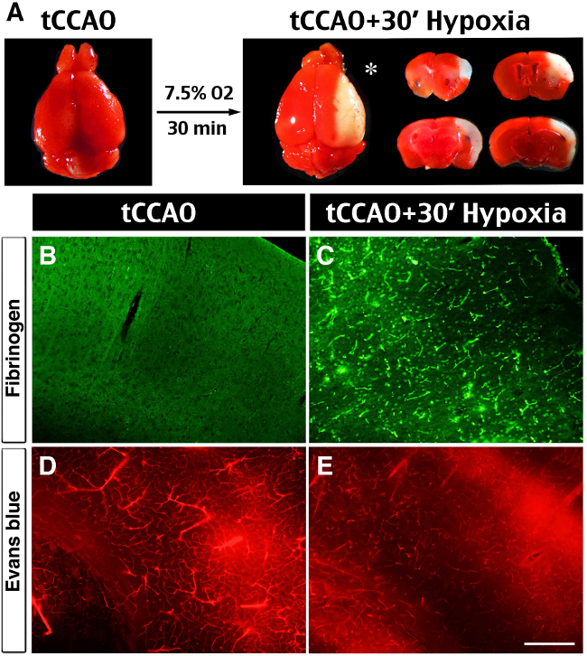
Figure 3: Brain infarct, spontaneous thrombosis and vessel obstruction after the tHI insult. (A) In vivo TTC-stain showed no visible infarction at 24 hr after 30 min transient ligation of the right common carotid artery (tCCAO), but the addition of 30 min hypoxia (7.5% oxygen) to tCCAO produced sizeable infarction in the ipsilateral hemisphere (asterisk), mostly in the middle cerebral artery-supplying area. (B, C) Anti-fibrin(ogen) immunostaining at 1 hr after the tHI insult showed widespread deposits in the ipsilateral hemisphere. In contrast, there was no fibrin(ogen) deposits at 1 hr after the tCCAo (30 min) insult (n >4 for each). (D, E) Cerebral perfusion was evaluated by tail-vein injection of Evans Blue dye at 4 hr after tCCAO (30 min) or tHI (30 min) insult. In post-tCCAO brain, Evans Blue dye filled most of the blood vessels in the ipsilateral hemisphere. In contrast, in post-tHI brain, Evan Blue dye filled fewer blood vessels and was leaked into the parenchyma (n >3). Scale bar: 250 μm.
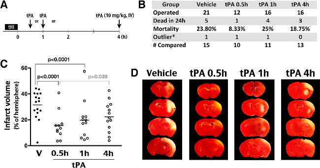
Figure 4: Effects of tPA thrombolysis in the tHI stroke model. (A) Outline of experiments to compare the effects of intravenous tPA administration (10 mg/kg) at 0.5, 1, or 4 hr after the tHI insult. (B) Summary of the number of operated animals, mortality in 24 hr after insults, outliers (the infarct size outside the mean +/- 2 SD), and the number of animals included for comparison of the infarct size. (C) Quantification showed an averaged 32% infarct volume in the vehicle group and significant reduction of infarction in the 0.5 hr (to 16%) and 1 hr (to 20%) groups (Shown are the mean and SEM for each group). The p-values are determined by t-test. (D) Representative TTC-stained brain from animals that were challenged by the tHI insult and received tPA treatment at the indicated time-point after injury. In TTC staining, live tissue showed red color; infarct tissue was pale.
Stroke is a major health issue of growing significance for any society with an aging population. Globally, stroke is the second-leading cause of death with an estimated 5.9 million fatal events in 2010, equivalent to 11.1% of all deaths18. Stroke is also the third-leading cause of disability-adjusted life years (DALYs) lost globally in 2010, rising from the fifth position in 199019. These epidemiological data highlight the need of more effective therapies of acute (ischemic) stroke. However, despite intense research in preclinical neuroprotective treatment, tPA-thrombolysis remains the only specific therapy of acute ischemic stroke that is approved by the Food and Drug Administration in the United States, while numerous once promising neuroprotective agents in animal studies failed in clinical trials. The difficulty in translating neuroprotective therapies into patients has many factors, and the current emphasis is on good laboratory practice, meta-analysis of multiple datasets, and international cooperation to improve preclinical stroke research20,21. However, there is a minority opinion suggesting that the translational difficulty is due to a poor choice of mechanical vascular occlusion models (e.g. intraluminal suture MCA occlusion) in the majority of preclinical stroke research to date2,3. Because mechanical vascular occlusion models rarely induce thrombosis and cerebral reperfusion occurs too fast upon the release of mechanical obstruction, these models neither respond to the real-word therapy (tPA fibrinolysis) nor provide a narrow therapeutic window as those in stroke patients. Consequently, the suggested remedy is to emphasize, at least include, thromboembolic stroke models in preclinical stroke research3.
This recommendation, however, has its restraints because current thromboembolic stroke models (exogenous emboli delivery, MCA-injection of thrombin, and photothrombosis) all have certain technical drawbacks4. For the exogenous emboli model, intravascular infusion of emboli results in significant variability in infarct size and location, as well as unpredictable responses to tPA thrombolysis due to differences in clot preparation4,5. Direct injection of thrombin into the MCA branch requires craniectomy, and its utility for optimizing the thrombolytic therapy is yet to be proven4,6. Chemically initiated thromboembolism based on systemic injection of a photosensitive dye (e.g. Rose Bengal or erythrosine B) and irradiation through the exposed skull often produces platelet-only aggregates that do not respond to thrombolysis4,7. Taken together, there is a need of simpler and tPA-responsive thromboembolic models for preclinical stroke research.
The tHI paradigm has four unique advantages as a thromboembolic stroke model. First, the tHI insult wielded endogenous components to form in situ thrombi without the help of exogenous chemicals or pre-formed emboli. Thus, thrombus formation in the tHI model is more relevant to physiological conditions. Second, the tHI model responds favorable to rapid tPA-treatment (at 0.5 and 1 hr post-injury) but not to delayed treatment (at 4 hr). This therapeutic window is similar to those observed in stroke patients. Thus, the tHI model may be utilized for research aimed to improve reperfusion therapy in acute ischemic stroke. Third, the surgical procedures in the tHI model are simple and straightforward, when compared to the intraluminal suture MCA occlusion model. The duration of hypoxia in the tHI model is also controllable. These attributes make the tHI model less susceptible to procedural variations among different laboratories. Finally, the tHI model may shed insights into the mechanisms of incomplete reperfusion despite recanalization of large arteries after thrombolysis, which is a unique challenge in stroke therapy when compared to cardiac ischemia1,22. Hence, the tHI model provides a unique system to study the mechanisms of the cerebral vascular bed-specific dysregulation of hemostasis23.
All experimental brain injury models have limitations, and the tHI model is no exception. Three major technical constraints of the tHI stroke model have been identified in the experiment. First, unlike other stroke models where the insult is limited to the brain, the combination of hypoxia and carotid occlusion leads to peripheral vasodilation and a greater demand for cardiac output12. Thus, when comparing the effects of mouse mutations or neuroprotective agents against the tHI insult, their impacts on the cardiovascular functions shall also be carefully compared. Second, we found that different mouse inbred strains have variable responses to the tHI model, which may be due to uneven patency of the posterior communicating artery24, dissimilar cardiac functions, or a combination of both. Hence, it is recommended that the sex, age, body weight, and mouse strains between two experimental groups be comparable in neuroprotection studies. Finally, the tHI stroke relies on animals to breathe in the hypoxic gas under a lightly anesthetized condition. The effects of anesthesia on stroke outcomes need to be minimized and kept consistent between animals. Nonetheless, as long as researchers are vigilant of these technical details and reduce the variables from animals to animals, the tHI stroke model can be quickly established to yield high consistency of brain infarction.
In summary, tHI is a simple and standardized stroke model that responds favorably to the real-world therapy (tPA thrombolysis) in a clinically relevant temporal window. This new model is a valuable addition to preclinical stroke research, and may help to improve thrombolysis therapy in acute ischemic stroke.
The authors have nothing to disclose.
This study was supported by the NIH grant NS074559 (to C. K.). We thank all collaborators who contributed to our research articles that the present methodology report is based upon.
adult male mice | Charles River | C57BL/6 | 10~13 weeks old (22~30 g) |
Mobile Laboratory Animal Anesthesia System | VetEquip | 901807 | anesthesia |
Medical air (Compressed) air tank | Airgas | UN1002 | anesthesia |
Isoflurane | Piramal Healthcare | NDC 66794-013-25 | anesthesia |
Multi-Station Lab Animal AnesthesiaSystem | Surgivet | V703501 | hypoxia system |
7.5% O2 balanced by 92.5% N2 tank | Airgas | UN1956 | hypoxia system |
Temperature Controller with heating lamp | Cole Parmer | EW-89000-10 | temperature controllers |
Rectal probe | Cole Parmer | NCI-00141PG | temperature controllers |
Dissecting microscope | Olympus | SZ40 | surgical setup |
Heat pump with warming pad | Gaymar | TP700 | surgical setup |
Fine curved forceps (serrated) | FST | 11370-31 | surgical instrument |
Fine curved forceps (smooth) | FST | 11373-12 | surgical instrument |
micro scissors | FST | 15000-03 | surgical instrument |
micro needle holders | FST | 12060-01 | surgical instrument |
Halsted-Mosquito hemostats | FST | 13008-12 | surgical instrument |
5-0 silk suture | Harvard Apparatus | 624143 | surgical supplies |
4-0 Nylon monofilament suture | LOOK | 766B | surgical supplies |
Tissue glue | Abbott Laboratories | NC9855218 | surgical supplies |
Puralube Vet ointment | Fisher | NC0138063 | eye dryness prevention |
MoorFLPI-2 blood flow imager | Moor | 780-nm laser source | Laser Speckle Contrast Imaging |
Mannitol | Sigma | M4125 | in-vivo TTC |
2,3,5-triphenyltetrazolium chloride (TTC) | Sigma | T8877 | in-vivo TTC |
Vibratome | Stoelting | 51425 | brain section for in-vivo TTC |
Digital microscope | Dino-Lite | AM2111 | whole-braina imaging |
O.C.T compound | Sakura Finetek | 4583 | |
goat anti-rabbit Alexa Fluro 488 | Invitrogen | A11008 | Immunohistochemistry |
Cryostat | Vibratome | ultrapro 5000 | brain section for IHC |
Evans blue | Sigma | E2129 | Detecting vascular perfusion |
Microtome | Electron Microscopy Sciences | 5000 | brain section for histology |
Avertin (2, 2, 2-Tribromoethanol) | Sigma | T48402 | euthanasia |
Fluorescent microscope | Olympus | DP73 |