MyoD is a myogenic transcription factor with a strong capacity to induce myogenic transdifferentiation of many fully differentiated non-muscle cell lines. The epigenetic mechanisms involved in this transdifferentiation are largely unknown. Here we describe a double-affinity purification method followed by mass spectrometry to exhaustively characterize MyoD partners.
Skeletal muscle terminal differentiation starts with the commitment of pluripotent mesodermal precursor cells to myoblasts. These cells have still the ability to proliferate or they can differentiate and fuse into multinucleated myotubes, which maturate further to form myofibers. Skeletal muscle terminal differentiation is orchestrated by the coordinated action of various transcription factors, in particular the members of the Muscle Regulatory Factors or MRFs (MyoD, Myogenin, Myf5, and MRF4), also called the myogenic bHLH transcription factors family. These factors cooperate with chromatin-remodeling complexes within elaborate transcriptional regulatory network to achieve skeletal myogenesis. In this, MyoD is considered the master myogenic transcription factor in triggering muscle terminal differentiation. This notion is strengthened by the ability of MyoD to convert non-muscle cells into skeletal muscle cells. Here we describe an approach used to identify MyoD protein partners in an exhaustive manner in order to elucidate the different factors involved in skeletal muscle terminal differentiation. The long-term aim is to understand the epigenetic mechanisms involved in the regulation of skeletal muscle genes, i.e., MyoD targets. MyoD partners are identified by using Tandem Affinity Purification (TAP-Tag) from a heterologous system coupled to mass spectrometry (MS) characterization, followed by validation of the role of relevant partners during skeletal muscle terminal differentiation. Aberrant forms of myogenic factors, or their aberrant regulation, are associated with a number of muscle disorders: congenital myasthenia, myotonic dystrophy, rhabdomyosarcoma and defects in muscle regeneration. As such, myogenic factors provide a pool of potential therapeutic targets in muscle disorders, both with regard to mechanisms that cause disease itself and regenerative mechanisms that can improve disease treatment. Thus, the detailed understanding of the intermolecular interactions and the genetic programs controlled by the myogenic factors is essential for the rational design of efficient therapies.
Eukaryotic multi-cellular organisms are composed of different organs and tissues. Each functional tissue has a specific gene pattern expression, which is determined at each differentiation step. Cellular differentiation involves activation of specific genes, maintenance of their expression and, generally, silencing of a set of genes such those involved in cell proliferation. Skeletal muscle differentiation, or myogenesis, is thus a multi-step process, that begins with the determination of mesodermal stem cells into myoblasts, and then leads to the terminal differentiation of these myoblasts into first mono-nucleated, and then multi-nucleated, myotubes. Thus, myoblasts are "determined" cells, that are still able to proliferate, but they are committed to the skeletal muscle lineage, and thus can differentiate solely into skeletal muscle cells either during embryonic development or in adult muscle regeneration. The process of skeletal muscle terminal differentiation is orchestrated by a specific genetic program that begins with the permanent exit from the cell cycle of myoblast precursor cells that leads to a definitive silencing of proliferation associated genes, such as E2F target genes1. Indeed, during the process of terminal differentiation, myoblast proliferation arrest is a crucial step that precedes the expression of skeletal muscle specific genes and the fusion of myoblasts into myotubes2. Such a program permits adult muscle stem cells, also called satellite cells, to differentiate during the regeneration process following skeletal muscle injury.
Mammalian myogenesis is critically dependent on a family of myogenic basic helix-loop-helix (bHLH) transcription factors MyoD, Myf5, MRF4 and Myogenin, frequently referred to as the family of skeletal muscle determination factors or MRFs (Muscle Regulatory Factors)3. Each of them plays an essential role in specification and differentiation of skeletal muscle cells and has a specific expression pattern45-7. The activation of Myf5 and MyoD constitutes the determinative step that commits cells to the myogenic lineage, and subsequent expression of Myogenin triggers myogenesis with activation of skeletal muscle specific genes, such as MCK (Muscle Creatine Kinase). Myogenic bHLH transcription factors cooperate with members of the MEF2 family in the activation of muscle genes from previously silent loci8. They also stimulate skeletal muscle gene transcription as heterodimers with ubiquitous bHLH proteins, E12 and E47, known as E proteins, which bind so-called E-boxes in various gene-regulatory regions8. Twist, Id (inhibitor of differentiation) and other factors negatively regulate this process, by competing with MyoD for E proteins binding8.
MyoD is considered as the major player in triggering muscle terminal differentiation9 since it has the capacity to induce a myogenic determination/differentiation (trans-differentiation) program in many fully differentiated non-muscle systems10-13. Indeed, forced expression of MyoD induces the trans-differentiation of different cellular types even those derived from another embryologic origin12. For example, MyoD can convert hepatocytes, fibroblasts, melanocytes, neuroblasts, and adipocytes into muscle-like cells. The trans-differentiative action of MyoD involves an abnormal activation of the myogenic genetic program (notably its target genes) in a non-muscular environment, concomitant to the silencing of the original genetic program (notably, proliferation genes).
In proliferating myoblasts, MyoD is expressed but is unable to activate its target genes even when it binds to their promoters14-16. Therefore, the requirement for MyoD to be continuously expressed in undifferentiated myoblasts remains quite elusive. MyoD could repress its target genes due to recruitment of repressive chromatin-modifying enzymes in proliferating myoblasts prior to loading of activating chromatin remodeling enzymes14,17. For example, in proliferating myoblasts, MyoD is associated with transcriptional co-repressor KAP-1, histone deacetylases (HDACs) and repressive lysine methyltransferases (KMTs), including histone H3 lysine 9 or H3K9 and H3K27 KMTs, and actively suppress its target genes expression by establishing a locally repressive chromatin structure14,17. Importantly, a recent report indicated that MyoD is itself directly methylated by the H3K9 KMT G9a resulting in inhibition of its transactivating activity16.
The epigenetic mechanisms involved in this trans-differentiation of non-muscle cells by MyoD are largely unknown. Notably, some cell lines are resistant to MyoD-induced trans-differentiation. Thus, in HeLa cells, MyoD is either inactive or even might function as repressor rather than activator of transcription due to lack of expression of the BAF60C subunit of the chromatin remodeling complex SWI/SNF18. This model can thus be of choice to better characterize the mechanisms of MyoD-induced gene repression. It is also suitable to assay the ability of MyoD to induce repressive chromatin environment at its target loci with its associated partners and therefore uncover the MyoD-dependent repressive mechanisms in proliferating myoblasts to fine-tune terminal differentiation.
Here we describe the protocol for the identification of MyoD partners by using Tandem Affinity Purification (TAP-Tag) coupled to mass spectrometry (MS) characterization. The use of HeLa-S3 cells stably expressing Flag-HA-MyoD permitted to get enough material to purify the MyoD complexes from fractionated nuclear extracts. The identification of MyoD partners in the heterologous system was followed by validation in a relevant system.
1. Preparation of HeLa-S3 Nuclear Salt-extractable and Chromatin-bound Fractions
2. Protein Complexes Purification
Note: Proceed in parallel extracts from each cell line (HeLa-S3 Flag-HA-MyoD and the control, HeLa-S3 Flag-HA).
3. Mass Spectrometry Analysis
Note: The following steps should be discussed with the Mass Spectrometry facility that will perform the analysis.
4. Data Analysis
Note: This is a general guidance for the analysis. The exact steps will depend on the particular mode of the data given by MS facility used to perform the analysis (e.g., 21,22).
5. Confirmation of Identified Interactors by Western Blot
To understand the regulation of MyoD activity, we undertook the exhaustive characterization of the MyoD complexes using biochemical purification, based on the immunopurification of a double tagged form of MyoD followed by mass spectrometry (MS). The use of HeLa-S3 cell line expressing Flag-HA-tagged MyoD and a control cell line expressing Flag-HA permits to get enough material to purify the MyoD complex by performing double-affinity purification of Flag-HA-MyoD.
We fractionated the cell extracts into cytoplasmic and nuclear fractions, then further fractionated the nuclear fraction to salt-extracted (nuclear salt-extractable, SE) and enriched for nucleosomes (nucleosome-enriched, NE) fractions. TAP-Tag purification from these separated nuclear fractions (Figure 1, Tables 1 and 2) permitted unravelling partners that have relatively low abundance when localized at one specific subnuclear compartment. Furthermore, such a strategy was exploited to uncover partners of unbound (SE) versus DNA-bound (NE) MyoD to get insights on MyoD activity regulation.
For TAP-Tag purification, the Flag-HA tandem epitope system was used. The small hydrophilic Flag and HA epitopes have minimal interference with protein function and are highly accessible for antibody-antigen interaction. Anti-Flag and anti-HA resin-based sequential immunopurification was performed, followed by elution of the immunopurified complexes using Flag and HA peptides. The eluted proteins were then run on an SDS-PAGE to allow all proteins to enter the gel. The pieces of gel containing all purified proteins were cut; the proteins were extracted, trypsin-digested and identified by mass spectrometry (MS).
As shown in Figure 2, MyoD complexes purified from NE fraction have higher enrichment in transcription factors and co-repressors. MS analysis unraveled a series of known partners of MyoD (such as Pbx, Id, E12/E47 (HTF4), BRG1 (SMCA4), MEIS1…) and new partners that were confirmed in the studies generated by this analysis (such as HP1, CBF, MBD3, BAF47 (SNF5/INI1) and all the other SWI/SNF complex subunits…) (Figure 2 and 3)26-28. This sheds light on DNA-bound MyoD partners and possible co-regulators that can establish a repressive-chromatin environment. For example, HP1 proteins, which were identified as MyoD partners by described methodology, are known to bind methylated H3K9 to maintain gene repression and heterochromatin structure. Indeed, HP1 inhibits MyoD transcriptional activity resulting in impaired MyoD target gene expression and muscle terminal differentiation26.
Further fractionation of the SE and NE MyoD complexes on glycerol gradient (as described in29) uncovered the MyoD sub-complexes in the two subnuclear compartments (Figure 1B). In particular, SE MyoD is distributed in three sub-complexes, while the chromatin-bound MyoD belongs mainly to one complex.
Some of the TAP-tag/MS revealed interactors were confirmed by western blot on MyoD complexes. These include the transcription factor CBF, EBB, MTG8R and the SWI/SNF subunit BAF47 (SNF5) (Figure 3A, left) and HP1 proteins (CBX1 and CBX3) (Figure 3A, right). Importantly, since HeLa cells are not muscle cells and do not express MyoD, it is necessary to confirm interactions between newly identified interactors and MyoD in myoblasts (Figure 3 B-D). Notably, for such validation, the total nuclear extracts (without separation on SE and NE) are usually sufficient, which permits reduction of the amount of myoblasts used for sample preparation. The in vitro interaction assays as in26,27, help to further validated these findings. Finally, the functional meanings of these interactions in muscle cells should be further addressed as in26-28.
Taken together, presented data show a global view of ubiquitously expressed MyoD partners and pave the way to further functional studies in a more relevant muscle model.
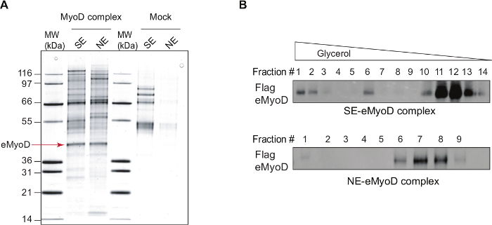
Figure 1: MyoD Complexes Isolated by Tandem Affinity Purification. (A) A silver staining of the double affinity-purified eMyoD complexes isolated from nuclear salt-extractable (SE) or nucleosome-enriched (NE) nuclear fractions of HeLa-S3 cell lines stably expressing Flag-HA-MyoD (MyoD complex) and control cell line (Mock). MW, molecular weight marker in kilo dalton (kDa). Arrow indicates Flag-HA-MyoD (eMyoD). This research was originally published in37. Copyright The American Society for Biochemistry and Molecular Biology. This figure has been modified from26: the lanes with mock purifications and eMyoD complex isolated from SE nuclear fractions are now shown. (B) Double affinity-purified eMyoD complexes as in (A) were fractionated on glycerol gradient ranging from 20% to 41%. Fractions were manually collected, concentrated and analyzed by western blot (WB) using anti-Flag antibodies. Note the presence of several eMyoD-containing sub-complexes in nuclear salt-extractable (SE) fraction. Please click here to view a larger version of this figure.
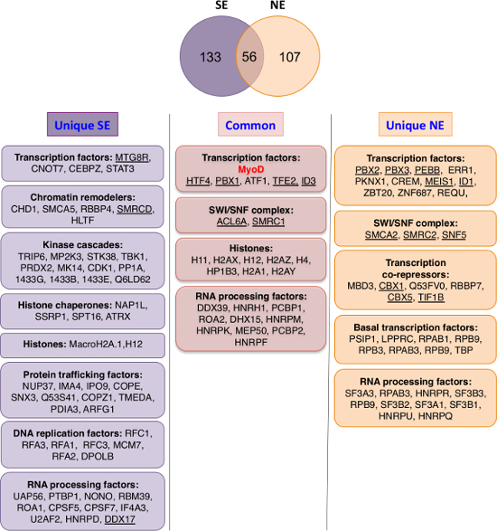
Figure 2: Comparison of Chromatin-bound (nucleosome enriched, NE) versus Nuclear Soluble (nuclear salt extractable, SE) MyoD Partners. Top: Venn diagram showing overlap between eMyoD interactors isolated from nuclear salt-extractable (SE) and nucleosome-enriched (NE) fraction. The ribosomal proteins, translation-initiation factors, DNA repair factors and the tubulin isoforms were excluded from the analysis as they are present in various different data-sets obtained by TAP and considered as non-specific. Bottom: The MyoD interactors found in both SE and NE fractions (common) or specific for one of the fractions (unique) were divided in groups based on their functional annotations. Cytoskeleton-related and other miscellaneous proteins are not depicted. The underlined proteins are MyoD interactors validated either in HeLa and/or in myoblasts by co-immunoprecipitation. Please click here to view a larger version of this figure.
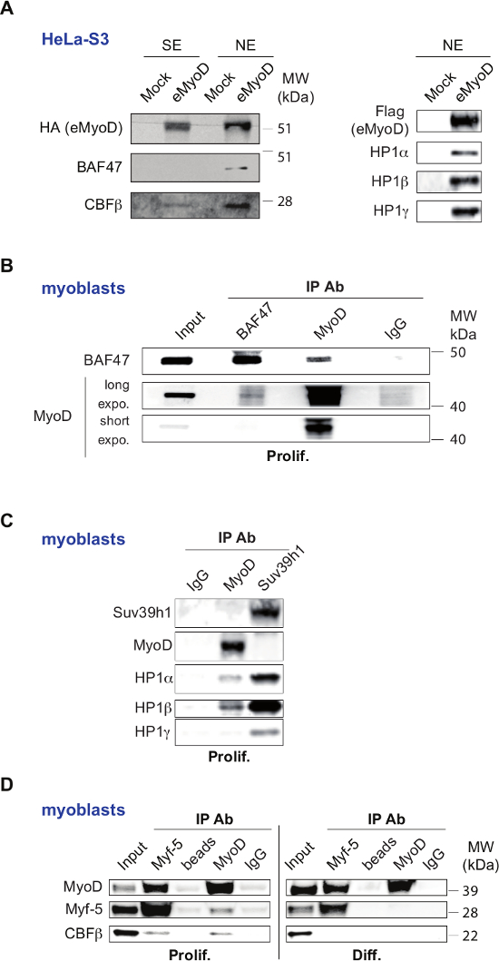
Figure 3: Validation of Selected Set of MyoD Interactors. (A) Validation of MyoD interactors, identified by mass spectrometry, in HeLa-S3 cell line stably expressing Flag-HA-MyoD (eMyoD). Left panel: Western blot analysis of double affinity-purified MyoD complexes isolated from nuclear salt-extractable (SE) or nucleosome-enriched (NE) fractions of HeLa-S3 cell line stably expressing eMyoD or control cell line (Mock) with the indicated antibodies. MW, molecular weight marker. Right panel: Western blot analysis of double affinity-purified MyoD complexes isolated from nuclear nucleosome-enriched fractions of HeLa-S3 cell line stably expressing eMyoD or control cell line (Mock) with indicated antibodies. This panel was originally published in The Journal of Biological Chemistry. Yahi H, Fritsch L, Philipot O, Guasconi V, Souidi M, Robin P, Polesskaya A, Losson R, Harel-Bellan A, Ait-Si-Ali S. J Biol Chem. 2008 Aug 29;283(35):23692-700. doi: 10.1074/jbc.M802647200. Epub 2008 Jul 2. Copyright The American Society for Biochemistry and Molecular Biology. This figure has been modified from26: police type and size was changed and the text was rotated to unify the labeling within the figure. MyoD was labeled as eMyoD to avoid the confusion with endogenous IP presented in the other panels. (B-D) Validation of MyoD interactors in C2C12 mouse myoblasts. (B) Nuclear total extracts from proliferating C2C12 myoblasts were used for immunoprecipitation (IP) with antibodies raised against BAF47 (SNF5) or MyoD, or with control IgG. The resulting precipitates were analyzed by WB with the indicated antibodies. For anti-MyoD antibodies longer (long expo.) and shorter (short expo.) exposure times are shown. Input extracts were loaded to show endogenous protein levels. This panel has been published in28 under the Creative Commons Attribution (CC BY) licence (http://creativecommons.org/licenses/by/4.0/). This figure has been modified from28: police type and size was changed and the test was rotated to unify the labeling within the figure. (C) Nuclear total extracts from proliferating C2C12 myoblasts were used for immunoprecipitation with antibodies raised against MyoD, Suv39h1 (positive control), or control IgG (negative control). The resulting precipitates were then subjected to WB with the indicated antibodies. This panel was originally published in The Journal of Biological Chemistry. Yahi H, Fritsch L, Philipot O, Guasconi V, Souidi M, Robin P, Polesskaya A, Losson R, Harel-Bellan A, Ait-Si-Ali S. J Biol Chem. 2008 Aug 29;283(35):23692-700. doi: 10.1074/jbc.M802647200. Epub 2008 Jul 2. Copyright The American Society for Biochemistry and Molecular Biology. This figure has been modified from26: police type and size was changed and the text was rotated to unify the labeling within the figure. (D) Nuclear total extracts from proliferating (prolif.) and differentiating C2C12 myoblasts (48 hr, indicated as Diff.) were used for immunoprecipitation (IP) with antibodies raised against MyoD and Myf5, or with normal rabbit IgG and with empty beads as negative control. The resulting precipitates were analyzed by WB with the indicated antibodies. Input extracts were loaded to show endogenous protein levels. This panel has been published in27 under the Creative Commons Attribution (CC BY) licence (http://creativecommons.org/licenses/by/4.0/). This figure has been modified from27: police type and size was changed and the text was rotated to unify the labeling within the figure. The panels with the results obtained from proliferating and differentiating C2C12 are separated in two as opposed to the original figure. Please click here to view a larger version of this figure.
Table 1: List of Proteins Identified by MS Analysis in Double Affinity-purified eMyoD Complexes Isolated from Nuclear Salt-extractable Fraction after the Subtraction of the Background Proteins. (Proteins identified by MS in eluates from control cell line were considered as non-specific background.) The data represent the sum of four independent purifications. Please click here to download this file.
Table 2: List of Proteins Identified by MS Analysis in Double Affinity-purified eMyoD Complexes Isolated from Nucleosome-enriched Fraction after the Subtraction of the Background Proteins. The data represent the sum of three independent purifications. Please click here to download this file.
Table 3. Buffer Compositions. Please click here to download this file.
The presented method permits exhaustive identification of partners of a transcription factor, MyoD. It revealed MyoD partners in a heterologous system resistant to MyoD-induced differentiation, namely HeLa-S3 cells. Thus, by definition, identified MyoD partners are ubiquitously expressed. These include general and sequence-specific transcription factors, chromatin-modifying enzymes, RNA processing proteins, kinases (Tables 1 and 2). Since HeLa-S3 cells are resistant to MyoD-induced trans-differentiation, MyoD activity is mainly repressive and the identified partners are likely to be MyoD co-repressors. This is highlighted by the presence of Id proteins (Tables 1 and 2, Figure 2), known to inhibit MyoD transactivation ability8. Importantly, such repressive state corresponds to the MyoD status in proliferating myoblasts. Indeed, we confirmed that MyoD interactions with CBFβ, a MyoD partner identified by the described method, can be detected in proliferating C2C12 myoblasts, but are lost when cells underwent differentiation (see Figure 3D). Nevertheless, it is important to note that one of the main limitations of the presented methodology is the lack of MyoD co-activators identification. Consistently, using this method none of the known MyoD co-activators, such as histone acetyltransferases32 were identified.
Purification of MyoD complexes either from the soluble fraction (nuclear salt extractable, SE) of the nucleus or the chromatin-enriched fraction (nucleosome enriched, NE) (Figure 1A) serves at least two major functions. Firstly, such fractionation increases the representation of the non-stoichiometric partners that would be masked if MyoD purification performed from total nuclear extracts. Secondly, this permits the separation of two functional subpopulations of MyoD: pre-deposited/evicted (SE) and chromatin-bound (NE). Among interactors specific for DNA-unbound MyoD, many kinases, transcription factors, trafficking proteins as well as some chromatin remodelers were identified (Figure 2). When fully validated, such a network could provide an insight into the mode of activity regulation of DNA-unbound MyoD. Additional search for MyoD post-translational modifications in these two nuclear compartments by mass spectrometry, could further unravel MyoD activity regulation. Finally, fractionation of soluble and chromatin-bound MyoD complexes on a glycerol gradient revealed that while the chromatin-bound MyoD is mainly contained in one complex, DNA-unbound MyoD is distributed in three sub-complexes (Figure 1B). Characterization of these sub-complexes by either MS and/or western blot against protein candidates should elucidate further the mode of MyoD regulation.
As stressed above, HeLa cells do not naturally express the muscle transcription factor MyoD. It was thus important to confirm the found interactions in a skeletal muscle model (Figure 3). Many reports confirmed in relevant models such functional interactions between MyoD and some of the partners identified in the presented TAP-tag assay. This is for example the case for prohibitin33, DDX1734 , Meis135, CBF27, HP126, and SWI/SNF chromatin-remodeling complex36,28,30.
Note that it is possible to perform such TAP-tag purification from low amount of cells, such as from myoblasts, when combined to sensitive MS. Indeed, a recent paper described MyoD partners characterization after inducible expression of Flag-tagged MyoD in myoblasts30. Since stable and continuous MyoD overexpression in myoblasts is deleterious, an alternative approach would be adding the Flag-HA tags into the endogenous MyoD allele(s) in myoblasts by using genome-editing methods, such as CRISPR-Cas931. Notably, addition of the tag(s) could potentially alter protein function and/or association with binding partners, therefore the place of the tag (N- or C-terminus) should be chosen with caution. A functional assay of the fusion protein in relevant system must be performed prior to TAP-tag purification to ensure the tagged protein is functional. Immunoprecipitation of endogenous proteins avoids these problems, however, it relies on the availability of a specific and high affinity antibody, which is rarely available.
Another added value of the described approach is the possibility to identify post-translational modifications (PTMs) of the purified protein itself and of its abundant partners. Thereby, with this feature the TAP-tag purification is suitable to identify not only new interaction partners but also new enzymatic functions associated to the protein of interest and/or its partners. In the case of a chromatin-binding protein (i.e., transcription factors, enzymes), this method is thus adapted for identification of the associated "histone code". Indeed, the amino-terminal histone tails, which are exposed on the nucleosome surface, are subject to multiple covalent PTMs. Histone PTMs confer a unique signature to the nucleosomes involved. A combination of different modifications on histone N-terminal tails can thus alter chromatin structure to allow gene expression regulation. Thus, characterizing such modifications associated to a given protein could provide insights into the roles and mechanisms of action of the studied chromatin-binding proteins22.
In summary, the presented methodology permits comprehensive identification of MyoD partners. The TAP-tag purification provides an alternative to other approaches such as GST pull-downs, yeast two-hybrid assays and phage display. Even if for practical reasons (production of large amount of nuclear extracts) we have to use a heterologous cellular system, we have been able to confirm the involvement of the identified MyoD partners in skeletal muscle differentiation. The obtained data shows that the MyoD myogenic factor seems to interact with a plethora of proteins ranging from transcriptional regulators to RNA binding proteins, suggesting the different mechanisms regulating the activity of a transcription factor.
In conclusion, the same methodological approach could be used to identify ubiquitously expressed partners of numerous nuclear factors that could be difficult to study in their specific cellular context.
The authors have nothing to disclose.
Work in the Ait-Si-Ali lab was supported by the Association Française contre les Myopathies Téléthon (AFM-Téléthon); Institut National du Cancer (INCa); Agence Nationale de la Recherche (ANR), Fondation Association pour la Recherche sur le Cancer (Fondation ARC); Groupement des Entreprises Françaises pour la Lutte contre le Cancer (GEFLUC); CNRS; Université Paris Diderot and the ”Who Am I?” Laboratory of Excellence #ANR-11- LABX-0071 funded by the French Government through its ”Investments for the Future” program operated by the ANR under grant #ANR-11-IDEX-0005-01. EB was supported by an INCa grant.
Cell lines | |||
HeLa-S3 | ATCC | CCL-2.2 | |
C2C12 | ATCC | CRL-1772 | |
Name | Company | Catalog Number | Comments |
Equipment | |||
Spinner | Corning | 778531 | |
Homogenizer : Dounce homogenizer | Wheaton | 432-1273 and 432-1271 | |
Agitating device for spinners | Bellco | 778531 | |
Sonicator | Diagenode | UCD 200 | |
Hemocytometer | Marienfeld Superior | 640610 | |
Low-binding tubes | Sorenson | 27210 | |
Empty spin column | Bio-Rad | 7326204 | |
Name | Company | Catalog Number | Comments |
Reagents | |||
SDS-polyacrylamide 4-12 % gel | Life technologies | NP0336BOX | |
4X loading buffer | Life technologies | NP0007 | |
10X reducing agent | Life technologies | NP0004 | |
Silver staining kit | Life technologies | A8592 | |
Centrifugal filter units, 10K | Millipore | UFC501024 | |
Protein G agarose beads | Sigma-Aldrich | P4691 | |
Protein A/G Resin | Thermo Scientific | 53132 | |
Flag resin (anti-Flag M2-agarose affinity gel) | Sigma-Aldrich | A2220 | |
HA resin (monoclonal Anti-HA agarose) | Sigma-Aldrich | A2095 | |
MNase | Sigma-Aldrich | N3755 | |
Flag free peptide (DYKDDDDK) | Ansynth Service BV | Custom synthesis | Resuspend up to 4 mg/mL in 50 mM Tris-HCl, pH 7.8 |
HA free peptide (YPYDVPDYA) | Ansynth Service BV | Custom synthesis | Resuspend up to 4 mg/mL in 50 mM Tris-HCl, pH 7.8 |
Bicinchoninic acid based protein assay kit : BCA kit | Thermo Scientific | 23225 | |
Protease inhibitors | Sigma-Aldrich | S8830 | |
Luminol-based enhanced chemiluminescence (ECL) HRP substrate | Life technologies | 34075 | |
BSA | Sigma-Aldrich | A9647 | |
Sheared salmon sperm DNA (Deoxyribonucleic acid sodium salt from salmon testes) | Sigma-Aldrich | D1626 | |
Spermine tetrahydrochloride | Sigma-Aldrich | S1141 | |
Spermidine | Sigma-Aldrich | S0266 | |
Glycerol | Sigma-Aldrich | G5516 | |
PBS | Sigma-Aldrich | D8537 | |
MOPS running buffer | Life technologies | NP0001 | |
DMEM (high glucose) | Sigma-Aldrich | D0822 | Pre-warm at 37 °C before use |
Trypsin-EDTA (0.05% phenol red | Life technologies | 25300-054 | |
Serum | GE healthcare life sciences | PAA A15-102 | Each lot of serum has to be first tested for your cells. |
Penicillin and Streptomycin | Life technologies | 15140-122 | |
Trypan Blue Solution, 0.4% | Life technologies | 15250-061 | |
Water (sterile-filtered) | Sigma-Aldrich | W3500 | |
Name | Company | Catalog Number | Comments |
Antibodies | |||
HA from rat (12CA5) | Roche | 11583816001 | |
Flag | Sigma-Aldrich | A8592 | |
MyoD | Santa Cruz | sc-32758 | To use for western blotting |
MyoD | Santa Cruz | SC-760 | To use for immunoprecipitation and western blotting |
CBFbeta | Santa Cruz | FL-182 | |
HP1alpha | Euromedex | 2HP2G9 | |
HP1beta | Euromedex | 1MOD1A9AS | |
HP1gamma | Euromedex | 2MOD1GC | |
Suv39h1 | Cell Signaling Technology | 8729 | |
BAF47 | BD Biosciences | 612111 | |
Myf5 | Santa Cruz | SC-302 | |
Tubulin | Sigma-Aldrich | T9026 | |
Actin | Sigma-Aldrich | A5441 | |
IgG Mouse | Santa Cruz | SC-2025 | |
IgG Rabbit | Santa Cruz | SC-2027 | |
Goat anti-rat -HRP | Sigma-Aldrich | A9037 | |
Goat anti-rabbit -HRP | Sigma-Aldrich | A6154 | |
Goat anti-mouse -HRP | Sigma-Aldrich | A4416 |