The social amoeba Dictyostelium discoideum undergoes a developmental transition into a multicellular organism when starved. The evolutionary conserved protein coronin A plays a crucial role in the initiation of development. Using aggregation assays as our main method, we aim to elucidate the role of coronin A in early development.
Dictyostelium discoideum amoeba are found in soil, feeding on bacteria. When food sources become scarce, they secrete factors to initiate a multicellular development program, during which single cells chemotax towards aggregation centers1-4. This process is dependent on the release of cyclic adenosine monophosphate (cAMP)5. cAMP is produced in waves through the concerted action of adenylate cyclase and phosphodiesterases, and binds to G protein-coupled cAMP receptors6,7. A widely used assay to analyze the mechanisms involved in the developmental cycle of the lower eukaryote Dictyostelium discoideum is based on the observation of cell aggregation in submerged conditions8,9. This protocol describes the analysis of the role of coronin A in the developmental cycle by starvation in tissue-culture plates submerged in balanced salt solution (BSS)10. Coronin A is a member of the widely conserved protein family of coronins that have been implicated in a wide variety of activities11,12. Dictyostelium cells lacking coronin A are unable to form multicellular aggregates, and this defect can be rescued by supplying pulses of cAMP, suggesting that coronin A acts upstream of the cAMP cascade10. The techniques described in these studies provide robust tools to investigate functions of proteins during the initial stages of the developmental cycle of Dictyostelium discoideum upstream of the cAMP cascade. Therefore, utilizing this aggregation assay may allow the further study of coronin A function and advance our understanding of coronin biology.
The coronin family of proteins is highly conserved throughout eukaryotes. These proteins are characterized by the presence of an amino-terminal tryptophan-aspartate (WD) repeat-containing region followed by a unique region connected to a carboxy-terminal coiled-coil domain13,14 (Figure 1). Coronins have been implicated in a variety of cellular functions, including cytoskeletal regulation and signal transduction12. In mammals, up to six short coronin molecules (coronin 1-6) as well as a 'tandem' coronin 7, can be co-expressed12,15. Coronin 1 is the most extensively studied family member, and was shown to be involved in pathogen destruction, T cell survival and neuronal signaling. How, exactly, coronin 1 carries out these activities remains unclear. While coronin 1 was shown to regulate Ca2+ and cAMP-dependent signaling as well as F-actin cytoskeleton modulation 16-18, the potential co-expression of up to 7 family members in mammals has made it challenging to study the molecular function of coronins in these systems, due to potential redundancies. Unlike mammalian organisms, the lower eukaryote Dictyostelium discoideum expresses only two coronin family members (coronin A, the ortholog of mammalian coronin 1 and coronin B, the ortholog of mammalian coronin 7) with apparently non-redundant functions15,19,20. This fact makes Dictyostelium discoideum a potent model to study the function of coronins.
To study the role of coronin A in Dictyostelium discoideum, we induced the developmental cycle by starvation in tissue-culture plates containing balanced salt solution (BSS) buffer using either wild type cells or cells lacking coronin A10. We found that cells lacking coronin A were unable to form multicellular aggregates upon starvation. For an accurate quantitative assessment of this phenotype the automated live cell imaging described in this protocol is a vital tool. The defect in the initiation of the early starvation response in cells lacking coronin A can be rescued by supplying pulses of cAMP, suggesting that coronin A acts upstream of the cAMP cascade. The exogenous application of cAMP pulses to simulate the initiation of development has been utilized by several laboratories in the past8,9. However, this procedure is also known to be highly dependent on cell densities and timing. Therefore, the protocol described here aims to reduce these variabilities in order to guarantee a high degree of reproducibility. Taken together, the techniques utilized in these studies provide robust tools to investigate functions of proteins during early stages of the developmental cycle of Dictyostelium discoideum and have the potential to identify up- as well as downstream effectors of coronin A function.
Cells deficient in coronin A show a defect in early development (Figure 2). In the absence of coronin A cells are unable to form multicellular aggregates, which is the initial step during the developmental cycle of Dictyostelium discoideum. Therefore, coronin A appears to play a role during the early starvation response and/or cAMP signaling. Indeed, the lack of multicellular aggregate formation in the absence of coronin A is accompanied by reduced cAMP signaling 10. However, the application of exogenous cAMP pulses fully restores the wild-type phenotype (Figure 3). This implicates that coronin A, rather than being directly involved in cAMP signaling, functions upstream of the cAMP cascade (Figure 5). When Dictyostelium cells are exposed to starving conditions they induce the secretion of early starvation factors22,23. One of the best characterized factors involved in the early starvation response is conditioned medium factor (CMF)22. Based on our results, we hypothesized that coronin A either regulates the secretion of these early starvation factors or is involved in the signal transduction induced by these factors. In order to be able to differentiate between these two effects we performed conditioned medium experiments. Wild-type or corA-deficient cells were exposed to supernatants from either starved wild-type or corA-deficient cells10 (Figure 4). These supernatants are potent initiators of multicellular aggregate formation and therefore contain factors that initiate downstream developmental signaling pathways, including cAMP signaling. Our results show that supernatant from corA-deficient cells was equally capable to induce development as was the case for supernatant from wild-type cells (Figure 4). This implicates that corA-deficient cells are able to produce and secrete early starvation factors at similar levels to their wild-type controls. However, corA-deficient cells are neither able to respond to their own supernatant nor to supernatant provided from wild-type cells (Figure 4). Therefore, coronin A appears to be involved in signaling pathways induced by early starvation factors, rather than their expression/secretion10 (Figure 5).
In general, to be able to further investigate the signals secreted by starving Dictyostelium cells that induce the developmental cycle, the aggregation assay under submerged conditions is a potent read-out. Cells seeded at higher densities will spontaneously aggregate due to an abundance of all required signals. However, at lower densities only the addition of conditioned medium will induce cellular aggregation (Figure 4)22,24. Therefore, this procedure can be utilized to fractionate conditioned medium and study distinct components for their activity during the early starvation response.
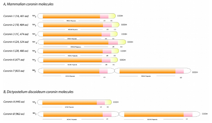
Figure 1: Coronin proteins present in mammals and Dictyostelium discoideum. The coronin family is characterized by the presence of N-terminal WD-repeat regions followed by a unique domain and a C-terminal coiled coil region. While mammals express 7 coronin proteins (A), Dictyostelium discoideum only contains two family members (B), making the risk of redundancy less likely when studying their function. Coronin A is the ortholog of mammalian coronin 1 and coronin B shows a similar domain structure as coronin 7. Abbreviations: CC, coiled-coil domain; UD, unique domain; WD repeats, tryptophan-aspartate repeats. Length is given for mouse sequences (A) in amino acids (aa). Modified from Pieters et al., 201312. Please click here to view a larger version of this figure.
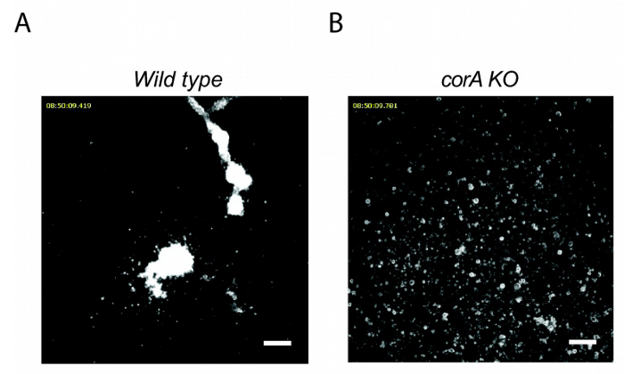
Figure 2: corA–Dictyostelium discoideum are unable to aggregate during the early starvation response. Wild-type (A) or corA-deficient (B) Dictyostelium cells were seeded into multiwell plates at a density of 2 x 10⁵ cells/cm2, starved in BSS, and imaged over a period of 20 hr. Scale bar = 100 µm. Please click here to view a larger version of this figure.
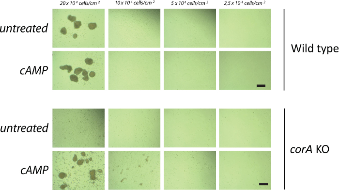
Figure 3: Externally applied cAMP pulses rescue the early developmental defect of corA–Dictyostelium discoideum. Vegetative wild-type (A and B) and corA-deficient cells (C and D) were washed and starved for 2 hr before being pulsed with (B and D) or without (A and C) 50 nM cAMP during 5 hr every 6.5 min in suspension. Cells were then washed, resuspended in BSS, and placed on a Petri dish at a density of 10 x 104 cells/cm2. Images were taken 20 hr after seeding of the cells. Scale bar = 100 µm. Please click here to view a larger version of this figure.
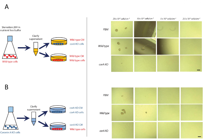
Figure 4: corA-deficient Dictyostelium discoideum are able to produce all required factors for developmental induction but are unable to respond to them. Exponentially growing wild-type and corA-deficient cells were washed in PBM and seeded into multiwell plates at a density of (5, 10, 20, or 40) x 104 cells/cm2, incubated in conditioned medium obtained from wild-type (A) or corA-deficient starving cells (B). The images were taken 16 hr after seeding the cells. Scale bar = 100 µm. Please click here to view a larger version of this figure.
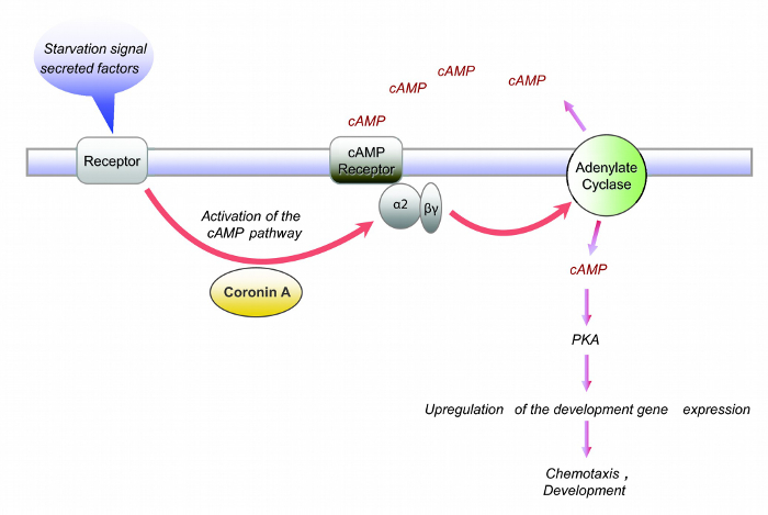
Figure 5: Current model of Coronin A function in Dictyostelium discoideum. Coronin A is involved in the signal transduction of factor(s) secreted during the early starvation response leading to the induction of the cAMP cascade and the progression through the developmental cycle. Modified from Vinet et al., 201410. Please click here to view a larger version of this figure.
The coronin proteins are found in most taxa of the eukaryotic clade. Dictyostelium discoideum coronin A, the homologue of mammalian coronin 1, is involved in the early starvation response, since coronin A-deficient cells are not able to form aggregation centers during the early developmental cycle10. To be able to quantitatively and accurately assess the delay in development between the strains, a microscope live cell imaging set-up with the automated stage controller is an essential tool.
D. discoideum utilizes cAMP as an essential signaling molecule during several stages of its life cycle1-4. One of the checkpoints during which cAMP is required is the transition from a single cell to the formation of multicellular structures. The onset of cAMP waves after ~6 hr of starvation is sensed by the cells and provides the chemotactic signal to move towards an aggregation center5. These cAMP waves can be mimicked experimentally by pulsing starving D. discoideum cells with cAMP every 6.5 min over a period of 5h8,9. Using this procedure it is important to starve the cells for up to 2h before applying the cAMP pulses, in order to obtain reliable results. Utilizing this experimental set up we were able to show that coronin A is acting upstream of cAMP signaling, as these externally applied pulses rescued the development in corA-deficient cells (Figure 3 and 5). These results also indicate that while coronin A appears to have an essential role in sensing the early starvation response, it is dispensable for processes such as chemotaxis and cell aggregation per se10.
The aggregation assay itself is a robust method for studying the early development of Dictyostelium discoideum. However, there are several pitfalls that can influence the reproducibility of these experiments, and it is therefore important that several factors are taken into account: the age of the cells plays an important role when analyzing corA-deficient cells. These cells tend to compensate the phenotype and re-aggregate approximately after 2 weeks from the point they are thawed. The density of the cells is a crucial factor as well, as cells that show a signaling defect and not a chemotactic defect might tend to aggregate at higher densities due to the increased concentration of early starvation factors. The importance of the precise counting and dilution of cells in these assays cannot be overstated. For different strains, an adjustment of multiple variables of the assay, such as cell densities, time of adherence, time of development, will be required. The aggregation assay as described here is a versatile technique that can be modified to suit different inquiries: the cells can be starved, or development can be stimulated or inhibited using different compounds, allowing for a specific modulation of aggregation. The here described set-up also allows for unbiased approaches to identify strains with early developmental defects, as well as for the identification of genes or factors which are important for early development. This is also the method's major drawback: the aggregation assay is a reduced version of the complete developmental cycle. Therefore, it is not suitable for the analysis of phenotypes affecting later stages of the developmental cycle. The main advantage is the possibility to adopt the aggregation assay to a large scale and automated format: in 48-well plates and with 16 hr of development recorded during automated imaging, the aggregation assay proves to be more time and resource-efficient than development on agar plates.
In experiments in which we bypassed the early starvation response, using externally applied cAMP pulses, we showed that coronin A is involved in the sensing of secreted factors by Dictyostelium cells upon starvation. The precise mechanism of coronin A-mediated signaling during the early starvation response remains unclear. Several factors involved during this stage of development have been studied, in particular conditioned medium factor (CMF)25,26. However, whether CMF acts on its own or with co-factors remains unclear and its signaling cascade is only partially understood. Using the aggregation capacity of Dictyostelium cells under submerged conditions here described as a positive read-out for development allows to conduct large-scale screening experiments in a time-efficient manner as compared to aggregation assays on agar surfaces. However, aggregation assays under submerged conditions do have the disadvantage of not fulfilling the complete developmental cycle as cells will be arrested in the initial aggregation stage. Therefore, the technique described here is not suitable for the examination of later developmental processes. In summary, the experimental approach documented here may allow further characterization of potential early starvation factor(s) that induce coronin A-dependent entry into the early starvation response.
The authors have nothing to disclose.
We thank the Dictyostelium Stock Center for strains and reagents. This study was financed by grants from the Swiss National Science Foundation and the Canton of Basel.
HL-5 media (for 1L: 5 g proteose peptone, 5 g thiotone E peptone, 10 g glucose, 5 g yeast extract, 0.35 g Na2HPO4∗2H2O, 0.35 g KH2PO4, 0.05 g dihydrostreptomycin-sulfate, pH 6.6) | |||
Proteose peptone | BD Bioscience | 211693 | |
Thiotone E peptone | BD Bioscience | 211684 | |
Yeast extract | BD Bioscience | 212750 | |
Glucose | AppliChem | A3666 | |
Na2HPO4∗2H2O | Fluka | 71643 | |
KH2PO4 | AppliChem | A1043 | |
dihydrostreptomycin-sulfate | Sigma-Aldrich | D1954000 | |
PBM (0.02 M potassium phosphate, 10 μM CaCl2, and l mM MgCl2, pH 6.1) | self made | ||
BSS (10 mM NaCl, 10 mM KCl, 2.5 mM CaCl2, pH 6.5) | self made | ||
0.45-μm Filtropure S filter | Sarstedt | 83.1826 | |
Falcon 24-well Tissue culture plate | Fisher Scientific | 08-772-1H | |
Cellobserver microscope | Zeiss | custom built | |
AxioVision software | Zeiss | ||
IPC Microprocessor–controlled dispensing pump | ISMATEC | ISM 931 | |
Axiovert 135M microscope | Zeiss | 491237-0001-000 | |
Incubation Shaker | Inforst HT Minitron |