Here, we present a protocol to detect the presence of neutrophils in fixed/permeabilized histology sections and assess the activation state of live purified neutrophils. In particular, the MUB40 peptide binds lactoferrin present in neutrophil-specific and tertiary granules. Exposure of the granule contents through either permeabilization or neutrophil activation allows for the marking of neutrophils.
Here, we provide a protocol involving the use of MUB40, a synthesized peptide with the ability to bind glycosylated lactoferrin stored at high concentrations in specific and tertiary granules of neutrophils. This protocol details how MUB40 conjugated directly to a fluorophore can be used to stain neutrophils in fixed/permeabilized tissues as well as how this can be used in live-cell imaging to assay for neutrophil activation and de-granulation. Neutrophil detection methods are limited to species-specific monoclonal antibodies, which are not always suitable for certain applications. MUB40 does not penetrate the cell membrane and is thus excluded from lactoferrin stored in non-activated/non-permeabilized neutrophils. MUB40 has the added benefit of recognizing lactoferrin from a broad host range, making it especially useful for comparing results in studies involving multiple research models, reducing the number of duplicate reagents, and simplifying protocols through single-step staining.
Neutrophils are one of the primary arms of the innate immune system and are routinely recruited to the sites of inflammation around the body. The study of neutrophils has been largely impaired by their short lifespan in vitro (less than 8 h) and by limited detection tools under basal conditions or after activation. Here, we present a well-tested protocol for the broad and specific detection of mammalian neutrophils in fixed/permeabilized samples. We also provide a detailed protocol for staining live neutrophils with MUB40. Using the live neutrophil staining protocol, the timing and location of neutrophil activation can be pinpointed. This protocol is ideal for researchers who wish to study neutrophil activation or granule release. Beyond their antimicrobial functions, neutrophils are now appreciated as immunomodulatory cells involved in a wide range of diseases and immune responses (innate and adaptive)1,2. Neutrophils are present in most inflammatory tissues, at high numbers in infected tissues, in inflammatory tumors3, during IBS and Crohn's flare-ups4, and in areas of non-infectious inflammation such as the synovia of Rheumatoid Arthritis (RA) patients5. Neutrophils contain four classes of pre-formed granules named azurophil (α), specific (β1), tertiary (β2) granules, and secretory vesicles (γ)6. Upon migration to an inflammatory site, recruited neutrophils become activated and sequentially secrete granule contents (composed of adhesion, antimicrobial compounds and immunomodulatory molecules), which promotes further recruitment and contributes to infection resolution (but also to host tissue damage)7. While neutrophils are considered a critical aspect of innate immunity, to date there are few detection reagents available to study them, and even fewer that can be used to assess the activation state of living neutrophils.
Current methods of detecting neutrophils rely on monoclonal antibodies generated against cell-surface exposed antigens, such as Ly-6G2 or proteins specifically stored in neutrophil granules under basal conditions (e.g., myeloperoxidase, lactoferrin). Advantages of monoclonal antibodies include their strong binding, sensitivity, and versatility under various assay conditions. However, there are several downsides to using monoclonal antibodies and anti-Ly-6G in particular. These downsides include the specificity, as Ly-6G is present on a majority of myeloid cells in bone marrow and on all granulocytes including eosinophils; thus, deciphering neutrophils from eosinophils with this marker requires more complexes approaches8. Another frequent tradeoff with monoclonal antibodies is their often-limited host specificity range, making comparison studies with more than one animal species difficult. A third drawback of antibody detection methods, especially when using live cells or in vivo, is their potential to disrupt cell function or lead to cell activation. For example, the administration of anti-Ly-6G to mice is commonly applied to neutrophil depletion and transient neutropenia9. It has additionally been demonstrated that antibody injection may stimulate neutrophil antitumor function10. Finally, antibody detection of neutrophils does not reveal the activation state of the cell.
We have identified a 40-amino acid peptide called MUB40, which can be used in a number of assays to label neutrophils under in vitro or in vivo conditions as well as assay the activation status of live neutrophils11. MUB40 is derived from MUB7012, a domain of the Lactobacillus reuteri surface mucus-binding protein originally described for its ability to bind mucus13,14. MUB40 interacts with glycosylated lactoferrin that is present at high concentrations in neutrophil-specific and tertiary granules. MUB40 can be exposed to these granules through standard permeabilization steps present in histopathology or fluorescence-activated cell sorting (FACS) protocols for robust staining of fixed neutrophils. When live neutrophils are kept in an inactivated state, the MUB40 peptide is excluded from the granule contents, and cells do not stain positive with the marker. Upon activation, MUB40 can bind to exposed lactoferrin and lead to robust staining with the peptide. Thus, MUB40 can be used to determine the activation state of purified neutrophils, making it an attractive marker to follow the infectious process. The ability to bind to live activated neutrophils also allows MUB40 to be used as a tool to detect areas of neutrophil inflammation in live animal models (e.g., a mouse arthritis model15). The protocols in this manuscript detail how fluorescently labeled MUB40 can be used to reveal neutrophils in histology tissues and how to assay the activation of live purified neutrophils in vitro.
All methods described here have been reviewed and approved by the French organizations CoRC (Comité de Recherche Clinique), CPP (Comité de Protection des Personnes), and CNIL (Commission Nationale Informatique et Liberté).
1. Staining Neutrophils in Histopathology Tissue with Fluorescently Labelled MUB40
2. Visualization of Neutrophil Activation with Retro-Inverso-MUB40-Cy5 Peptide
Results of MUB40-stained tissues from histopathology slides typically reveal individual cells scattered throughout the tissue. MUB40 stains lactoferrin, which is present in neutrophil granules and compartmentalized. Thus, typically seen are punctate staining or several large separated areas of signal coming from individual neutrophils (Figure 1). It is helpful to add a second cell marker such as DAPI to help co-localize the MUB40 signal with the stained cell. The total number of detected cells depends upon the number of neutrophils present in the field of view and can vary dramatically depending upon the source and strength of the immune response. Shown here are representative images from purified fixed human neutrophils (Figure 1A) and from a tissue biopsy of an ulcerative colitis patient (Figure 1B). Also shown are images taken from a relatively low level of neutrophils in the lungs of mice infected with Klebsiella pneumoniae (Figure 1C) and a high level of neutrophils in the colon of a guinea pig infected with Shigella sonnei (Figure 1D).
Results using live, purified neutrophils typically show little-to-no cell staining at early time points, and gradually increase in RI-MUB40 staining over time as more neutrophils become activated. Shown here is a time course series of images from an experiment using purified human neutrophils infected with fluorescent S. sonnei (Figure 2). Depending on the strength of the activating signal, neutrophils may begin staining with MUB40 rapidly, so it is recommended to start acquiring images prior to the addition of activators. When cells become activated and RI-MUB40 positive, they should exhibit a similar staining profile to that of fixed and permeabilized cells, which give a punctate staining. The optimal concentration of MUB40 for both live and fixed cell staining is 1 μg/mL (Figure 3). Lower concentrations (0.1-0.01 μg/mL) can be used but result in lower signal intensity. It is also recommended to use a DIC image or other live-cell stain to differentiate which cells are displaying an RI-MUB40 signal.
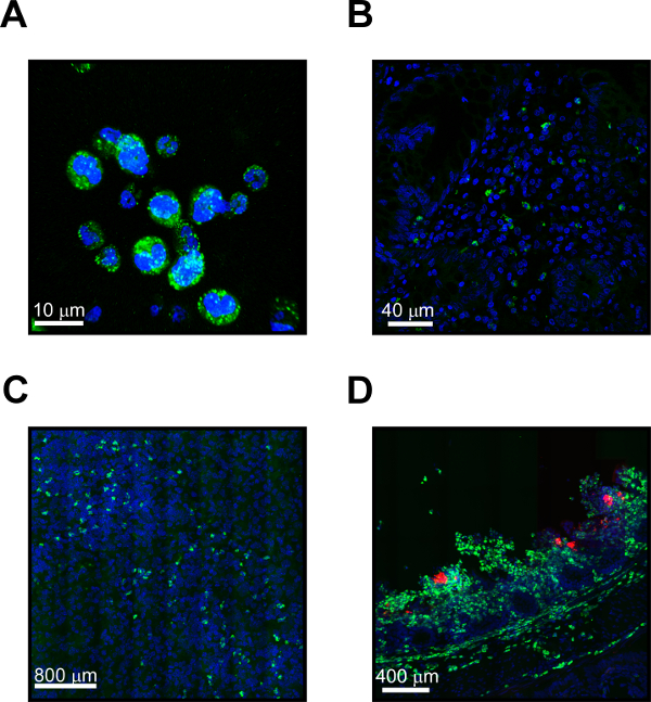
Figure 1: MUB40-stained neutrophils of human, mouse, and guinea pig origin. (A) Representative MUB40 staining (green) of neutrophils purified from healthy human donors. Cell nuclei are stained with DAPI (blue). (B) Staining of human neutrophils in tissue from an ulcerative colitis biopsy. Neutrophils are revealed with MUB40 (green) and cell nuclei are stained with DAPI (blue). (C) Histopathology from the lung of a mouse infected with Klebsiella pneumoniae. Mouse neutrophils are revealed in the tissue using MUB40 (green) and cell nuclei are stained with DAPI (blue). (D) Histopathology from the colon of a guinea pig infected with Shigella sonnei. Neutrophils responding to the infection are revealed in the tissue with MUB40 (green). S. sonnei bacteria (red) express the fluorescent dsRED protein. Cell nuclei are stained with DAPI (blue). Please click here to view a larger version of this figure.
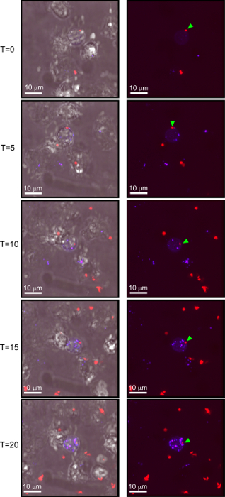
Figure 2: Live neutrophils stain with MUB40 only when activated. Selected images from a time lapse series involving purified human neutrophils infected with S. sonnei in the presence of RI-MUB40. In the first set of images (top) a neutrophil encounters a S. sonnei bacterium (red) shown with a green arrow. Over the time course, the bacterium is internalized by the neutrophil and digested. As the neutrophil becomes activated from the bacteria, it stains progressively stronger with RI-MUB40. Images on the left are fluorescent channels overlaid with DIC images. Images on the right are fluorescent channels only. Images were taken at 5-min intervals. Please click here to view a larger version of this figure.
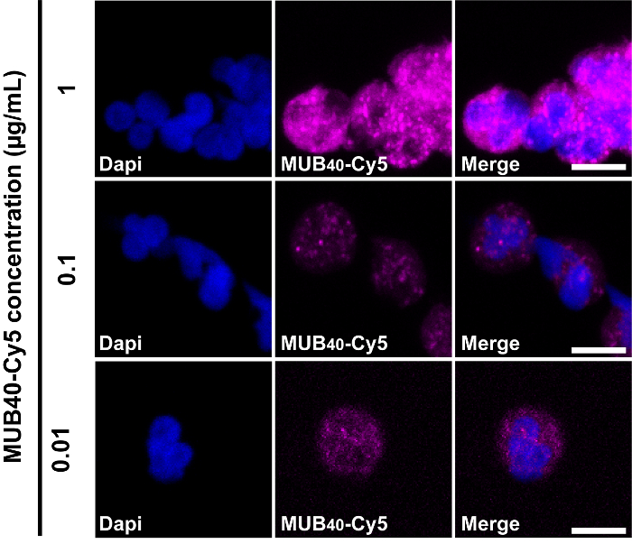
Figure 3: Effect of different MUB40 concentrations on neutrophil staining. Representative staining of fixed/permeabilized neutrophils with various concentrations of MUB40-Cy5. Purified human neutrophils were fixed and stained with the indicated concentrations of MUB40-Cy5. Cell nuclei are stained with DAPI (blue). Please click here to view a larger version of this figure.
Here, two assays are described in which the MUB40 peptide is used to study neutrophil inflammation and activation. We show how MUB40 can reveal neutrophils present in histopathology sections or show their activation state using live purified neutrophils. The critical steps for using MUB40 as a staining reagent are the same as with any other fluorescent detection method. Care must be taken to ensure compatibility of fluorescent signals and that adequate washing steps have been taken to remove background staining. The most important considerations to achieve good staining results are the quality of the microscope, glass slides/cover slips, and the tissue sections. When viewing live purified neutrophils, the most important steps are in the purification process itself. Neutrophils are sensitive and can be activated by a number of different signals. In order to ensure that the experiment produces meaningful results, care must be taken to keep the purified neutrophils inactive until they are ready to be used. This can be achieved by performing the neutrophil purification protocol in an anoxic chamber to limit neutrophil exposure to oxygen.
Both a limitation and advantage of MUB40, depending upon the experimental question being addressed, is that MUB40 only stains activated neutrophils unless they are somehow permeabilized. This can be a huge advantage if the experiment calls for following inflammation in a live animal or purified cell, but it may be a disadvantage if the experiment requires detection of all live neutrophils regardless of activation state. A second potential limitation to this protocol is the fact that lactoferrin is present in various bodily secretions such as tears, milk, and mucus. For histopathology staining in which these secretions are maintained (e.g., colonic biopsies in which the mucus layer is intact), MUB40 will also stain the mucus layer along with any present neutrophils. In practice, this has yet to affect our analysis, but it should be noted as a potential signal source.
Currently, MUB40 is the only neutrophil biomarker that can differentiate between activated and non-activated live neutrophils. Also, MUB40, unlike antibody detection methods, does not appear to alter the function or survival of neutrophils in vitro or in vivo. For example, addition of specific antibodies against live neutrophils can be an activating signal affecting neutrophil function or survival in the host. This makes MUB40 a very useful reagent for research involving neutrophil activation or granule release in vitro or in vivo. Additionally, MUB40 has a broad host specificity range and can be directly conjugated to a number of different fluorophores, allowing for use in single-step staining assays across multiple hosts. Thus, MUB40 staining is comparable between mouse, human, and other mammalian targets, and it simplifies and reduces the number of reagents needed to detect neutrophils.
While this protocol focuses specifically on histopathology and live cell imaging using MUB40, we have also successfully used the peptide in FACS sorting and live animal in vivo imaging. We anticipate that MUB40 will be amenable to many different assays involving the detection of neutrophils or visualization of inflammatory events in the body, and that future studies and protocols will be developed to further leverage the spectrum of MUB40 uses, particularly using non-invasive imaging technics.
The authors have nothing to disclose.
This work was supported by the Fondation Laurette Fugain (LF-2015-15) (B.S.M.) and ANR JCJC grants (ANR-17-CE15-0012) (B.S.M.).
MUB40-Cy5 | Benoit Marteyn | benoit.marteyn@pasteur.fr | Fluorescent MUB40 peptide (available with other conjugated fluorophores) |
RI-MUB40-Cy5 | Benoit Marteyn | benoit.marteyn@pasteur.fr | Retro-inverso fluorescent MUB40 peptide. Synthesized with D-amino acids and resistant to proteases (available with other conjugated fluorophores) |
Parformaldehyde | Sigma-Aldrich | 16005 | Fixative for histology |
Sucrose | Sigma-Aldrich | S7903 | Solution used to remove excess PFA |
Optimal Cutting Temperature Compound (OCT) | Sakura Finetek USA | 4583 | Used to freeze tissue before cryostat sectioning |
2-Methylbutane | Sigma-Aldrich | M32631 | Used to freeze tissue before cryostat sectioning |
Ethanol | Sigma-Aldrich | 1.00974 | Used for dry ice bath to freeze tissue |
Leica Cryostat | Leica Biosystems | CM1520 | Used to section tissues |
Glass microscopy slides | Fisher Scientific | 12-518-101 | |
Cover slips | Thor Labs | CG15CH | Hi precision coverslip |
Dako pen | Sigma-Aldrich | Z377821 | Used to prevent liquid loss during tissue staining |
Saponin | Sigma-Aldrich | 47036 | Used to permeabilize cells for IF staining |
DAPI | Sigma-Aldrich | 10236276001 | Stains DNA |
Alexa fluor 488 Phalloidin | Fisher Scientific | A12379 | Binds actin |
Prolong Gold antifade Mountant | Fisher Scientific | P36930 | Prolongs IF signal and resistance to photobleaching |
Fluorescent microscope | Various | Various | Used to image IF slides |
Anoxic Cabinet | Various | Various | Used for the purification of live inactivated neutrophils |
Sodium Chloride NaCL | Sigma-Aldrich | S7653 | |
EDTA | Sigma-Aldrich | E9884 | Washing buffer component |
MACS BSA buffer | Miltenyi Biotec | 130-091-376 | Washing buffer component |
Percoll | Sigma-Aldrich | P4937 | Gradient for neutrophil purification |
CD235a glycophorin magnetic microbeads | Miltenyi Biotec | 130-050-501 | Used to remove contaminating RBCs |
LS columns | Miltenyi Biotec | 130-042-401 | Used in the removal of RBCs from neutrophils |
RPMI-1640 without Phenol Red | Sigma-Aldrich | R8755 | Used for neutrophil assays |