Decellularized cartilage-derived scaffolds can be used as a scaffold to guide cartilage repair and as a means to regenerate osteochondral tissue. This paper describes the decellularization process in detail and provides suggestions to use these scaffolds in in vitro settings.
Osteochondral defects lack sufficient intrinsic repair capacity to regenerate functionally sound bone and cartilage tissue. To this extent, cartilage research has focused on the development of regenerative scaffolds. This article describes the development of scaffolds that are completely derived from natural cartilage extracellular matrix, coming from an equine donor. Potential applications of the scaffolds include producing allografts for cartilage repair, serving as a scaffold for osteochondral tissue engineering, and providing in vitro models to study tissue formation. By decellularizing the tissue, the donor cells are removed, but many of the natural bioactive cues are thought to be retained. The main advantage of using such a natural scaffold in comparison to a synthetically produced scaffold is that no further functionalization of polymers is required to drive osteochondral tissue regeneration. The cartilage-derived matrix scaffolds can be used for bone and cartilage tissue regeneration in both in vivo and in vitro settings.
Articular cartilage defects in the knee caused by traumatic events can lead to discomfort, and above all can have a large impact on the lives of the young and active population1,2,3. Moreover, cartilage damage at a young age may lead to a more rapid onset of osteoarthritis later in life4. Currently, the only salvage therapy for generalized osteoarthritis of the knee is joint replacement surgery. As cartilage is a hypocellular, aneural, and avascular tissue, its regenerative capacity is severely limited. Therefore, regenerative medicine approaches are sought after to aid and stimulate the regenerative capacity of the native tissue. For this purpose, scaffolds are designed and used as either a cell-carrier or as an inductive material that incites differentiation and regeneration of tissue by the body's native cells5.
Decellularized scaffolds have been widely studied within regenerative medicine6. It has had some success, for example, in aiding the regeneration of skin7, abdominal structures8, and tendons9. The advantage of using decellularized scaffolds is their natural origin and their capacity to retain bioactive cues that both attract and induce cell differentiation into the appropriate lineage required for tissue repair6,10. Moreover, since extracellular matrix (ECM) is a natural biomaterial, and decellularization prevents a potential immune response by removing cellular or genetic content, issues regarding biocompatibility and biodegradability are overcome.
Cartilage-derived matrix (CDM) scaffolds have shown great chondrogenic potential in in vitro experiments when seeded with mesenchymal stromal cells11. In addition, these scaffolds have shown the potential to form bone tissue through endochondral ossification on ectopic locations in in vivo settings12. As CDM scaffolds guide the formation of both bone and cartilage tissue, these scaffolds may hold potential for osteochondral defect repair in addition to cartilage repair.
This article describes a protocol adapted from Yang et al. (2010)13 for the production of decellularized CDM scaffolds from equine stifle cartilage. These scaffolds are rich in collagen type II and devoid of cells, and do not contain any glycosaminoglycans (GAGs) after decellularization. Both in vitro and in vivo experiments on (osteo)chondral defect repair can be conducted using these scaffolds.
For this protocol, equine stifle cartilage was obtained from horses that had died from other causes than osteoarthritis. Tissue was obtained with permission of the owners, in line with the institutional ethical regulations.
NOTE: This protocol describes the fabrication of scaffolds from decellularized equine cartilage, which can be used for applications such as in vitro tissue culture platforms or for in vivo implantation in regenerative medicine strategies. The enzymatic treatment steps must be performed in the described chronological order.
1. Harvesting of Articular Cartilage from Donor (Cadaveric) Joints
2. Creating Decellularized Cartilage Particles
3. Enzymatic Decellularization with Trypsin 0.25%-EDTA
4. Enzymatic Decellularization with Nuclease Treatment
5. Detergent Decellularization
6. Creating Scaffolds from the Decellularized Particles
7. Characterization of the Decellularized Scaffolds with Histological Stainings
NOTE: To ensure complete decellularization and to visualize the remaining natural character of the cartilage, perform several histological stainings before using the scaffolds in any experiment, including hematoxylin and eosin (H&E) staining to ensure decellularization, Safranin-O staining to visualize residual GAG presence, collagen type I immunohistochemistry to differentiate between collagen content, and collagen type II immunohistochemistry to differentiate between collagen content.
8. Characterization of the Decellularized Scaffolds with Quantitative Analyses
9. Seeding of the Decellularized Scaffolds
Decellularization of CDM scaffolds must always be confirmed using histological stainings as well as using DNA quantification to measure the amount of DNA remnants. Insufficient decellularization might lead to undesired immunological responses that influence the results in in vivo settings15,16,17. For this specific decellularization method, DNA was below the detection range, which started at 13.6 ± 2.3 ng/mg DNA/dry weight (n = 3). Full decellularization using this protocol will lead to the production of a scaffold that is rich in collagen type II (Figure 4C) and has no cells (Figure 4A) or GAGs (Figure 4B). Healthy equine cartilage is displayed as comparison, stained for Safranin-O (Figure 5A) and collagen II (Figure 5B).
The scaffold must display a macroscopically homogeneous porosity. Air bubbles will lead to easily detectable large holes in the scaffold and are, therefore, carefully removed. These large holes in the scaffold may have a detrimental impact on the mechanical properties and lead to inhomogeneous cell attachment upon seeding. Successful production of the scaffold also involves a freeze-drying step lasting for at least 24 h; this will lead to a scaffold that has a white appearance (Figure 3). In case of insufficient lyophilization, the scaffolds will have a yellowish color and no clear pores can be observed.
In order for the scaffold to be used for in vivo applications and in vitro cell-seeding, cell integration with the scaffold, as well as cellular functionality, must be shown. Here, scaffolds were seeded with MSCs that produced ECM after 4 (Figure 6A-D) and 6 (Figure 6E–H) weeks of in vitro culturing. Formation of GAG, collagen II, and a peripheral collagen I, as well as the presence of cells, was shown. Additionally, the specificity of collagen II is displayed in Figure 5B, where cartilage but not bone is stained positive for collagen II in an osteochondral plug.
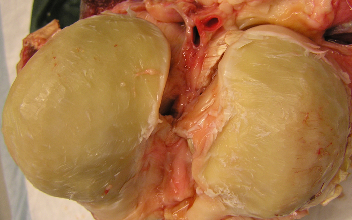
Figure 1: Equine knee after removing full-thickness cartilage. The cartilage is removed from the condyles using a scalpel until the calcified cartilage layer that cannot be cut using a scalpel is reached. Please click here to view a larger version of this figure.
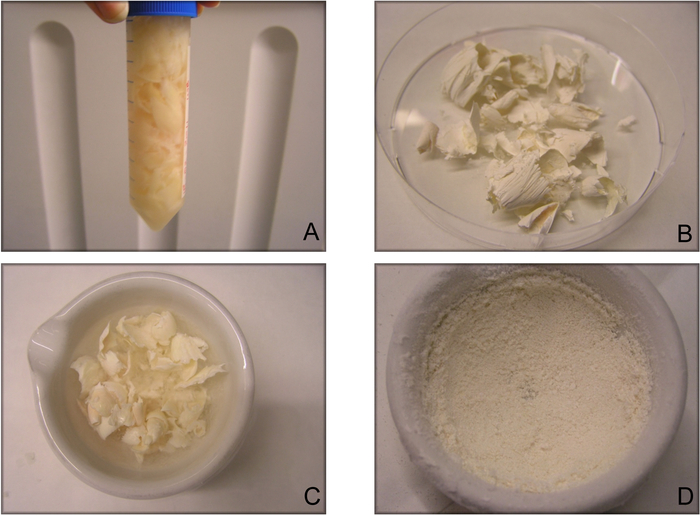
Figure 2: Sequential steps in creating decellularized cartilage-derived matrix particles. (A) Cartilage slices that have been removed from the condyles are washed in an antibiotic-infused solution. (B) Cartilage slices are lyophilized, and now have a white and paper-like appearance. (C) Snap-freezing of the lyophilized cartilage is performed right before (D) pulverizing cartilage particles by hand-milling using a mortar and pestle. Step D can also be done with automatic milling. This figure has been modified from Benders et al.11. Please click here to view a larger version of this figure.
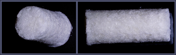
Figure 3: The final product, a decellularized cartilage-derived matrix scaffold. This cylindrical scaffold is 2 cm high and has a diameter of 8 mm. The scaffold has a clear porous structure. The left and right picture display a scaffold from two different angles. Note that no large holes are present at the surface of the scaffold as all of the air bubbles were removed prior to lyophilization. This figure has been modified from Benders et al.11. Please click here to view a larger version of this figure.
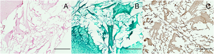
Figure 4: Histological characterization of the scaffold. (A) H&E staining shows ECM particles of different sizes and the absence of cells. (B) Safranin-O staining shows that no GAGs have been retained in the decellularization process. (C) Collagen type II immunolocalization reveals that the decellularized particles are rich in collagen type II. Scale bars = 500 µm. This figure has been modified from Benders et al.11. Please click here to view a larger version of this figure.
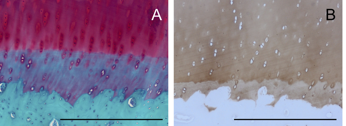
Figure 5: Histological representation of healthy equine cartilage. (A) An osteochondral plug stained with Safranin-O and Fast Green shows bone without GAGs (green), cartilage with GAGs (red), and the calcified cartilage layer in between. (B) A collagen II-stained osteochondral plug stains cartilage but not bone. Scale bars = 500 µm. Please click here to view a larger version of this figure.
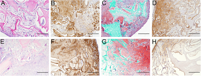
Figure 6: Neo-matrix formation on the scaffold after 4 and 6 weeks of culture using mesenchymal stromal cells. After 4 (A-D) and 6 (E-H) weeks of culture, newly formed matrix is rich in cells (A+E), collagen II (B+F), and GAGs (C+G), as can be observed with H & E, collagen II, and Safranin-O stainings, respectively. IN addition, collagen I is present after both 4 (D) and 6 (H) weeks in the periphery of the scaffold. Cell density as well as the amount of matrix deposition is higher at the periphery. Scale bars = 500 µm. This figure has been modified from Benders et al.11. Please click here to view a larger version of this figure.
The ECM of articular cartilage is very dense and quite resilient to different enzymatic treatments. The multi-step decellularization protocol described in this article addresses such resistance and successfully generates decellularized matrices. To achieve that, the process spans over several days. Many decellularization processes have been proposed for different types of tissues18, and this article describes a protocol suitable for the decellularization of cartilage. In this protocol, it is, however, necessary to follow the enzymatic treatment with the detergent steps in order to remove all cells. The amount of DNA is diminished remarkably in the first few steps involving the treatment with trypsin, and leaving out these steps will not result in proper decellularization11.
Note that this protocol is based on the decellularization of equine cartilage tissue. The activity of enzyme solutions used was found sufficient for the adequate removal of the equine chondrocytes. However, despite the conservation of the matrix composition across species, the protocol may have to be adjusted for decellularization of cartilage from other animals due to the differences in the amount of naturally residing chondrocytes19. For example, cartilage of smaller animals is known to have a higher cell content, and may, therefore, require a more aggressive decellularization process. A particular reason for choosing equine cartilage to create decellularized scaffolds is that equine and human cartilage show clear resemblance in thickness, cell density, and biochemical make-up20.
To ensure a reproducible product, several assessment criteria may be important to determine whether complete decellularization has been achieved. In this protocol, both an H&E staining and biochemical quantification were used to evaluate the residual amount of DNA in the end product. Other researchers have also proposed to determine the size of the remaining DNA, with a maximum of 200 bp in length for quality control21, or a residual DNA amount of less than 50 ng/mg dry tissue weight18,22. Regardless, alterations to the protocol must always be followed up with histological evaluation and quantitative assays to determine the effect of decellularization, as well as the remaining ECM products.
The main limitation of this protocol is that, the thorough decellularization procedure involving the exposure to trypsin leads to extensive loss of GAGs. Even though trypsin does not cleave GAGs, the reason for the loss of GAGs can be the opening up of the cartilage tissue by trypsin cleaving proteins that anchor or encapsulate GAGs. ECM components like GAGs are important for retaining water in articular cartilage, and therefore play a significant role in the biomechanical resilience of the tissue4. Protocols that aim to reduce the loss of GAGs throughout the decellularization process will affect the thoroughness of the decellularization process.
After decellularization, cell integration and function have been shown in cell-seeded scaffolds. Previous research has shown that matrix production by chondrocytes on this scaffold is unsatisfactory, especially when compared to the abundant matrix deposition by mesenchymal stromal cells11. As cartilage-like tissue is deposited on the scaffold, this new matrix is generally first deposited in the periphery of the scaffold before invading the rest of the scaffolds. This can be clearly observed on the histological slices where a cell-rich periphery, rather than a cell-rich center, is often seen (Figure 6). However, this effect may be reduced when using perfusion bioreactors for cell seeding and to enhance nutrient exchange. As matrix deposition occurs, the scaffolds will also assume a glossier appearance, becoming more mechanically consistent and less brittle. As such, they can be easily cut using a scalpel without falling apart. The properties of the newly formed matrix can be evaluated using both histological stainings and quantitative assays. As no GAGs are left after the decellularization process, all of the GAGs that can be quantitatively measured will be a product of neosynthesis.
The scaffolds produced using this decellularization protocol provide an off-the-shelf solution and can be implanted without the necessity of cell-seeding prior to implantation. However, when applied as a treatment for (osteo)chondral defects, the biomechanical properties will have to be enhanced to diminish the chance of indentation of the construct in the early phases of articular loading. Co-implantation with protective layers on top, or other reinforcement strategies may need to be implemented. In the future, further refinement of the protocol may enhance the regenerative potential of the scaffold. For instance, the retention of GAGs, the preservation of collagen fiber orientation, or combination with other biomaterials to reinforce these scaffolds may be beneficial to allow for an improved and smoothly regenerated articular surface. Consequently, these scaffolds may play a role as components of the next generation of regenerative grafts for the treatment of (osteo)chondral defects.
The authors have nothing to disclose.
The authors would like to acknowledge W. Boot for assistance in the production of the scaffolds. K.E.M. Benders is supported by the Alexandre Suerman Stipendium from the University Medical Center. R. Levato and J. Malda are supported by the Dutch Arthritis Foundation (grant agreements CO-14-1-001 and LLP-12, respectively).
Cadaveric joint | This can be obtained as rest material from the local butcher or veterinary center. | ||
Sterile phosphate-buffered saline (PBS) | |||
Penicillin-Streptomycin | Gibco | 15140 | |
Amphotericin B | Thermo Fischer Scientific | 15290026 | |
Liquid nitrogen | |||
Trypsin-EDTA (0.25%), phenol red | Thermo Fischer Scientific | 25200072 | |
Tris-HCl pH 7.5 | |||
Deoxyribonuclease I from bovine pancreas | Sigma-Aldrich | DN25 | |
Ribonuclease A from bovine pancreas | Sigma-Aldrich | R6513 | |
Triton X-100 (octoxynol-1) | Sigma-Aldrich | X100 | |
Papain | Sigma-Aldrich | P3125 | |
Trisodium citrate dihydrate | Sigma-Aldrich | S4641 | |
Alginate | Sigma-Aldrich | 180947 | |
Formalin | |||
CaCl2 | |||
Ethanol | |||
Xylene | |||
Paraffin | |||
Ethylene oxide sterilization | Synergy Health, Venlo, the Netherlands | ||
Multipotent Stromal cells/chondrocytes from equine donors | MSCs and chondrocytes can be isolated from donor joints that are rest material, coming from the local butcher or veterinary center. | ||
MEM alpha | Thermo Fischer Scientific | 22561 | |
L-ascorbic acid 2-phosphate | Sigma-Aldrich | A8960 | |
DMEM | Thermo Fischer Scientific | 41965 | |
Heat inactivated bovine serum albumin | Sigma-Aldrich | 10735086001 | |
Fibroblast growth factor-2 (FGF-2) | R & D Systems | 233-FB | |
DNA quantification kit (Quant-iT PicoGreen dsDNA Reagent) | Thermo Fischer Scientific | P7581 | |
1,9-Dimethyl-Methylene Blue zinc chloride double salt | Sigma-Aldrich | 341088 | |
Freeze-dryer | SALMENKIPP | ALPHA 1-2 LD plus | |
Analytical mill | IKA | A 11 basic | |
mortar/pestle | Haldenwanger 55/0A | ||
Roller plate | CAT | RM5 | |
Centrifuge (for 50 mL tubes) | Eppendorf | 5810R | |
Capsule (cylindric mold) | TAAB | 8 mm flat | |
Superlight S UV | Lumatec | 2001AV | |
Incubator | |||
Microtome | |||
Sieve (mesh size 0.71 mm) | VWR | 34111229 | |
Scalpel | |||
Scalpel holder | |||
Small laddle |