This article describes the encapsulation of falcarindiol in lipid-coated 74 nm nanoparticles. The cellular uptake of the nanoparticles by human stem cells into lipid droplets is monitored by fluorescent and confocal imaging. Nanoparticles are fabricated by the rapid injection method of solvent shifting, and their size is measured with the dynamic light scattering technique.
Nanoparticles are the focus of an increased interest in drug delivery systems for cancer therapy. Lipid-coated nanoparticles are inspired in structure and size by low-density lipoproteins (LDLs) because cancer cells have an increased need for cholesterol to proliferate, and this has been exploited as a mechanism for delivering anticancer drugs to cancer cells. Moreover, depending on drug chemistry, encapsulating the drug can be advantageous to avoid degradation of the drug during circulation in vivo. Therefore, in this study, this design is used to fabricate lipid-coated nanoparticles of the anticancer drug falcarindiol, providing a potential new delivery system of falcarindiol in order to stabilize its chemical structure against degradation and improve its uptake by tumors. Falcarindiol nanoparticles, with a phospholipid and cholesterol monolayer encapsulating the purified drug core of the particle, were designed. The lipid monolayer coating consists of 1,2-distearoyl-sn-glycero-3-phosphocholine (DSPC), cholesterol (Chol), and 1,2-distearoyl-sn-glycero-3-phosphoethanolamine-N-[methoxy(polyethylene glycol)-2000] (DSPE PEG 2000) along with the fluorescent label DiI (molar ratios of 43:50:5:2). The nanoparticles are fabricated using the rapid injection method, which is a fast and simple technique to precipitate nanoparticles by good-solvent for anti-solvent exchange. It consists of a rapid injection of an ethanol solution containing the nanoparticle components into an aqueous phase. The size of the fluorescent nanoparticles is measured using dynamic light scattering (DLS) at 74.1 ± 6.7 nm. The uptake of the nanoparticles is tested in human mesenchymal stem cells (hMSCs) and imaged using fluorescence and confocal microscopy. The uptake of the nanoparticles is observed in hMSCs, suggesting the potential for such a stable drug delivery system for falcarindiol.
Lipid-coated nanoparticles are seeing an increased interest regarding their function as drug delivery systems for cancer therapy1. Cancers have an altered lipid-metabolic reprogramming2 and an increased need for cholesterol to proliferate3. They overexpress LDLs1 and take in more LDLs than normal cells, to the extent that a cancer patient's LDL count can even go down4. LDL uptake promotes aggressive phenotypes5 resulting in proliferation and invasion in breast cancer6. An abundance of LDL receptors (LDLRs) is a prognostic indicator of metastatic potential7. Inspired by the LDL and its uptake by cancer cells, a new strategy has been called: Make the drug look like the cancer's food8. Thus, these new nanoparticle drug delivery designs8,9,10 have been inspired by the core- and lipid-stabilized design of the natural LDLs11 as a mechanism for delivering anticancer drugs to cancer cells. This passive targeting delivery system supports the encapsulating of, especially, hydrophobic drugs, which are usually given in oral dosage form but provide only a small amount of the drugs to the bloodstream, so limiting their expected efficacy12. As with the stealth liposomes13, a polyethylene glycol (PEG) coating helps to reduce any immunologic response and extends the circulation in the bloodstream for optimum tumor uptake by the purported enhanced permeation and retention (EPR) effect14,15. However, in addition to, in some instances, instability in the circulation and undesirable distribution in the system16, some obstacles remain unsolved, such as how and to what extent such nanoparticles are taken in by cells and what is their intracellular fate. It is here that this paper addresses the nanoparticle uptake of a particular hydrophobic anticancer drug falcarindiol, using confocal and epifluorescence imaging techniques.
The aim of the study is to fabricate lipid-coated nanoparticles of falcarindiol and to study their intracellular uptake in hMSCs. Thereby, potentially stabilizing its administration, overcoming the challenges associated with the delivery, and improving the bioavailability. Thus assessing a new delivery system for this anticancer drug. Previously, falcarindiol has been administrated orally via a high concentration purified falcarindiol as a food supplement17. However, there is need for a more structured approach to deliver this promising drug. Therefore, falcarindiol nanoparticles, with a phospholipid and cholesterol encapsulating monolayer with the purified drug constituting the core of the particle, were designed. The rapid injection method of solvent shifting, as recently developed by Needham et al.8, is used in this study to encapsulate the polyacetylene falcarindiol.
The method has previously been used for the fabrication of lipid nanoparticles to encapsulate diagnostic imaging agents18,19, as well as test molecules (triolein)27 and drugs (orlistat, niclosamide stearate)8,27,28. It is a relatively simple technique when carried out with the right molecules. It forms nanosized particles, at the limit of their critical nucleation (~20 nm diameter), of highly insoluble hydrophobic solutes dissolved in a polar solvent. The solvent exchange is accomplished by a rapid injection of the organic solution into an excess of antisolvent (usually, an aqueous phase in a 1:9 organic:aqueous volume ratio)20,21.
The compositional design of the nanoparticles give rise to multiple advantages. The DSPC:Chol components provide a very tight, almost impermeable, biocompatible, and biodegradable monolayer.The PEG provides a sterically stabilizing interface which acts as a shield from opsonization by the immune system, slowing any uptake by the reticuloendothelial system (liver and spleen) and protecting against the mononuclear phagocyte system, preventing their retention and degradation by the immune system, and hence, increasing their circulation half-time in blood22. This allows the particles to circulate until they extravasate at diseased sites, such as tumors, where the vascular system is leaky, allowing EPR-effect to give rise to passive accumulation of the particles. Additionally, the lipid coat allows one to have better control over the nanoparticles' size by kinetically trapping the core at its critical nucleus dimension27,28. Lipids induce various surface properties (including peptide targeting, which was not yet available for this project), a pure drug core, and a low polydispersity22,27,28. The method used for particle size analysis is DLS, a technique that allows researchers to measure the size of a large number of particles at the same time. However, this method can bias the measurements to bigger sizes, if the nanoparticles are not monodispersed23. This issue is assessed with the lipid coat as well. More details of these fundamental designs and the quantification of all characteristics are given in other publications27,28.
The drug encapsulated in the nanoparticles is falcarindiol, a dietary polyacetylene found in plants from the Apiaceae family. It is a secondary metabolite from the aliphatic C17polyacetylenes type that has been found to display health-promoting effects, including anti-inflammatory activity, antibacterial effects, and cytotoxicity against a wide range of cancer cell lines. Its high reactivity is related to its ability to interact with different biomolecules, acting as a very reactive alkylating agent against mercapto and amino groups24. Falcarindiol has previously been shown to reduce the number of neoplastic lesions in the colon17,25, although the biological mechanisms are still unknown. However, it is thought that it interacts with biomolecules such as NF-κB, COX1, COX-2, and cytokines, inhibiting their tumor progression and cell proliferation processes, leading to arresting the cell cycle, endoplasmic reticulum (ER) stress, and apoptosis17,26 in cancer cells. Falcarindiol is used in this study as an example anticancer drug due to its anticancer potential and mechanism are being studied currently, and because it shows promising anticancer effects. The cellular uptake of the nanoparticles is tested in hMSCs and imaged using epifluorescence and confocal microscopy. This cell type was chosen due to its large size, making them ideal for microscopy.
1. Nanoparticle synthesis by rapid solvent shifting technique
2. Particle size analysis using the DLS technique
NOTE: Size measurements were carried out by using a DLS analyzer which determines particle size distributions. It is equipped with a 100 mW laser that operates at a wavelength of 662.2 nm and with an avalanche photodiode detector placed at a 90° angle to the incident angle. The beam is scattered by the nanoparticles and detected by the photodetector.
3. Cell treatment
4. Microscopy
Two different types of nanoparticles were fabricated, namely pure falcarindiol nanoparticles and lipid-coated falcarindiol nanoparticles. Various concentrations of lipids and cholesterol were tested. As shown in Table 1, uncoated nanoparticles formed in water and measured in PBS had a diameter of 71 ± 20.3 nm with a polydispersity index (PDI) of 0.571. Those parameters were measured on a DLS analyzer. The lipid-coated nanoparticles of falcarindiol used in the experiments, and so including the fluorescent dye, DiI, were of a similar size, namely 74.1 ± 6.7 nm; however, they were found to be relatively monodispersed and had a lower PDI of 0.182, which indicates a smaller distribution of particle sizes, since PDI describes the size distribution of the nanoparticle population. Generally, a PDI below 0.3 is desired when fabricating nanoparticles for pharmaceutical purposes.
Following the fabrication, the particle size was measured after 3 h and 24 h, times based on the delay required for the addition of nanoparticles to the cell culture. No aggregation was observed after 24 h, however data is not shown in this manuscript as it will be reported in another study, and it is recommended to test for particle stability after 24 h. After confirming the size stability of the lipid-coated nanoparticles, DiI-labeled, lipid-coated nanoparticles were fabricated by following the protocol and, eventually, used for the uptake study. For every study, a fresh nanoparticle sample was prepared. A schematic of the final falcarindiol nanoparticles’ structure is shown in Figure 3, and the particle’s size data after fabrication is shown in Table 1, as well as the measurement taken 3 h after fabrication.
As a first observation of the nanoparticles inside the cells, epifluorescence microscopy images were acquired after 24 h of treatment. The nanoparticles were visualized as white bright dots, and it could be hypothesized that nanoparticles were located inside the cells, surrounding the nucleus (Figure 4A).
To verify that falcarindiol nanoparticles had entered the cells, confocal microscopy was performed on hMSCs treated for 24 h. Confocal images confirmed that nanoparticles had entered the cells, and a large number of nanoparticles were scattered in the cytoplasm in every cell (Figure 4B-D). These results show that nanoparticles act as a stable drug delivery system for falcarindiol.
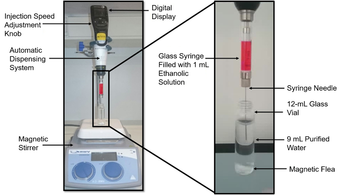
Figure 1: Nanoparticles preparation setup showing assembly for injection under stirring27. The setup consists of the autopipette with a 1 mL glass syringe filled with 1 mL of the ethanolic solution containing the nanoparticles' components. The glass vial contains 9 mL of water and the magnetic flea is placed on the magnetic stirrer. Please click here to view a larger version of this figure.
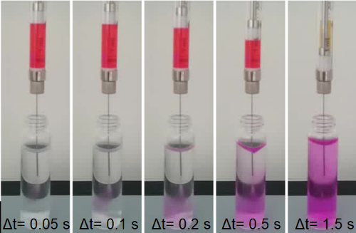
Figure 2: Schematic of the mixing of solvents in the rapid injection method of solvent shifting27. Panels show the injection of 1 mL of ethanolic phase containing nanoparticles' components at a speed of 833 µL/s into 9 mL of water while stirring at 500 rpm. The rapid injection with chaotic mixing of the ethanolic solution containing the nanoparticles' components (falcarindiol, DSPC, cholesterol, DSPE PEG 2000, DiI) into the antisolvent (water), leadd to the formation of the nanoparticles. The color is given by DiI. It can be observed how the ethanolic solution is mixed, rapidly increasing its concentration and the nanoparticles are formed. Please click here to view a larger version of this figure.
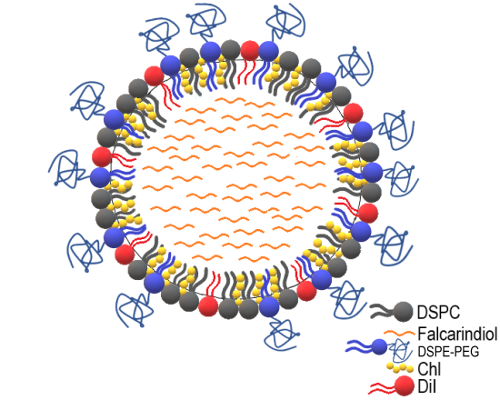
Figure 3: Schematic of the final falcarindiol nanoparticles' structure. Nanoparticle structure, including DSPC, DSPE PEG 2000, cholesterol, DiI, and falcarindiol. The different components are scaled according to their concentrations. Please click here to view a larger version of this figure.
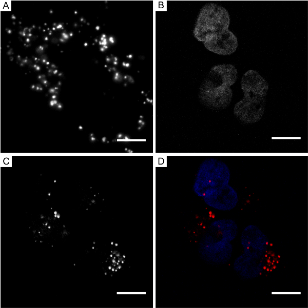
Figure 4: Images of lipid-coated falcarindiol nanoparticles in human mesenchymal stem cells. (A) Epifluorescence microscopy image of hMSCs treated with falcarindiol nanoparticles for 24 h. The following panels show confocal microscopy images of the hMSCs treated with falcarindiol nanoparticles for 24 h: (B) DAPI stain of nuclei, (C) DiI nanoparticles, and (D) an overlay of both images, nuclei are shown in blue and the nanoparticles in red. The scale bars are 10 µm. Nanoparticles are visualized as white bright dots in the cell cytoplasm. The images show that a large number of nanoparticles have entered the cells after 24 h of incubation. Please click here to view a larger version of this figure.
Type of nanoparticles | Solvent | Nanoparticle size (nm) | Polydisperisty (nm) | Polydispersity Index (PDI) |
Uncoated Falcarindiol | Water | 83.9 | ± 23,9 | 0.571 |
PBS | 71 | ± 20,3 | 0.571 | |
Lipid coated Falcarindiol | PBS | 91.6 | ± 6,4 | 0.141 |
PBS after 3h | 93.5 | ± 5,7 | 0.122 | |
Lipid coated Falcarindiol + DiI | PBS | 74.1 | ± 6,7 | 0.182 |
Table 1: Different designs of the fabricated nanoparticles. Size and polydispersity index of the synthesized falcarindiol nanoparticles, depending on the solvent and nanoparticle type. Falcarindiol nanoparticles with and without a lipid coat were fabricated. Various concentrations of the coat components were tested. The particle aggregation was tested after 3 h.
A detailed protocol for fabricating lipid-coated nanoparticles for drug delivery with the simple, fast, reproducible, and scalable rapid injection method of solvent shifting was followed27,28 and is presented in this paper, as applied to falcarindiol. By controlling the speed of the injection of the organic phase into aqueous phase and by using coating lipids at appropriate concentrations to coat the falcarindiol core, particle in the sub-100 nm range could be obtained successfully. The possibility of induced polydispersity due to the involvement of turbulent mixing for the falcarindiol precipitation alone was controlled by the presence of coating lipids. The structure of these lipid-coated nanoparticles resembled the low-density lipoproteins with exception of the exclusion of the native 500,000 kDa ApoB100 and the additional presence of PEG-lipids for steric stability). This passively targeted drug delivery system allows the encapsulation of a broad range of especially hydrophobic drugs and diagnostic materials18,19, reducing the immunologic response and increasing the accumulation in cancerous tissues16,18. Furthermore, depending on the drug degradation reactions (e.g., hydrolysis, enzymolysis), the drug delivery system protects the drug from degradation during its circulation in vivo.
Therefore, falcarindiol nanoparticles, with a lipid-encapsulating monolayer containing a pure core of the purified drug, were designed and fabricated. The lipid monolayer coating consisted of DSPC, cholesterol, and DSPE-PEG 2000, with the fluorescent label DiI. The fabrication of the particles was carried out using the rapid injection method of solvent shifting, which consists of a rapid injection of an ethanolic solution containing the nanoparticles components into an excess of aqueous phase (1:9). The size of the nanoparticles was measured using DLS, and the uptake of the nanoparticles was examined in hMSCs and imaged using fluorescence and confocal microscopy.
Uncoated nanoparticles can also be obtained, with the sizes of 71 ± 20.3 nm. However, after following the protocol described above, nanoparticles of 74.1 nm ± 6.7 nm, with polydispersity values of 0.182, were fabricated. Thus, after modifying the nanoparticles by adding the lipid coat, the size and PDI of the nanoparticles was reduced, , making them more suitable for drug delivery.
It is important to be highly aware of the critical steps in the protocol, such as the importance of the syringe position when injecting the organic phase, the preparation of the nanoparticles on the same day of treatment to avoid aggregation, and the seeding of the cells the day before to ensure adequate confluence level. In fact, all the steps in the first part of the protocol can be considered critical as they either affect the size of the nanoparticles or the final concentration of the active compound and coating lipids. Considering ‘concentration’ as an important parameter, steps 1.2, 1.4, 1.6, 1.15, and 1.16 are critical. Considering ‘nanoparticle size’ as an important parameter, steps 1.7, 1.11, and 1.12 are critical.
Fluorescence and confocal microscopy showed that nanoparticles had entered the cells, and a large number of nanoparticles were scattered in the cytoplasm of every cell. These results suggest that nanoparticles designed in this study can function as a new, and stable drug delivery system for falcarindiol.
This technique provides a simple, fast, and reproducible approach to encapsulate different cancer drugs, and the limitations of the method are assessed with the lipid coat.
The authors have nothing to disclose.
The authors thank Dr. Moustapha Kassem (Odense University Hospital, Denmark) for the human mesenchymal stem cells. The authors thank the Danish Medical Bioimaging Center for access to their microscopes. The authors thank the Carlsberg and Villum foundations for financial support (to E.A.C.). The authors acknowledge the financial support provided by the Niels Bohr Professorship award from the Danish National Research Foundation.
12 mL Screw Neck Vial (Clear glass, 15-425 thread, 66 X 18.5 mm) | Microlab Aarhus A/S | ML 33154LP | |
6 well plates | Greiner Bio One International GmbH | 657160 | |
Absolute Ethanol | EMD Millipore (VWR) | EM8.18760.1000 | |
Chloroform | Rathburn Chemicals Ltd. | RH1009 | |
Cholesterol | Avanti Polar Lipids, Inc. | 700000P | |
Confocal Microscope | Zeiss LSM510 | ||
Cover Slips thickness #1.5 | Paul Marienfeld GmbH & Co | 117650 | |
Desiccator | Self-build | ||
DiI | Invitrogen | D282 | |
DLS | Beckman Coulter | DelsaMAXpro 3167-DMP | |
DSPC (Chloroform stock) | Avanti Polar Lipids, Inc. | 850365C | |
DSPE PEG 2000 (Chloroform stock) | Avanti Polar Lipids, Inc. | 880120C | |
eVol XR | SGE analytical science, Trajan Scientific Australia Pty Ltd. | 2910200 | |
Fetal Bovine serum | Gibco | 10270-106 | |
Fluorescence Miccroscope | Olymous IX81 | With Manual TIRF and Andor iXon EMCCD | |
Incubator | Panasonic | MCO-18AC | |
Magnetic flea | VWR Chemicals | 15 x 4.5 mm | Cylindrical shape with PTFE coating |
Magnetic stirrer | IKA | RT-10 | |
Minimum Essential Media | Gibco | 32561-029 | |
PBS tablets for cell culture | VWR Chemicals | 97062-732 | |
Pen/strep | VWR Chemicals | 97063-708 | |
Phosphate Buffer Saline (PBS, pH 7.4) | Thermo Fisher | 10010031 | |
Rotary Evaporator | Rotavapor, Büchi Labortechnik AG | R-210 | |
Sample concentrator | Stuart, Cole-Parmer Instrument Company, LLC | SBHCONC/1 |