Presented here is a method for the 3D bioprinting of gelatin methacryloyl.
Gelatin methacryloyl (GelMA) has become a popular biomaterial in the field of bioprinting. The derivation of this material is gelatin, which is hydrolyzed from mammal collagen. Thus, the arginine-glycine-aspartic acid (RGD) sequences and target motifs of matrix metalloproteinase (MMP) remain on the molecular chains, which help achieve cell attachment and degradation. Furthermore, formation properties of GelMA are versatile. The methacrylamide groups allow a material to become rapidly crosslinked under light irradiation in the presence of a photoinitiator. Therefore, it makes great sense to establish suitable methods for synthesizing three-dimensional (3D) structures with this promising material. However, its low viscosity restricts GelMA’s printability. Presented here are methods to carry out 3D bioprinting of GelMA hydrogels, namely the fabrication of GelMA microspheres, GelMA fibers, GelMA complex structures, and GelMA-based microfluidic chips. The resulting structures and biocompatibility of the materials as well as the printing methods are discussed. It is believed that this protocol may serve as a bridge between previously applied biomaterials and GelMA as well as contribute to the establishment of GelMA-based 3D architectures for biomedical applications.
Hydrogels are thought to be a suitable material in the field of biofabrication1,2,3,4. Among them, gelatin methacryloyl (GelMA) has become one of the most versatile biomaterials, initially proposed in 2000 by Van Den Bulcke et al.5. GelMA is synthesized by the direct reaction of gelatin with methacrylic anhydride (MA). The gelatin, which is hydrolyzed by the mammal collagen, is composed of target motifs of matrix metalloproteinase (MMP). Thus, in vitro three-dimensional (3D) tissue models established by GelMA can ideally mimic the interactions between cells and extracellular matrix (ECM) in vivo. Furthermore, arginine-glycine-aspartic acid (RGD) sequences, which are absent in some other hydrogels such as alginates, remain on the molecular chains of GelMA. This makes it possible to realize the attachment of encapsulated cells inside the hydrogel networks6. Additionally, the formation capability of GelMA is promising. The methacrylamide groups on the GelMA molecular chains react with the photoinitiator under mild reaction conditions and form covalent bonds upon exposure to light irradiation. Therefore, the printed structures can be rapidly crosslinked to maintain the designed shapes in a simple way.
Based on these properties, a series of fields utilize GelMA to carry out various applications, such as tissue engineering, basic cytology analysis, drug screening, and biosensing. Accordingly, various fabrication strategies have been also demonstrated7,8,9,10,11,12,13,14. However, it is still challenging to carry out 3D bioprinting based on GelMA, which is due to its fundamental properties. GelMA is a temperature-sensitive material. During the printing process, the temperature of the printing atmosphere has to be strictly controlled in order to maintain the physical state of the bioink. Besides, the viscosity of GelMA is generally lower than other common hydrogels (i.e., alginate, chitosan, hyaluronic acid, etc.). However, other obstacles are faced when building 3D architectures with this material15.
This article summarizes several approaches for the 3D bioprinting of GelMA proposed by our lab and describes the printed samples (i.e., the synthesis of GelMA microspheres, GelMA fibers, GelMA complex structures, and GelMA-based microfluidic chips). Each method has specialized functions and can be adopted in different situations with different requirements. GelMA microspheres are generated by an electroassisted module, which forms extra external electric force to shrink the droplet size. In terms of GelMA fibers, they are extruded by a coaxial bioprinting nozzle with the help of viscous sodium alginate. In addition, the establishment of complex 3D structures is achieved with a digital light processing (DLP) bioprinter. Finally, a twice crosslinking strategy is proposed to build GelMA-based microfluidic chips, combining GelMA hydrogel and traditional microfluidic chips. It is believed that this protocol is a significant summary of the GelMA bioprinting strategies used in our lab and may inspire other researchers in relative fields.
1. Cell culturing
2. Fabrication of GelMA microspheres
3. Fabrication of GelMA fibers
4. Fabrication of complex 3D GelMA structures
NOTE: Figure 3A shows the fabrication sketch of the complex 3D GelMA structures.
5. Fabrication of GelMA-based microfluidic chips
NOTE: Figure 4A shows the fabrication sketch of the GelMA-based microfluidic chip.
During the fabrication of GelMA microspheres, the GelMA droplets were separated by the external electric field force. When the droplets fell into the receiving silicon oil, they remained standard spheroid shape without tails. This is because the GelMA droplets were in an aqueous phase, while the silicon oil was in an oil phase. The surface tension that formed between the two phases caused the GelMA droplets to maintain a standard spheroid shape. In terms of the cell-laden microspheres, cells experienced the high voltage electric field force in this process. From the morphology of the stained MDA-MB-231s (Figure 1B–E), it was found that the encapsulated MDA-MB-231s maintained its spreading capability, verifying the biocompatibility of this electroassisted fabrication method.
In terms of the GelMA fibers, GelMA and sodium alginate solution flowed in the inner and outer nozzles of the coaxial nozzle, respectively. As the sodium alginate had higher viscosity than GelMA, GelMA was restricted in the sodium alginate solution and maintained a line shape. The irradiation by light (405 nm wavelength) caused the inner GelMA to become crosslinked, forming the GelMA fibers (Figure 2B). Besides, BMSCs were encapsulated in the GelMA fibers (Figure 2C,D). As shown, the encapsulated BMSCs maintained spreading capability in the GelMA hydrogel networks after the fabrication process (Figure 2E).
A DLP bioprinter was chosen to fabricate GelMA structures with more complex shapes. As shown in Figure 3B–D, the structures of “nose”, “ear”, and “multichamber” were established. On the surface of the crosslinked GelMA structures, the seeded HUVECs attached to the GelMA materials and spread (Figure 3F). This demonstrated the possibility that the establishment of GelMA complex 3D structures with the help of a DLP bioprinter holds great potential in applications in the field of tissue engineering.
Unlike the traditional microfluidic chip that is based on materials without biodegradation properties16,17,18,20 (i.e, resin, glass, polydimethylsiloxane [PDMS], and polymethyl methacrylate [PMMA]), a GelMA-based microfluidic chip was fabricated here using a twice cross-linking strategy. Two components in the bioink were crosslinked successively. Chips with various microchannels were built by designing different molds on demand (Figure 4B,C). Besides, it was verified that HUVECs were seeded in the channels and attached to the channel wall, forming the macroscopic vessel shape (Figure 4D,E).
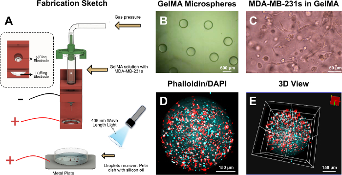
Figure 1: GelMA microspheres. (A) Fabrication sketch of the GelMA microspheres. (B) Optical microscope image of the GelMA microspheres. (C) Optical microscope image of the MDA-MB-231s in GelMA. (D) 2D view of the F-actin and nucleus of the encapsulated MDA-MB-231s. (E) 3D view of the F-actin and nucleus of the encapsulated MDA-MB-231s. Please click here to view a larger version of this figure.
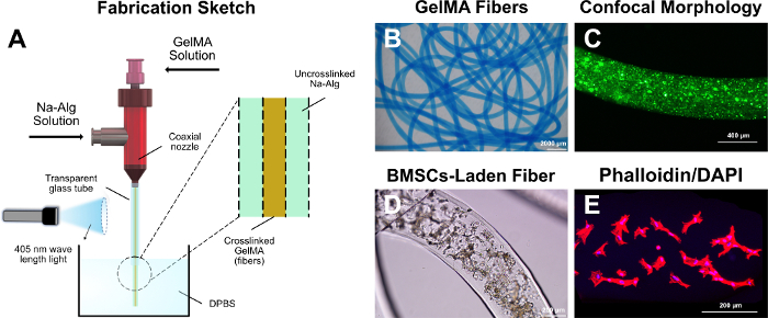
Figure 2: GelMA fibers. (A) Fabrication sketch of the GelMA fibers. (B) Optical microscope image of the GelMA fibers (with blue ink). (C) Confocal fluorescence microscope image of the GelMA fibers (with green fluorescence particles). (D) Optical microscope image of the BMSCs in GelMA fibers. (E) The F-actin and nucleus of the encapsulated BMSCs. Please click here to view a larger version of this figure.
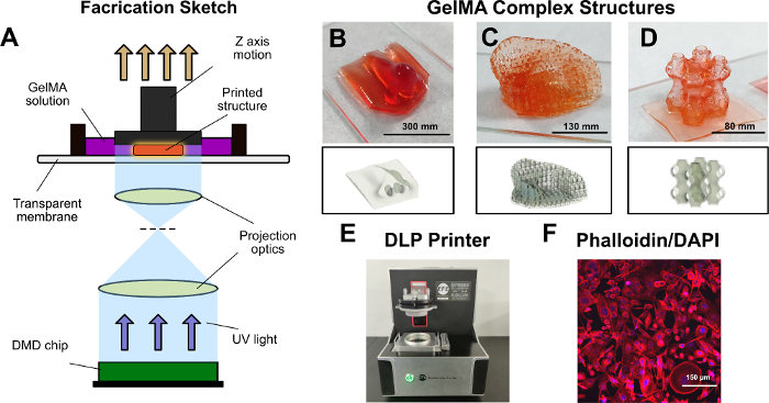
Figure 3: GelMA complex 3D structures. (A) Fabrication sketch of the complex GelMA 3D structures. (B) Optical microscope image of the GelMA “nose”. (C) Optical microscope image of the GelMA “ear”. (D) Optical microscope image of the GelMA “multichamber”. (E) The applied DLP bioprinter. (F) The F-actin and nucleus of the seeded MDA-MB-231s. Please click here to view a larger version of this figure.
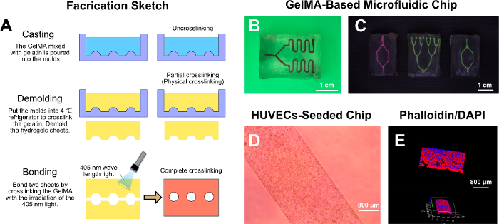
Figure 4: GelMA-based microfluidic chip. (A) Fabrication sketch of the GelMA-based microfluidic chip. (B,C) Optical microscope images of the GelMA-based microfluidic chip. (D) Optical microscope image of the seeded HUVECs on the channel wall. (E) The F-actin and nucleus of the seeded HUVECs on the channel wall. Please click here to view a larger version of this figure.
This article describes several strategies to fabricate GelMA 3D structures, namely GelMA microspheres, GelMA fibers, GelMA complex structures, and GelMA-based microfluidic chips. GelMA has promising biocompatibility and formation capability and is widely used in the field of biofabrication. Microsphere structures are suitable for controlled drug release, tissue culturing, and injection into organisms for further therapy21,22,23,24,25. Because the viscosity of GelMA solution is low, its formation is challenging. Thus, during the fabrication of the GelMA microspheres, the electrohydrodynamic (EHD) principle was chosen to solve this problem. The voltage applied was relatively low, and the microdroplets were generated one-by-one. To fabricate microspheres of a smaller size, the applied voltage can be increased, and the fluid would be in another state with the Taylor cone26.
Because of the Coulomb explosion phenomenon, the dropping droplets were further separated by their excessive electric density, resulting in smaller GelMA microspheres. Furthermore, monocomponent GelMA fibers were fabricated with the help of a coaxial nozzle and sodium alginate solution. A coaxial nozzle was applied here. As mentioned above, because of the low viscosity of GelMA, sodium alginate provided resistance to help maintain the shape of fiber. Hydrogel fiber structures are suitable for mimicking the fiber-shaped tissues in vivo (i.e., muscles, vessels, etc.27,28,29,30,31,32). For GelMA fibers with more complicated components, the applied bioprinting nozzle can be further modified. For example, a three-layer coaxial nozzle can be assembled to generate multilayer GelMA fibers.
In the establishment of complex GelMA 3D structures, it was found that the DLP bioprinter breaks through the printing obstacle caused by the low viscosity of GelMA. With the help of CAD software, GelMA 3D structures were fabricated on demand. Finally, a new GelMA fabrication method, the twice cross-linking strategy, was demonstrated and applied to the combination of GelMA and a traditional microfluidic chip. The hydrogels have higher biocompatibility, and researchers can encapsulate cells inside the chip body. The proposed GelMA-based microfluidic chip can be further improved by encapsulating cells in the chips to serve as suitable models in vitro for drug screening, cellular interaction studies, etc. We believe that the methods for fabrication of GelMA described here will increase the rate of development in this field and can be applied in further biomedical research.
The authors have nothing to disclose.
This work was sponsored by the National Key Research and Development Program of China (2018YFA0703000), the National Nature Science Foundation of China (No.U1609207, 81827804), the Science Fund for Creative Research Groups of the National Natural Science Foundation of China (No. 51821093).
0.22 μm filter membrane | Millipore | ||
2-(4-amidinophenyl)-6-indolecarbamidine dihydrochloride (DAPI) | Yeasen Biological Technology Co., Ltd., Shanghai, China | ||
3D bioprinter | SuZhou Intelligent Manufacturing Research Institute, SuZhou, China | ||
405nm wavelength light | SuZhou Intelligent Manufacturing Research Institute, SuZhou, China | ||
co-axial nozzle | SuZhou Intelligent Manufacturing Research Institute, SuZhou, China | ||
confocal fluorescence microscope | OLYMPUS FV3000 | ||
digital light processing (DLP) bioprinter | SuZhou Intelligent Manufacturing Research Institute, SuZhou, China | ||
DLP printer | SuZhou Intelligent Manufacturing Research Institute, SuZhou, China | ||
Dulbecco's Phosphate Buffered Saline (DPBS) | Tangpu Biological Technology Co., Ltd., Hangzhou, China | ||
Dulbecco's Modified Eagle Medium (DMEM) | Tangpu Biological Technology Co., Ltd., Hangzhou, China | ||
Dulbecco's Modified Eagle Medium with L-glutamine (DMEM/F-12) | Tangpu Biological Technology Co., Ltd., Hangzhou, China | ||
EFL Software | SuZhou Intelligent Manufacturing Research Institute, SuZhou, China | ||
fetal bovine serum (FBS) | Tangpu Biological Technology Co., Ltd., Hangzhou, China | ||
gelatin | Sigma-Aldrich, Shanghai, China | ||
gelatin methacryloyl (GelMA) | SuZhou Intelligent Manufacturing Research Institute, SuZhou, China | ||
high voltage power | SuZhou Intelligent Manufacturing Research Institute, SuZhou, China | ||
lithium phenyl-2, 4, 6-trimethylbenzoylphosphinate (LAP) | SuZhou Intelligent Manufacturing Research Institute, SuZhou, China | ||
paraformaldehyde | Tangpu Biological Technology Co., Ltd., Hangzhou, China | ||
penicillin/streptomycin | Tangpu Biological Technology Co., Ltd., Hangzhou, China | ||
sodium alginate (Na-Alg) | Sigma-Aldrich, Shanghai, China | ||
TRITC phalloidin | Yeasen Biological Technology Co., Ltd., Shanghai, China | ||
Triton X-100 | Solarbio Co., Ltd., Shanghai, China |