Iron oxide nanoparticles are synthesized via a nonaqueous sol gel procedure and coated with anionic short molecules or polymer. The use of magnetometry for monitoring the incorporation and biotransformations of magnetic nanoparticles inside human stem cells is demonstrated using a vibrating sample magnetometer (VSM).
Magnetic nanoparticles, made of iron oxide, present a peculiar interest for a wide range of biomedical applications for which they are often internalized in cells and then left within. One challenge is to assess their fate in the intracellular environment with reliable and precise methodologies. Herein, we introduce the use of the vibrating sample magnetometer (VSM) to precisely quantify the integrity of magnetic nanoparticles within cells by measuring their magnetic moment. Stem cells are first labeled with two types of magnetic nanoparticles; the nanoparticles have the same core produced via a fast and efficient microwave-based nonaqueous sol gel synthesis and differ in their coating: the commonly used citric acid molecule is compared to polyacrylic acid. The formation of 3D cell-spheroids is then achieved via centrifugation and the magnetic moment of these spheroids is measured at different times with the VSM. The obtained moment is a direct fingerprint of the nanoparticles’ integrity, with decreasing values indicative of a nanoparticle degradation. For both nanoparticles, the magnetic moment decreases over culture time revealing their biodegradation. A protective effect of the polyacrylic acid coating is also shown, when compared to citric acid.
There is increased interest in the magnetic features of iron oxide nanoparticles for a wide range of biomedical applications. Their response to magnetic resonance makes them reliable contrast agents for magnetic resonance imaging (MRI), an advantage in regenerative medicine where cells labeled with magnetic nanoparticles can be tracked in vivo following implantation1. Using magnetic fields, cells can also be guided at a distance; this way, cellular spheroids2,3, rings4, or sheets5 can be engineered magnetically and also remotely stimulated6, an asset in the development of scaffold-free tissues. The range of possibilities for these nanoparticles also includes drug delivery systems7,8 and magnetic and photoinduced hyperthermal treatment to kill cancerous cells9,10,11. For all these applications, the nanoparticles are integrated in the biological environment either by intravenous injection or via direct internalization in cells and are then left within, which brings into question their intracellular fate.
In vivo analyses conveyed a general understanding of the nanoparticles’ fate in the organism: upon injection in the blood stream, they are first captured mostly by the macrophages of the liver (Kupffer cells), spleen and bone marrow, are progressively degraded, and join the iron pool of the organism12,13,14,15,16,17,18,19. Qualitative observations are only possible due to the circulation of the nanoparticles throughout the organism. Typically, transmission electronic microscopy (TEM) can be used to directly observe the nanoparticles and the presence of iron in the organs can be determined via dosage. More recently, their fate has been assessed directly on a pool of cells, meaning in close circuit with no iron escape, allowing a quantitative measurement of their biotransformations at the cell-level20,21,22. Such measurements are possible via the analysis of the magnetic properties of the nanoparticles that are tightly linked to their structural integrity. Vibrating sample magnetometry (VSM) is a technique where the sample is vibrated periodically so that the coil-measurement of the flux induced provides the magnetic moment of the sample at the applied magnetic field. Such synchronous detection allows for a rapid measurement, which is an asset for determining the magnetic moments of a large number of samples20,21,22,23. The macroscopic magnetic signature retrieved by VSM then gives a quantitative overview of the entire biological sample directly correlated to the nanoparticles' size and structure. In particular, it provides the magnetic moment at saturation (expressed in emu) of the samples, which is a direct quantification of the number of magnetic nanoparticles present in the sample, respectively to their specific magnetic properties.
It has been shown that the intracellular processing of magnetic nanoparticles is tightly linked to their structural features20. These features can be controlled via optimal synthesis protocols. Each protocol presents advantages and limitations. Iron oxide nanoparticles are commonly synthesized in aqueous solutions via coprecipitation of iron ions24. To overcome the limitations of nanoparticles size polydispersity, other synthesis methods such as polyol-mediated sol-gel methods have been developed25. Nonaqueous approaches by thermal decomposition leads to the production of very well-calibrated iron oxide nanoparticles26. However, the use of massive amounts of surfactants like oleylamine or oleic acid complicates their functionalization and water transfer for biomedical applications. For this reason, we synthesize such magnetic nanoparticles through a nonaqueous sol gel route leading to high crystallinity, purity and reproducibility27. This protocol produces well-controlled size nanoparticles that can be tuned through temperature variation28. Nevertheless, the microwave-assisted non-aqueous sol-gel route has an upper size limit of the obtained nanoparticles of around 12 nm. This procedure would not be adapted for applications using ferromagnetic particles at room temperature. In addition to the core synthesis, another main feature to be considered is the coating. Lying at the surface of the nanoparticle, the coating acts as an anchoring molecule, helping the targeted internalization of the nanoparticles, or it can protect the nanoparticle from degradation. Since benzyl alcohol acts as an oxygen source and a ligand at the same time, bare nanoparticles are produced without the need for additional surfactants or ligands. The nanoparticles are then easily surface functionalized after synthesis without a surfactant exchange process.
Herein, two types of nanoparticles are assessed that possess the same core and differ in the coating. The core is synthesized using a fast and highly efficient microwave based technique. The two coatings compared consist of citric acid, one of the most used as coating agent in biomedical applications29,30, and polyacrylic acid (PAA), a polymeric coating with a high number of chelating functions. VSM magnetometry measurements are then used first to quantify the nanoparticle uptake by the cells, and then as a direct assessment of the nanoparticle structural integrity upon internalization in stem cells. Results demonstrate that the incubation concentration impacts nanoparticle uptake and that the coating influences their degradation, with the large number of anchoring molecules of PAA protecting the core from degradation.
1. Synthesis of magnetic nanoparticles
2. Culture and magnetic labeling of stem cells
3. Formation of stem cell-spheroids
4. Quantification of magnetic nanoparticles in solution and in cellulo using a vibrating sample magnetometer (VSM)
5. Transmission Electron Microscopy (TEM) analysis
Using the microwave-assisted synthesis, magnetic nanoparticles with a monodisperse 8.8 ± 2.5 nm core size are produced and coated with either citrate or PAA (Figure 1A). Stem cells are then incubated with these nanoparticles dispersed in culture medium at a given concentration for 30 minutes, resulting in their endocytosis and confinement within the cellular endosomes (Figure 1B). The magnetic stem cells are then suspended in medium, centrifuged, and the cell pellet formed is cultured for up to 21 days (Figure 1C). The spheroids obtained are fixed, such as stopping biological processes, and kept in PBS until being measured via VSM.
First, the magnetic moment of the nanoparticle solution is measured using the VSM: 10 µL of the nanoparticle aqueous dispersion containing 7 µg of iron is measured, and the obtained curve is displayed in Figure 2A. Due to the presence of water in this solution, and to the sample holder, a diamagnetic signal is captured in addition to the superparamagnetic signal of the nanoparticles. A diamagnetic constant (Mdia) corresponding to the slope of the second part of the curve can be measured, as shown in Figure 2A, and this constant can be subtracted such as obtaining the magnetic moment of the nanoparticles only (Msample = M – Mdia). The saturation magnetization of the nanoparticles (Ms) can then be extracted; it corresponds to 518 µemu for the aqueous dispersion, meaning that the solution is at 74 emu/g of iron (Fe) corresponding to 52 emu/g of nanoparticles (Fe3O4).
The magnetic moment of cell spheroids can similarly be obtained. In this case, a cell-spheroid (made of 200,000 cells) is inserted in the sample holder, placed in the VSM, and measured (Figure 2B). The magnetic moment values obtained are here much lower than the ones of the initial nanoparticle solution; however, they remain within the detection range. The saturation magnetization of this particular sample is of 69 µemu. Besides, this spheroid contains 1.3 µg of nanoparticles (6.7 pg of nanoparticles per cell), consistent with the saturation value at 52 emu/gFe3O4. This value can thus be used to determine the amount of nanoparticles in cellular samples. In Figure 2C, spheroids corresponding to cells labelled with three concentrations of citrate-coated nanoparticles are measured one day after labelling. Results clearly show that the uptake of the nanoparticles depends on the incubation concentration, with concentrations of 0.125, 0.25 and 0.5 mM leading to uptakes of 0.3, 0.7 and 1.3 µg of iron per spheroid, meaning 1.3, 3,3 and 6.7 pg of iron per cell, respectively.
Attention has been brought on the importance of the surface coating, which directly interacts with the biological environment20. Two coatings have here been produced: a citrate coating, commonly used for biomedical applications, and a PAA coating, with a higher number of chelating functions. Stem cells are labeled with these two types of nanoparticles and centrifuged such as forming a cell pellet that then becomes a cohesive cell-spheroid (Figure 3A). Magnetism of these cell-spheroids is measured via VSM at day 1 and day 21 (Figure 3B and Figure 3C). Results demonstrate a decrease in magnetism upon the 21 days of culture, indicating the biodegradation of the nanoparticles, this degradation being more important for the citrate-coated nanoparticles (Figure 3B) than the PAA-coated ones (Figure 3C). TEM images confirm the degradation of the nanoparticles and show the appearance of smaller (6 nm in size) light grey dots, typical of ferritin loaded with iron. Some nanoparticles remaining intact can also be observed, particularly with the PAA coating.
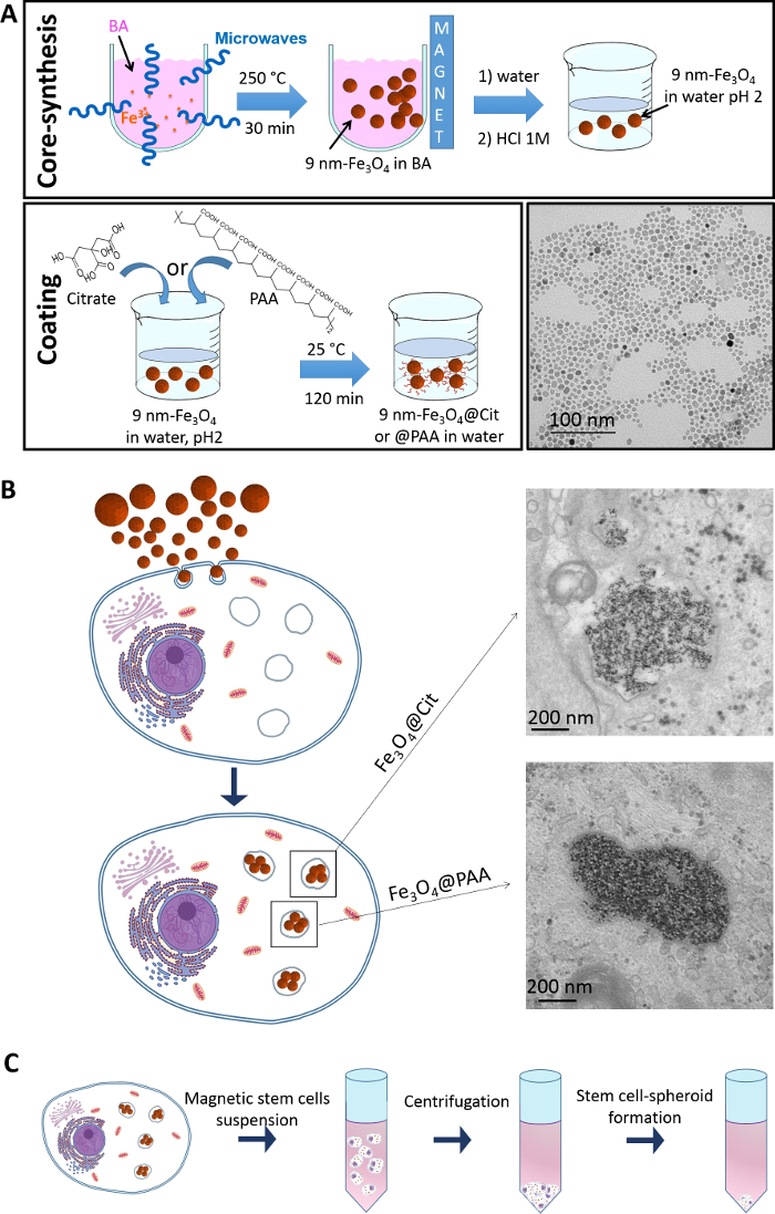
Figure 1: Microwave-assisted synthesis of iron oxide (Fe3O4) nanoparticles, their internalization in stem cells and the subsequent culture of the cells as spheroids. (A) Schematic of the various steps of the nanoparticle synthesis. First, the core is synthesized via a non-aqueous sol gel procedure. The coating molecule, either Citrate (Cit) or polyacrylic acid (PAA), is then grafted at the surface of the iron oxide core. A representative TEM image shows the synthesized nanoparticles with a citrate coating. (B) Schematic of the nanoparticle internalization within stem cell, showing the nanoparticles confined in the endosomes upon internalization. Representative TEM images also show the citrate and PAA coated nanoparticles inside the cells, confined in the endosomes. (C) Schematic of stem cell-spheroids formation. Please click here to view a larger version of this figure.
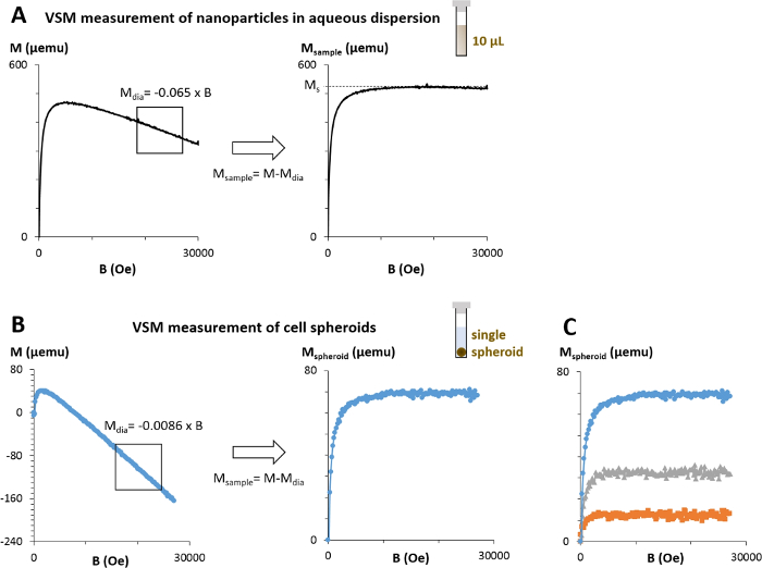
Figure 2: Measurement of the samples magnetic moment via VSM. (A) 10 μL of nanoparticles dispersed in an aqueous solution are measured via the VSM. The signal obtained represents the magnetic moment of this nanoparticle solution in function of the magnetic field (B). The diamagnetism coming from the presence of water and the sample holder can then be measured as it corresponds to the slope of the second part of the curve and subtracted such as obtaining the magnetic moment of the nanoparticles only. The magnetic moment at saturation (Ms) can then be determined. (B) The magnetic moment of cell spheroids can similarly be obtained; In this case a single cell-spheroid is measured at a given time period. (C) Curves of spheroids corresponding to cells labeled with citrate-coated nanoparticles at three independent concentrations of 0.125 mM (orange), 0.25 mM (grey) and 0.5 mM (blue). Please click here to view a larger version of this figure.
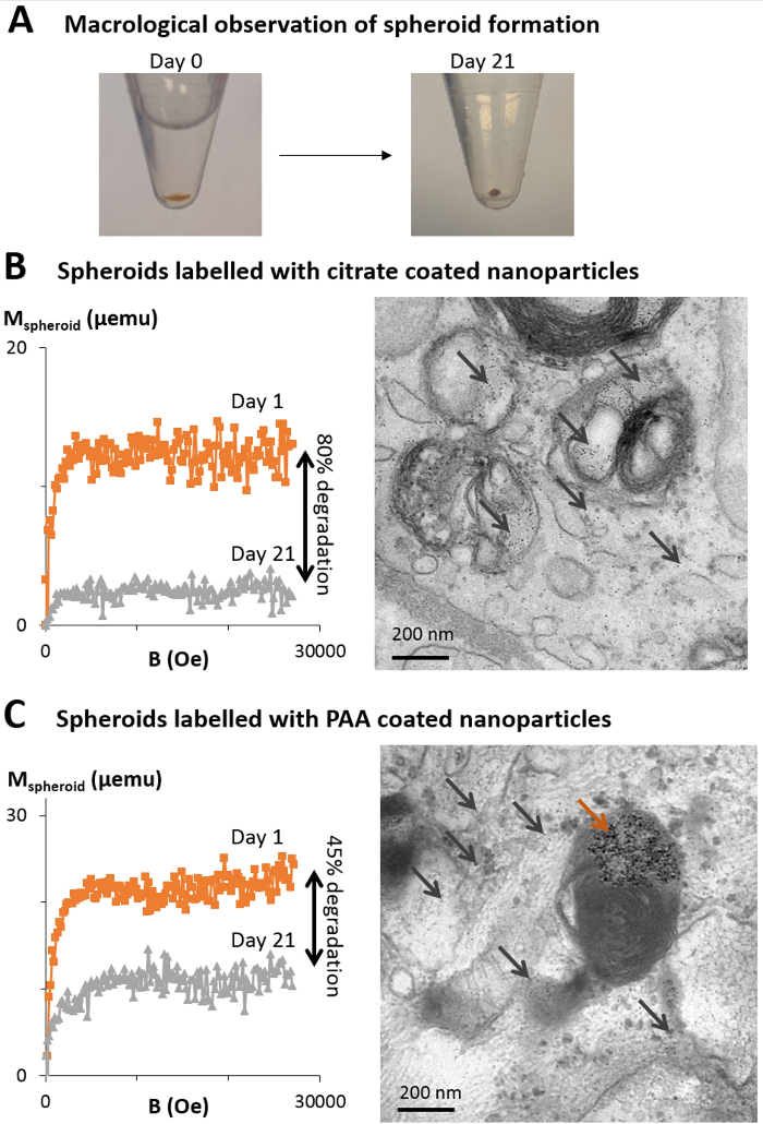
Figure 3: Quantification of magnetic nanoparticle degradation in cellulo via VSM. (A) Upon cell labeling with magnetic nanoparticles, a cell pellet is formed by centrifugation (day 0). The cells then form a cohesive structure resulting in an easy to handle cell-spheroid that can be kept in culture without cell loss for extended time periods (months). (B, C) Herein, two types of magnetic nanoparticles, coated with citrate (B) or PAA (C), are internalized in stem cells and the cells are cultured as spheroids for up to 21 days. Magnetism of spheroids cultured for 1 day (orange curves) and 21 days (grey curves) are measured with the VSM, with a decrease in magnetism indicating a degradation of the nanoparticles. Representative TEM images taken at day 21 show light grey dots about 6 nm is size within the endosomes and the cytoplasm of the cells, typical size and shape of ferritin, the iron storage protein (black arrows). Some intact nanoparticles can also be observed, mostly for the PAA-coated nanoparticles (brown arrow). Please click here to view a larger version of this figure.
Using a fast and efficient microwave-based synthesis, magnetic nanoparticles can easily be synthesized, with controlled size, and further coated with given molecules. A critical step is to stock the iron salt and the benzyl alcohol under vacuum to keep a small dispersion in size. The benzyl alcohol acts as both as solvent and ligand at the same time allowing to directly obtain calibrated bare iron oxide without the need of additional ligands. After nanoparticles transfer in water the bare magnetic nanoparticles can be easily coated with a large variety of ligands. This coating confers inter-nanoparticle and nanoparticle-cell interacting properties that can influence their cellular internalization, a parameter that needs to be tightly controlled as a minimum internalization is required for biomedical applications while a too high internalization could be damaging to the cells and potentially originate cytotoxic events31. Magnetometry is a powerful tool to assess this internalization as well as the fate of magnetic nanoparticles in cellulo. Upon the incorporation of magnetic nanoparticles into stem cells, spheroids can be formed via a simple centrifugation followed by culture in a medium that stops the proliferation of the cells and drives extracellular matrix production. The cells become highly cohesive and start creating a tissue. The spheroids are then very easy to handle and can be cultured for extended time periods (months) that allows for long-term tracking of magnetic nanoparticles’ fate in a biological environment. By stopping cell division, there is no dilution of the nanoparticles from mother to daughter cell; moreover, it has been verified that, with this cell-spheroid model, there is no iron escape over a month of culture21,22,23. As a consequence, a decrease in magnetism values can only correspond to a degradation of the nanoparticles and not to iron being exported out of the cells.
Magnetism of the cell spheroids is here measured via VSM that provides a fast and precise quantification. Other methods have also been explored to measure cellular magnetism, such as using a Superconducting Interface Device (SQUID), a machine typically more precise than the VSM with a sensitivity around 10-7 emu compared to 10-6 emu for the VSM, but operational cost is higher32. The sensitivity of the VSM allowing measurements of magnetic iron dose inferior to 2 pg/cell in the experimental setup is herein sufficiently precise as iron doses inferior to 2 pg/cell would be too low for the aimed applications, such as cell attraction toward a magnetic for cell guidance and tissue engineering purposes. Both VSM and SQUID magnetometry, in addition to allowing a quantification of cell magnetism, can give additional details on the nanoparticles’ features. By analysis of the signal obtained, the size of the nanoparticles can be deducted22,23 and also general notion of their magnetic features can be obtained, hysteresis and coercivity of nanomaterials can for example be revealed. Alternative magnetometry approaches also exist, such as magnetophoresis that consists in measuring the velocity of individual cells when attracted toward a magnet and the magnetism of single cells is extrapolated33,34. The magnetism at the single cell level can this way be obtained; however, no additional nanoparticle features can be determined. Additionally, a small magnetic sensor that provides a signal proportional to the sample magnetism has recently been used with a similar cell spheroid model. This magnetic sensor allowed the tracking of magnetic nanoparticles’ biodegradation in real-time, continuously, over 7 days35. However, the signal of this magnetic sensor cannot be directly translated into a magnetic moment as it depends on the size of the nanoparticles. For this reason, a calibration curve needs to be performed for each nanoparticle design, curve that can be realized using the VSM.
Measurements performed herein with the VSM demonstrate a progressive degradation of the magnetic nanoparticles in the cellular environment, indicated by a decrease in magnetism. They also evidence that a same core coated with different molecules is degraded at varying rates depending on the coating. When comparing a PAA and a citrate coating, PAA leads to higher core protection to degradation, most probably due to its strong anchoring to the magnetic core20. The fate of magnetic nanoparticles in the intracellular environment is assessed on cell spheroids; however, the method is not limited to it. Indeed, it can be extrapolated to cell suspensions, which has already been used to assess the effect of stem cell differentiation on the fate of the nanoparticles22. It revealed that, depending on the differentiation pathway, the magnetic nanoparticles are processed differently by the cells, with, in some cases, the neo-crystallization of iron upon its dissolution. VSM magnetometry is thus a useful tool to further explore the influence of both nanoparticle and cell features on the intracellular processing of iron oxide nano-objects.
The authors have nothing to disclose.
This work was supported by the European Union (ERC-2014-CoG project MaTissE #648779). The authors would like to acknowledge the CNanoMat physico-chemical characterizations platform of University Paris 13.
0.05% Trypsin-EDTA (1x) | Life Technologies | 25300-054 | |
Benzyl alcohol for synthesis | Sigma Aldrich | 8.22259 | |
Dexamethasone | Sigma | D4902 | Prepare a 1 mM stock solution diluted in Ethanol 100% and store at -20°C |
Dichloromethane ≥99% stabilised, GPR RECTAPUR | VWR Chemicals | 23367 | |
DMEM with Glutamax I | Life Technologies | 31966-021 | No sodium pyruvate, no HEPES |
Ethanol absolute | VWR | 20821.310 | |
Fetal Bovine Serum | Life Technologies | 10270-106 | |
Formalin solution 10% neutral buffered | Sigma | HT5012 | |
Hydrochloric acid, 1.0N Standardized Solution | Alfa Aesar | 35640 | |
Iron(III) acetylacetonate (> 99.9%) | Sigma Aldrich | 517003 | |
ITS Premix Universal Culture Supplement (20x) | Corning | 354352 | |
L-Ascorbic Acid 2-phosphate | Sigma | A8960 | Prepare a fresh concentrated solution (25 mM) diluted in distilled water |
L-Proline | Sigma | P5607 | Prepare a 175 mM stock solution diluted in distilled water and store at 4°C |
Mesenchymal Stem Cell (MSC) | Lonza | PT-2501 | |
Monowave glass vial | Anton Paar | 82723_us | |
Microwave reactor | Anton Paar | Monowave 300 | |
MSCGM BulletKit medium | Lonza | PT-3001 | For the complete medium, add the provided BulletKit (containing serum, glutamine and antibiotics) to the MSCGM medium |
PBS w/o CaCl2 w/o MgCl2 | Life Technologies | 14190-094 | |
Penicillin (10.000U/mL)/Streptomicin (10.000µg/mL) | Life Technologies | 15140-122 | |
Poly(acrylic acid, sodium salt) | Sigma Aldrich | 416010 | MW = 1200 g/mol |
RPMI medium 1640, no Glutamine | Life Technologies | 31870-025 | No sodium pyruvate, no HEPES |
Sodium hydroxide, 1.0N Standardized Solution | Alfa Aesar | 35629 | |
Sodium pyruvate solution 100mM | Sigma | S8636 | |
Sterile conical centrifuge tube | Falcon | 352097 | 15 mL tubes |
Trypsin-EDTA (0.05%), phenol red | Thermo Fisher Scientific | 25300054 | |
Tri-sodium citrate | VWR | 33615.268 | Prepare a 1 M stock solution diluted in distilled water and store at 4°C |
Tri-Sodium Citrate Dihydrate, Certified AR for Analysis | Sigma Aldrich | 10396430 | |
Ultra centrifugal filter | Amicon | AC S510024 |