In the present protocol, a mouse heart transplantation model is used for investigating the mechanism of cardiac allograft rejection. In this heterotopic heart transplantation model, operation efficiency is improved, and the survival of cardiac grafts is ensured by a cervical end-to-end anastomosis of heart implantation using a modified Cuff technique.
Cardiac allograft rejection limits the long-term survival of patients after heart transplantation. A mouse heart transplantation model is ideal for investigating the mechanism of cardiac allograft rejection in preclinical studies because of their high homology with human genes. This understanding would help develop unique approaches to improving patients' long-term survival treated with cardiac allografts. In a mouse model, abdominal donor heart implantation is commonly performed with an end-to-end anastomosis to the recipient's aorta and inferior vena cava using stitches. In this model, the donor's heart is implanted by end-to-end anastomosis to the recipient's carotid artery and jugular vein by the modified-Cuff technique. The transplantation surgery is performed without stitching and thus may increase the survival of the recipient since there is no interference with the blood supply and venous reflux of the lower body. This mouse model would help investigate the mechanisms underlying the immunological and pathological (acute/chronic) rejection of cardiac allografts.
Heart transplantation has become the standard treatment for terminal heart failure. More than 5,500 heart transplantations per year are performed in the organizations registered under the International Society for Heart and Lung Transplantation. Among the allogeneic heart transplant recipients, the 1-year rejection rate is still >10%, while the 3-year rejection rate increased to 36%1,2. However, effective prophylactic treatments for patients with cardiac allograft rejection are lacking. Therefore, animal model studies are warranted that elucidate the physiological mechanisms underlying the immunological and pathological rejection of cardiac allografts. Such studies would contribute to the investigation of novel targets required to develop efficacious drugs, which would help prevent cardiac allograft rejection and improve survival rates in those patient populations.
Some potential immunological and pathophysiological mechanisms of cardiac allograft rejection have been proposed recently in mouse model studies of heterotopic heart transplantation3,4,5. Consequently, mouse heterotopic heart transplantation became an ideal preclinical model to investigate the mechanisms of immune rejection and pathological injury occurring in cardiac allografts after heart transplantation because of their high homology with human genes. The prevalent concept is to perform heterotopic transplantation in a mouse model by an abdominal end-to-end anastomosis in the recipient aorta and inferior vena cava using stitches, similar to the normal human anatomy. However, this procedure may interfere with the recipient's blood supply and venous reflux of the lower body6. Therefore, a modified heterotopic heart transplantation procedure in a mouse model is proposed here.
The donor's heart is implanted with the recipient's carotid artery and jugular vein by an end-to-end cervical anastomosis using a modified Cuff technique. This modified procedure facilitated the operative feasibility and ensured the survival of the cardiac graft without interfering with the blood supply and venous reflux of the lower body.
All Animal experiments were performed in compliance with the Guide for the Care and Use of Laboratory Animals, Eight Edition, National Research Council (US) 2011. Procedures involving animal subjects have been approved by the Animal Care and Use Committee of the Chongqing University Cancer Hospital, Chongqing, China. Male BALB/c and C57BL/6 mice weighing 20-30 g, obtained from commercial sources (see Table of Materials), were used for allogeneic heart transplantation study. The C57BL/6 mice were used as donors and syngeneic recipients, while the BALB/c mice served as allogeneic recipients. A schematic of the protocol is shown in Figure 1.
NOTE: All supplies used during surgery, including surgical instruments and solutions, are sterile. The surgical procedure adheres to the principle of aseptic operation technique.
1. Recipient procedure
2. Donor procedure
3. Implantation
In this mouse cervical heterotopic heart transplantation model, the survival rate of recipient mice was approximately 95.2% (20 out of 21 mice survived). The primary cause of death was postoperative bleeding. The fast heartbeat with a regular rhythm serves as an indicator of the survival of the implanted donor heart.
C57BL/6 and BALB/c mice were MHC (H-2b) and MHC (H-2d) types in this model, respectively7,8. These two strains differ by the H-2, which causes acute T-cell-mediated rejection9. Of all the cardiac allografts, 62.5% were lost within 7 days after transplantation, as assessed by palpating the heartbeat. All cardiac allografts were lost within 8 days after transplantation. In contrast, all the isogeneic heart transplants survived beyond 4 weeks (Figure 2). Mice that survived beyond 4 weeks were euthanized by CO2 inhalation.
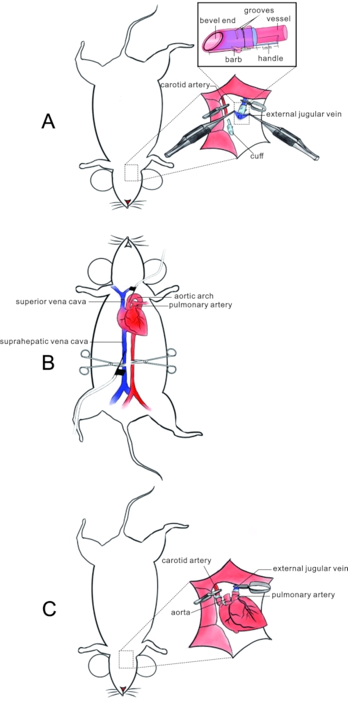
Figure 1: Schematic of the mouse cervical heterotopic heart transplantation model. (A) Protocol for preparing the recipient: after clipping the common carotid artery and external jugular vein at the proximal end, the vascular lumen of vessels is everted and fixed after passing through the barbed cuff with a bevel end and grooves on the surface. The dashed square shows the structure and usage of the cuff. (B) Donor heart resection: after the donor heart's perfusion with heparin and HTK solution from the inferior vena cava and aorta, the superior and inferior vena cava and pulmonary vein are ligated with sutures. The donor's heart is then resected by incising the vascular vessels. (C) Implantation of the donor's heart. The donor pulmonary artery and aorta is anastomosed to the recipient's external jugular vein, and carotid artery via the cuff with the recipient's vasculature turned inside out in an end-to-end pattern. Please click here to view a larger version of this figure.
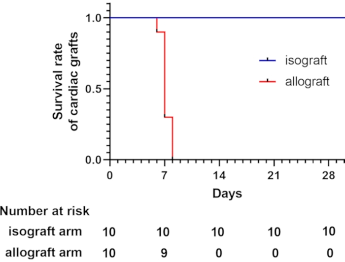
Figure 2: Survival curve for cardiac grafts. The survival curve of cardiac grafts shows that allogeneic heart transplants are lost within 8 days after transplantation, which was assessed by palpation of the heartbeat. A total of 10 recipient mice underwent the modified cervical heterotopic heart transplantation in each group. All the isogeneic heart transplants survived more than 4 weeks. Please click here to view a larger version of this figure.
Rupert Oberhuber et al.10 | Xin Mao et al (present work) | |||
Anaesthesia | xylazine and ketamine | isoflurane (Safe, continuous and stable) | ||
Right lobe of the submandibular gland | removal | preservation (Reduce twirling of grafts) | ||
Right sternocleidomastoid | removal | preservation (Reduce twirling of grafts) | ||
Cardiac perfusion | Retrograde perfusion with 4 °C HTK solution from the aortic arch | 1. Anterograde perfusion with ice-cold physiological saline supplemented with 100 IU/mL heparin solution from the suprahepatic vena cava. 2. Retrograde reperfusion with ice-cold HTK solution from the aortic arch. (Reduce coagulation and increase myocardial protection) | ||
Cuff | blunt end, with handle | bevel end, with handle, barb and grooves on the surface (Facilitate eversion and fixation) |
Table 1: Comparison of heart transplantation techniques. The current mouse cervical heterotopic heart transplantation technique is modified from Oberhuber, R. et al.10 and possesses additional advantages for cardiac graft survival.
The mouse heart transplantation model contributes to the investigation of rejection mechanisms after heart transplantation, contributing to the development of unique approaches to improve the long-term survival of cardiac allograft recipients. However, heart transplantation in mice is a complex and challenging task, requiring a high level of microsurgery techniques, especially in vascular anastomosis11,12,13. The mouse abdominal heterotopic heart transplantation model is performed using stitches by anastomosis of the donor aorta and pulmonary artery to the recipient aorta and inferior vena cava. The recipient's aorta and inferior vena cava need to be blocked in this operation. Therefore, ischaemia of the lower body and thrombosis of the inferior vena cava can increase the disability and death of recipient mice. To reduce the difficulties of vascular anastomosis during transplantation, Matsuura et al. first introduced a cervical heart transplantation model in mice using the cuff technique in 199114. In this model, the oversleeve-like everted anastomosis of vessels by ligation with cuff increased the anastomosis efficiency. In contrast to the anastomosis of vessels with sutures in an abdominal heart transplantation mouse model, it reduced the bleeding probability post-procedure. Therefore, the improvement of the anastomosis efficiency reduced the ischaemia time of cardiac muscle and increased the survival rate of cardiac grafts. Additionally, cervical implantation of the donor's heart does not interrupt the circulation of the recipient aorta and inferior vena cava compared with abdominal implantation15; therefore, the survival of recipient mice is increased.
A unique experimental mouse heterotopic heart transplantation model is described here, established by Rupert Oberhuber et al.10. The procedure involves an end-to-end cervical anastomosis of the donor aorta and pulmonary artery to the recipient carotid artery and jugular vein, following a modified Cuff technique. In this model, the systemic circulation of the recipient mice does not interfere with10, and the donor's heart was perfused from the inferior vena cava and aorta with heparin and HTK solutions for better myocardial protection. However, the critical component of this model differed from that of Oberhuber et al.10, which employed the modified barbed cuff with a bevel end and grooves similar to that of Finsterer et al.16. The bevel end facilitates an oversleeve-like evert of the vascular lumen. The grooves on the surface facilitate the fixation of everted vessel walls with a cuff using sutures, and the barb outside the cuff reduces the slippage of anastomosed vessel walls from the cuff (Figure 1). These modifications shorten the surgical time by 20% and improve cardiac grafts' implantation efficiency and survival. Furthermore, the modified barbed cuff is produced from the most common polyurethane catheter used for scalp acupuncture, thus significantly reducing the cost of the procedure. A comparison of the current technique with that of Oberhuber et al.10 is shown in Table 1.
The unique features of this model need to be noted. First, the length and calibre of the cuff are essential for successful anastomosis. The suitable length of the cuff was ~3 mm with a handle (1 mm) (Figure 1). The suitable calibre of the cuff is 26 G and 22 G for artery and veins, respectively. The unsuitable length and calibre of the cuff would result in twisting or excessive tension of the anastomosed vessels. Second, the suitable lengths of the recipient's vessels are 1.5 to 2-folds of the cuff. Third, the donor's heart is not perfused with excessive pressure, potentially damaging the graft. Fourth, the cardiac graft is fixed, and the cuff is anastomosed in a suitable position by suturing after implantation to avoid movement or twisting of anastomosed vessels or grafts. Fifth, preservation of the submaxillary gland and sternocleidomastoid contributes to reducing twirling or twisting of the anastomosed vessels or graft when resecting the right sublingual gland to produce a fossa for cardiac graft. Sixth, to facilitate the oversleeve-like evert of the vascular lumenand reduce thrombosis after surgery, heparin solution (100 IU/mL) can be provided to the anastomosed vessels while performing anastomosis.
This cuff technique facilitates the anastomosis of donor and recipient vessels during implantation; however, the hardness of the cuff may, in turn, increase the risk of twisting the anastomosed vessels, resulting in an increase in thrombosis after transplantation. Optimization of the cuff material is warranted to reduce complications, increase graft survival, and increase the utilization rate of the models in subsequent experiments. Furthermore, the fibrous scar of the cut may limit the space for the cardiac graft and affect its long-term survival. In addition, ejection of the cardiac graft may interfere with recipient mice's normal haemodynamic blood flow. Finally, this model is non-functional and cannot be used to evaluate the cardiac function of grafts. Nevertheless, this study provides knowledge regarding heart transplantation's immunological and pathological functions.
The authors have nothing to disclose.
This work was supported by the National Natural Science Foundation of China (81870304) to Jun Li.
5-0 Polyamide Monofilament suture | B.Braun Medical Inc. | C3090954 | |
8-0 Polyamide Monofilament suture | B.Braun Medical Inc. | C2090880 | |
10-0 Polyamide Monofilament suture | B.Braun Medical Inc. | G0090781 | |
22 G polyurethane cuff | B.Braun Medical Inc. | 4251628-02 | |
26 G polyurethane cuff | Suzhou Linhua Medical Instrument Co., LTD | REF383713 | |
Anesthesia induction chamber | RWD Life Science Co., LTD | V100 | |
Atraumatic microvascular clamp | Beyotime | FS500 | |
BALB/c and C57BL/6 mice (20–30 g) | Centre of Experimental Animals (Army Medical University, Chongqing, China) | ||
Buprenorphine | US Biological life Sciences | 352004 | |
Electrocoagulator | Guangzhou Runman Medical Instrument Co., LTD | ZJ1099 | |
Gauze | Henan piaoan group Co., LTD | 10210402 | |
Heating pad | Guangzhou Dewei Biological Technology Co., LTD | DK0032 | |
Heparin | North China Pharmaceutical Co., LTD | 2101131-2 | |
HTK solution | Shenzhen Changyi Pharmaceutical Co., LTD | YZB/Min8263-2013 | |
Injection syringe (10 mL) | Shandong weigao group medical polymer Co., LTD | 20211001 | |
Isoflurane | RWD Life Science Co., LTD | 21070201 | |
Physiological saline | Southwest pharmaceutical Co., LTD | H50021610 | |
Scalp needle | Hongyu Medical Group | 20183150210 | |
Shaver | Beyotime | FS600 | |
Small animal anesthesia machine | RWD Life Science Co., LTD | R500 | |
Surgical operation microscope | Tiannuoxiang Scientific Instrument Co. , Ltd, Beijing, China | SZX-6745 | |
Swab | Yubei Medical Materials Co., LTD | 21080274 |