Here, an easy-to-follow method to culture primary porcine retinal pigment epithelial cells in vitro is presented.
The retinal pigment epithelium (RPE) is a monolayer of polarized pigmented epithelial cells, located between the choroid and neuroretina in the retina. Multiple functions, including phagocytosis, nutrient/metabolite transportation, vitamin A metabolism, etc., are conducted by the RPE on a daily basis. RPE cells are terminally differentiated epithelial cells with little or no regenerative capacity. Loss of RPE cells results in multiple eye diseases leading to visual impairment, such as age-related macular degeneration. Therefore, the establishment of an in vitro culture model of primary RPE cells, which more closely resembles the RPE in vivo than cell lines, is critical for the characteristic and mechanistic studies of RPE cells. Considering the fact that the source of human eyeballs is limited, we create a protocol to culture primary porcine RPE cells. By using this protocol, RPE cells can be easily dissociated from adult porcine eyeballs. Subsequently, these dissociated cells attach to culture dishes/inserts, proliferate to form a confluent monolayer, and quickly re-establish key features of epithelial tissue in vivo within 2 wks. By qRT-PCR, it is demonstrated that primary porcine RPE cells express multiple signature genes at comparable levels with native RPE tissue, while the expressions of most of these genes are lost/highly reduced in human RPE-like cells, ARPE-19. Moreover, the immunofluorescence staining shows the distribution of tight junction, tissue polarity, and cytoskeleton proteins, as well as the presence of RPE65, an isomerase critical for vitamin A metabolism, in cultured primary cells. Altogether, we have developed an easy-to-follow approach to culture primary porcine RPE cells with high purity and native RPE features, which could serve as a good model to understand RPE physiology, study cell toxicities, and facilitate drug screenings.
The retinal pigment epithelium (RPE) is located between photoreceptors and choriocapillaris in the outer layer of the retina1 with multiple functions, including forming the blood-retinal barrier, transporting and exchanging nutrients and retinal metabolites, recycling vitamin A to maintain a normal visual cycle, and phagocytosis and clearance of shed photoreceptor outer segments (POSs)2,3. Since POSs require constant self-renewal to generate vision, the RPE cells need to continuously engulf detached POSs to maintain retinal homeostasis4. Therefore, RPE dysfunction results in many blinding eye diseases, such as age-related macular degeneration (AMD)4, retinitis pigmentosa (RP)5, Leber congenital amaurosis6, diabetic retinopathy7, etc. Till now, the exact pathogenesis of most of these diseases remains elusive. As a result, RPE cell culture is established to study RPE cell biology, pathological changes, and underlying mechanisms.
As the simplest model to study cell biology, the culture of RPE cells was started as early as the 1920s8. Although ARPE-19 is widely used as RPE cells, loss of pigmentation, cobblestone morphology, and, especially, the barrier functions in this cell line raise lots of concerns9. In comparison, the culture of primary human RPE cells offers a more realistic scenario for physiological and pathological studies9. However, the relatively limited availability restricts their usage and ethical issues always exist. In addition, several groups used mouse models to culture RPE cells. However, the size of the mouse eye is small, and a single culture usually requires many mice, which is not convenient9. Recently, scientists have developed new methods to use human embryonic stem cells or induced pluripotent stem cells to derive RPE cells. Although this technique has particular potential for the treatment of inherited RPE disorders, it is time-consuming and usually requires several months to generate mature RPE cells10. To overcome these problems, here we introduce an easy-to-follow protocol to isolate and culture high-purity RPE cells in the laboratory routinely. Under suitable culture conditions, these cells can display typical RPE functions and exhibit typical RPE morphologies. Therefore, this culture method can provide a good model to understand RPE physiology, study cytotoxicity, investigate pathological mechanisms of related ocular diseases, and conduct drug screenings.
The use of experimental animals complied with the regulations of the Association for Research in Vision and Ophthalmology (ARVO) and was approved by the Ethics Committee of Experimental Animal Management of Xiamen University.
1. Preparation of experimental surgical devices, tissue digestion enzyme, and cell culture buffer
2. Dissection of porcine eyeball RPE cells
3. Isolation and culture of porcine eyeball RPE cells
4. Characterization of primary porcine RPE cells
The primary porcine RPE (pRPE) cells were cultured in DMEM/Basic media with 10% FBS, and cell morphology under light microscope was photographed at 2 days (Figure 2A), 6 days (Figure 2B) and 10 days (Figure 2C) after seeding. After 1 wk, a confluent monolayer of pigmented pRPE cells with cobblestone morphologies was observed.
To better characterize the primary pRPE cells, primary human RPE cells (hRPE) at Passage 3 (P3)18 and ARPE-19 cells were cultured in DMEM/Basic media with 1% FBS for another wk, and then the total mRNA and protein of the cultured cells, as well as porcine RPE/choroid tissues, were harvested. The expression levels of key characteristic genes19 and protein markers of mature RPE were evaluated by qRT-PCR and Western blot. Compared with ARPE-19 cells, pRPE cells retained significantly higher expression levels of genes functioning in native RPE secretion (Figure 3A(c)), phagocytosis (Figure 3A(i)), transportation (Figure 3A(a),(d)), tight junctions (Figure 3A(j)), barrier formation (Figure 3A(b)), as well as visual cycle (Figure 3A(e),B). However, the expression levels of Krt8 and Krt18, two cytoskeleton marker genes of the RPE, were significantly lower in primary pRPE cells in comparison with primary hRPE and ARPE-19 cells (Figure 3A(g),(h)). Moreover, itgav, which participates in phagocytosis, was lower in primary pRPE cells as well (Figure 3A(f)). Since the expression levels of these three genes in pRPE cells were similar with the porcine RPE/choroid tissue, this may indicate the gene expression differences between the species. Moreover, the expression level of Tyr, which is responsible for melanin production, was highly reduced in both primary pRPE and hRPE cells (Figure 3A(k)), which may explain the loss of pigmentation in long-term cultured cells. The dissimilarities in the qRT-PCR results observed in the data of human pRPE and porcine pRPE cells might be because of the longer storage and higher passage number (P3) of human pRPE, thus indicating a loss of RPE characters on sub-culturing. In addition, Western blot showed that RPE65 protein, which is a key enzyme in the visual cycle featuring RPE cells, was expressed in pRPE cells, while its expression level was significantly reduced in primary hRPE cells and ARPE-19 cells (Figure 3B,C).
To further characterize the polarity and barrier functions of RPE cells, the cultured confluent monolayers of pRPE and ARPE-19 cells were cultured in transwell inserts with DMEM/Basic, DMEM/F12, and MEM-Nic media for 1 wk (Figure 4). Under these culture conditions, Na+-K+-ATPase (Figure 4A–F) and ZO-1 (Figure 4G–L) fluorescent staining results revealed that pRPE cells had higher expression levels of both Na+-K+-ATPase and ZO-1 than ARPE-19 cells. The equal distribution of Na+-K+-ATPase at the apical and basal surface of pRPE cells indicated that a longer culture time was required to restore cell polarities in vitro.
The RPE forms tight junctions near its apical surface to tightly regulate the exchange of metabolites between the inner retina and choroids20. ZO-1 staining suggested that tight junction proteins were normally localized at the plasma membrane of primary pRPE cells with regular cobblestone morphologies, but not of ARPE-19 cells. Among the three culture medias, the best ZO-1 staining patterns were observed in cells cultured in DMEM/Basic, while DMEM/F12 failed to maintain the cobblestone morphology of pRPE cells. A higher TER value serves as an indicator of tight junction formation and better barrier functions of RPE cells21. To confirm the function of tight junctions, transepithelial resistance (TER) was measured. However, only slightly higher TER was detected in comparison with empty transwell inserts (Figure 4M,N).
In the retina, Bruch's membrane exists between the RPE and the choroid. The main components of Bruch's membrane are collagen type IV, proteoglycans, and laminin22. In order to simulate the supporting effect of Bruch's membrane on RPE, laminin was spread on the surface of the transwell membrane to facilitate the maturation of cultured RPE cells. Based on the results obtained from Figure 4, the confluent pRPE cells were cultured on transwell inserts in DMEM/Basic media with 1% FBS for 2 wks. Immunofluorescent staining results showed that RPE-specific proteins RPE65 were expressed in all pRPE cells (Figure 5A,B). Na+-K+-ATPase was distributed on the apical surface of pRPE cells, suggesting the re-establishment of cell polarities (Figure 5C,D). Both ZO-1 staining and TER results indicated that tight junctions were well formed in primary pRPE cells when they were cultured on laminin (Figure 5E,F,H). Figure 5G depicts cells stained with Hoechst dye.
Moreover, Western blot demonstrated that both pRPE and ARPE-19 cells produced growth factors, including pigment epithelium-derived factor (PEDF) and vascular endothelial growth factor (VEGF) (Figure 6A). After confluent monolayers of pRPE were cultured in transwell inserts with DMEM/Basic, DMEM/F12, and MEM-Nic media for 1 wk, culture media from the top and bottom chambers of the transwell inserts were collected, and the secreted VEGF was quantified by ELISA. When DMEM/Basic and MEM-Nic media were used for cell culture, pPRE cells secreted more VEGF than cells in DMEM/F12 media (Figure 6B). However, no differences could be detected in VEGF amounts between the top and bottom chambers of the transwell inserts in all tested conditions (Figure 6B). In addition, after primary pRPE cells were cultured on the inserts coated with laminin and in DMEM/Basic media for 2 wks, the media from the top and bottom chambers were collected. Western blot analysis showed higher levels of PEDF in the top chamber than the bottom chamber, while the protein levels of secreted VEGF were similar at both chambers (Figure 6C). These results further supported the re-establishment of cell polarity when primary pRPE cells were cultured for another 2 wks after they reached confluency on laminin.
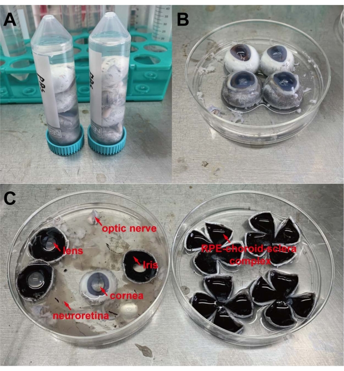
Figure 1: Basic steps of pRPE cell isolation. (A) Wash porcine eyeballs with 1x PBS in 50 mL sterile centrifuge tubes. (B,C) Prepare RPE-choroid-sclera complexes for enzymatic digestion. Please click here to view a larger version of this figure.
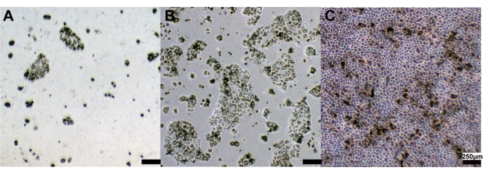
Figure 2: Primary pRPE cell morphologies after cell culture. Representative images of primary pRPE cells at (A) Day 2, (B) Day 6, and (C) Day 10. Scale bar 250 µm. Please click here to view a larger version of this figure.
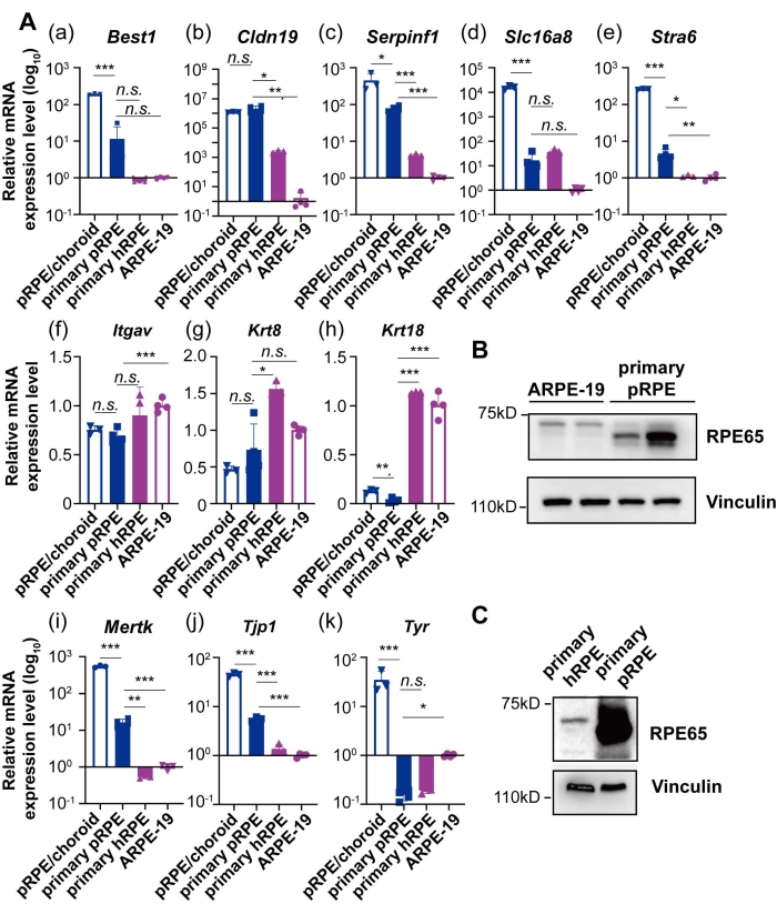
Figure 3: Expression levels of key signature genes and proteins in cultured RPE cells. (A) qRT-PCR analysis of mRNA levels of signature genes in primary pRPE cells, primary hRPE cells, ARPE-19 cells, and pRPE/choroid tissue. Gapdh was used as the house keeping gene for qRT-PCR. Four biological replicates were used for primary pRPE cells and ARPE-19 cells, while three biological replicates were used for primary hRPE cells and pRPE/choroid tissue. The gene expression levels in ARPE-19 cells were set as controls. (B) Western blot analysis of RPE65 proteins in ARPE-19 and pRPE cells. Vinculin was used as a loading control. (C) Western blot analysis of RPE65 proteins in primary hRPE cells and pRPE/choroid tissue. Vinculin was used as a loading control. For comparison, student t-test was used, *p < 0.05, **p < 0.01, ***p < 0.001. Please click here to view a larger version of this figure.
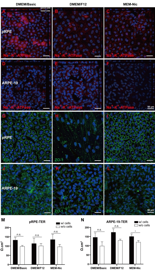
Figure 4: One-wk culture of primary pRPE and ARPE-19 cells in DMEM/Basic, DMEM/F12, and MEM-Nic media with 1% FBS. (A) Na+-K+-ATPase fluorescent staining (Red) of primary pRPE cells in DMEM/Basic, (B) DMEM/F12, and (C) MEM-Nic media. (D) Na+-K+-ATPase fluorescent staining (Red) of ARPE-19 cells in DMEM/Basic media, (E) DMEM/F12 media, and (F) MEM-Nic media. (G) ZO-1 fluorescent staining (Green) of primary pRPE cells in DMEM/Basic media, (H) DMEM/F12 media, and (I) MEM-Nic media. (J) ZO-1 fluorescent staining (Green) in ARPE-19 cells in DMEM/Basic media, (K) DMEM/F12 media, and (L) MEM-Nic media. TER measurements after primary pRPE (M) and ARPE-19 (N) cells were cultured in DMEM/Basic, DMEM/F12, and MEM-Nic media for 1 wk. For each type of cells, data were obtained from at least three different wells. For comparison, student t-test was used, *p < 0.05, **p < 0.01, ***p < 0.001. Scale bar 50 µm. Please click here to view a larger version of this figure.
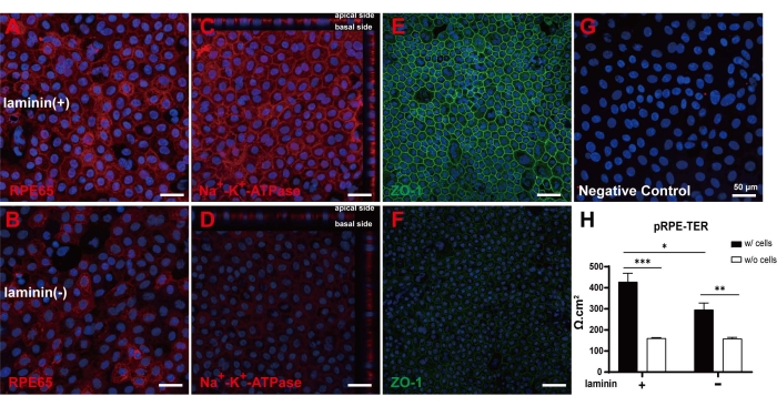
Figure 5: Two-wk culture of pRPE cells in DMEM/Basic media with 1% FBS. (A–F) Cells cultured with or without laminin were stained with RPE65 (A,B, Red), Na+-K+-ATPase (C,D, Red), ZO-1 (E,F, Green), and (G) Hoechst (Blue) . (H) TER measurements in cell sheets cultured with or without laminin. For different treatments, data were obtained from four different wells. For comparison, student t-test was used, *p < 0.05, **p < 0.01, ***p < 0.001. Scale bar = 50 µm. Please click here to view a larger version of this figure.
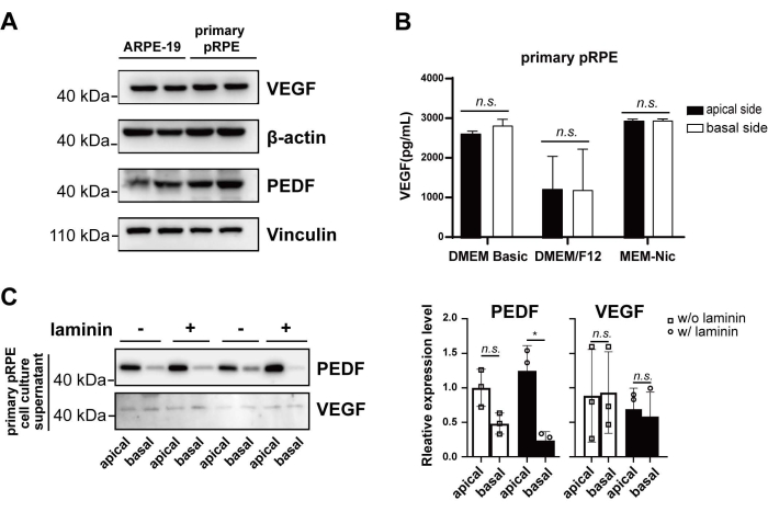
Figure 6: Secretory functions of cultured primary pRPE and ARPE-19 cells. (A) Western blot analysis of VEGF and PEDF in primary pRPE and ARPE-19 cells. (B) Secreted VEGF in the top and bottom chamber of transwell inserts with primary pRPE cells when confluent cells were cultured in DMEM/Basic, DMEM/F12, and MEM-Nic media for 1 wk. (C) Secreted PEDF and VEGF at the top and bottom chamber of transwell inserts with primary pRPE in DMEM/Basic media for 2 wks. For different treatments, data were obtained from three different wells. For comparison, student t-test was used, *p < 0.05, **p < 0.01, ***p < 0.001. Please click here to view a larger version of this figure.
Homo sapiens | Sus scrofa | |
Best1 | F: GAAGGCAAGGACGAGCAAG | F: GGACACCTGTATGCCTACGA |
R: TCCAACTGCTTGTGTTCTGC | R: GGAACGTGAAGAGGGGTACA | |
Cldn19 | F: GGTGACCCAGGAGTTCTTCA | F: TCGTGACCCAGGAGTTCTTC |
R: CTGTTGGGTCTCTCTGGCTC | R: GCTGCTGTTGGATCGCTC | |
Itgav | F: CGCAGTCCCATCTCAAATCC | F: GCTTTCTTCAGGACGGAACA |
R: GGCCCTGTATAAGATAGCTCGA | R: GAAATGAGCTGACCTTGCCA | |
Krt8 | F: GGAGCAGATCAAGACCCTCA | F: CCCAGGAGAAGGAGCAGATC |
R: GCCGCCTAAGGTTGTTGATG | R: ATGTTGTCGATGTTGCTCCG | |
Krt18 | F: CTTGGAGAAGAAGGGACCCC | F: GCTGATAATCGGAGGCTGGA |
R: GGCCAGCTCTGTCTCATACT | R: GAAGTCATCAGCAGCGAGAC | |
Mertk | F: GTGTGCAGCGTTCAGACAAT | F: GCGGCTATTTCTTGGTGGAA |
R: AAAATGTTGACGGGCTCAGG | R: ACGTAGATGGGGTCAGACAC | |
Serpinf1 | F: CAGATGAAAGGGAAGCTCGC | F: AAGACGTCGCTGGAGGATTT |
R: TTAGGGTCCGACATCATGGG | R: GGTCACTTTCAGAGGCAGGA | |
Slc16a8 | F: GGGTGTCCTCCATCATGCTA | F: CAGTTCGAGGTGCTCATGG |
R: GTCAGGTAGAGCTCCAGGAG | R: GACAGCCATGAAGACACCAG | |
Stra6 | F: CTTTGCAGGAAGAAGCTGGG | F: CTAGCCGTGTTGTCGATCCT |
R: TAAATGGCCGTCCCTGTCAG | R: GACCATGAAGACAGCAGCAG | |
Tjp1 | F: ACAGGAAAATGACCGAGTTGC | F: AAGACTTGTCAGCTCAGCCA |
R: TGGTTCAGGATCAGGACGAC | R: CCAGCATCTCGAGGTTCACT | |
Tyr | F: ACTCAGCCCAGCATCATTCT | F: ATCTACTCAGCCCAGCATCC |
R: ACATCAGCTGAAGAGGGGAG | R: GAGCCTTGGAGTCCTGGATT | |
Gapdh | F: CAGCCTCAAGATCATCAGCA | F: CATCCTGGGCTACACTGAGG |
R: ATGATGTTCTGGAGAGCCCC | R: GGGGCTCTTACTCCTTGGAG |
Table 1: Primers for qRT-PCR.
Here, a detailed and optimized protocol for the isolation, culture, and characterization of RPE cells from porcine eyeballs, which generates a good model for in vitro characterization of RPE cells and RPE-related disorder studies has been described. Methods for the isolation of the RPE from human, mouse, and rat eyes have been described previously23,24,25. However, it is difficult to obtain human eyeballs in some laboratories, and it usually raises ethical issues. The RPE tissues of mice and rats are relatively small, the cells are easily damaged during separation, and only a few cells can be obtained from each animal. In contrast, porcine eyeballs are much easier to obtain and handle, which could stably generate a relatively large number of primary cells from a single eyeball. In some studies, tweezers are used to mechanically separate RPE, which can easily lead to cell death. Therefore, hyaluronic acid, dispase, and Trypsin/EDTA solution have been employed to dissociate RPE cells previously. Previous studies have shown that Trypsin/EDTA solution has the ability to help RPE detach from the choroid21. Therefore, Trypsin/EDTA solution was used to digest the porcine tissues, which generate optimum results. After Trypsin/EDTA neuralization, RPE tissues could be dissociated into single cell suspension by gently pipetting with a pipette tip. One trick is that fresh Trypsin/EDTA solution is recommended for each experiment to fully dissociate RPE cells. Due to the large area of the RPE tissues of porcine eyeballs, the half bowl-shaped RPE-choroid-sclera complex needs to be cut into the shape of a four-leaf clover, which is conducive to the full contact between the RPE tissues and the Trypsin/EDTA solution so as to facilitate their separation from the choroid. However, the digestion time should not exceed half an hour. Within this time, other tissues except RPE will not be digested, which could effectively reduce the contaminations from other types of cells.
Laminin was spread on the surface of the transwell membrane to simulate the supportive effect of Bruch's membrane on RPE. The results showed that laminin was beneficial for the growth of pRPE cells, which stimulated the expression of transporter and tight junction proteins. However, the results also indicated that a longer culture time, of about 2 wks, was required for primary pRPE cells to restore certain features of native RPE tissues, including cell polarity and tight junctions.
One shortcoming for primary RPE culture is that it is difficult to maintain visual cycle enzyme expressions in vitro. Therefore, it is critical to examine whether cultured RPE cells could express RPE-specific proteins RPE65. Compared with ARPE-19 cells, pRPE cells expressed significantly more RPE65 protein. In contrast, only a weak band of RPE65 protein was observed in ARPE-19 cell lysates in Western blot. It is interesting to observe that the molecular weight of RPE65 was slightly different between human and pigs (Figure 3B,C). The loss of RPE65 expression in cultured RPE cells remains as a mystery. Even in cultured primary cells, RPE65 was gradually lost with passages (data not shown). Till now, how to maintain the expression of RPE65 in vitro requires further investigation, considering the fact that visual cycle is a critical function for native RPE tissues. Secondly, a major limitation of this protocol is that in vitro cultured RPE cells cannot be passaged. With prolonged culture, pRPE cells tend to lose their hexagonal shape and TER as well as pigmentation, so that only passage 0 cells can be used. Another limitation of this culture protocol is that genetic mutations, which lead to inherited retinal diseases, are difficult to reproduce in these cells. Therefore, virus-mediated gene editing may be employed to study hereditary RPE-disorders such as Leger congenital amaurosis in the future.
The authors have nothing to disclose.
The authors would like to show their gratitude and respect to all animals contributing their cells in this study. This study was supported in part by grants from the National Key R&D Program of China (2019YFA0111200, Yi Liao & Yuan Gao and Grant nos. 2018YFA0107301, Wei Li). The authors thank Jingru Huang and Xiang You from Central Lab, School of Medicine, Xiamen University for technical support in confocal imaging.
ARPE-19 cells | CCTCC | GDC0323 | |
Bovine serum albumin | Yeasen | 36101ES60 | |
Confocal microscopy | Zeiss | LSM 880 with Airyscan | |
ChemiDoc Touch | Bio-Rad | 1708370 | |
Cell scraper | Sangon | F619301 | |
10 cm culture dish | NEST | 121621EH01 | |
12-well culture plate | NEST | 29821075P | |
DMEM F12 Medium | Gibco | C11330500BT | |
DMEM basic Medium | Gibco | C11995500BT | |
EVOM2 | World Precision Instruments | EVOM2 | For TER measurement |
Fetal bovine serum | ExCell Bio | FSP500 | |
Goat anti-Rabbit IgG (H+L) Highly Cross-Adsorbed Secondary Antibody, Alexa Fluor 488 | ThermoFisher Scientific | A-11034 | |
Goat anti-Rabbit IgG (H+L) Cross-Adsorbed Secondary Antibody, Alexa Fluor 594 | ThermoFisher Scientific | A-11012 | |
Goat anti Mouse IgG (H/L):HRP | Bio-Rad | 0300-0108P | |
Goat anti Rabbit IgG (H/L):HRP | Bio-Rad | 5196-2504 | |
hydrocortisone | MCE | HY-N0583/CS-2226 | |
Hoechst 33342 solution (20 mM) | ThermoFisher Scientific | 62249 | |
LightCycler 96 Instrument | Roche | 5815916001 | |
Liothyronine | MCE | HY-A0070A/CS-4141 | |
laminin | Sigma-Aldrich | L2020-1MG | |
MEM(1X)+GlutaMAX Medium | Gibco | 10566-016 | |
MEM NEAA(100X) | Gibco | 11140-050 | |
Millex-GP syringe filter unit | Millipore | SLGPR33RB | |
N1 | Sigma-Aldrich | SLCF4683 | |
NcmECL Ultra | New Cell&Molecular Biotech | P10300 | |
Non-fat Powdered Milk | Solarbio | D8340 | |
Nicotinamide | SparkJade | SJ-MV0061 | |
Na+-K+ ATPase antibody | Abcam | ab76020 | Recognize both human and porcine proteins |
PAGE Gel Fast Preparation Kit(10%) | Epizyme | PG112 | |
primary Human RPE cells | – | – | Generous gift from Shoubi Wang lab |
Pierce BCA Protein Assay Kit | ThermoFisher Scientific | 23225 | |
Prism | GraphPad by Dotmatics | version 8.0 | |
Protease Inhibitor Cocktails | APExBIO | K1024 | |
PRE65 antibody | Proteintech | 17939-1-AP | Recognize both human and porcine proteins |
PEDF antibody | Santa Cruz Biotechnology | sc-390172 | Recognize both human and porcine proteins |
100 x penicillin/streptomycin | Biological Industries | 03-031-1BCS | |
Phosphate buffered saline (PBS) | RARBIO | RA-9005 | |
ReverTra Ace qPCR RT Master Mix | Toyobo | FSQ-201 | |
RIPA buffer | ThermoFisher Scientific | 89900 | |
15 mL sterile centrifuge tubes | NEST | 601052 | |
50 mL sterile centrifuge tubes | NEST | 602052 | |
0.25% Trypsin-EDTA | Gibco | 25200-056 | |
Taurine | Damas-beta | 107-35-7 | |
Trizol | Thermo-Fisher | 15596026 | RNA extraction solution |
TB Green Fast qPCR Mix | Takara | RR430A | |
12-well transwell inserts | Labselect | 14212 | |
VEGF antibody | Proteintech | 19003-1-AP | Recognize both human and porcine proteins |
VEGF ELISA kit | Novusbio | VAL106 | |
ZO-1 antibody | ABclonal | A0659 | Recognize both human and porcine proteins |