This protocol describes methodologies to establish mouse endometrial epithelial organoids for gene expression and histological analyses.
Endometrial tissue lines the inner cavity of the uterus and is under the cyclical control of estrogen and progesterone. It is a tissue that is composed of luminal and glandular epithelium, a stromal compartment, a vascular network, and a complex immune cell population. Mouse models have been a powerful tool to study the endometrium, revealing critical mechanisms that control implantation, placentation, and cancer. The recent development of 3D endometrial organoid cultures presents a state-of-the-art model to dissect the signaling pathways that underlie endometrial biology. Establishing endometrial organoids from genetically engineered mouse models, analyzing their transcriptomes, and visualizing their morphology at a single-cell resolution are crucial tools for the study of endometrial diseases. This paper outlines methods to establish 3D cultures of endometrial epithelium from mice and describes techniques to quantify gene expression and analyze the histology of the organoids. The goal is to provide a resource that can be used to establish, culture, and study the gene expression and morphological characteristics of endometrial epithelial organoids.
The endometrium – the inner lining mucosal tissue of the uterine cavity – is a unique and highly dynamic tissue that plays critical roles in a woman's reproductive health. During the reproductive lifespan, the endometrium holds the potential to undergo hundreds of cycles of proliferation, differentiation, and breakdown, coordinated by the concerted action of the ovarian hormones – estrogen and progesterone. Studies of genetically engineered mice have uncovered basic biological mechanisms underpinning the endometrial response to hormones and control of embryo implantation, stromal cell decidualization, and pregnancy1. In vitro studies, however, have been limited due to difficulties in maintaining non-transformed primary mouse endometrial tissues in traditional 2D cell cultures2,3. Recent advances in the culture of endometrial tissues as 3D organ systems, or organoids, present a novel opportunity to investigate biological pathways that control endometrial cell regeneration and differentiation. Mouse and human endometrial organoid systems have been developed from pure endometrial epithelium encapsulated in various matrices4,5, while human endometrium has been cultured as scaffold-free epithelial/stromal co-cultures6,7, and more recently as collagen-encapsulated epithelial/stromal assembloids8. The growth and regenerative potential of epithelial organoid cultures is supported by a defined cocktail of growth factors and small molecule inhibitors that have been empirically determined to maximize growth and regeneration of the organoids4,5,9. Furthermore, the ability to freeze and thaw endometrial organoids permits the long-term banking of endometrial organoids from mice and humans for future studies.
Genetically engineered mice have revealed the complex signaling pathways that control early pregnancy and decidualization, and have been used as models of pregnancy loss, endometrial cancer, and endometriosis. These genetic studies have been largely achieved with cell-specific deletion of loxP flanked alleles ("floxed") using cre recombinases that are specifically active in female reproductive tissues. These mouse models include the widely used progesterone receptor-cre10, which has strong recombinase activity in the endometrial epithelial and stromal tissues, lactoferrin i-cre, which induces endometrial epithelial recombination in adult mice11, or Wnt7a-cre, which triggers epithelial-specific deletion in Müllerian-derived tissues12. Culturing endometrial tissues from genetically engineered mouse models as 3D organoids has provided an excellent opportunity to investigate endometrial biology and facilitate the identification of growth factors and signaling pathways that control endometrial cell renewal and differentiation13,14. Methods for the isolation and culture of mouse endometrial tissue are described in the literature and report the use of various enzymatic strategies for the isolation of uterine epithelium for subsequent culturing of endometrial epithelial organoids4. While previous literature provides a critical framework for endometrial epithelial organoid culture protocols4,5,6, this paper provides a clear, comprehensive method for generating, maintaining, processing, and analyzing these organoids. Standardization of these techniques is important for accelerating advancements in the field of women's reproductive biology. Here, we report a detailed methodology for the enzymatic and mechanical purification of mouse endometrial epithelial tissue for the subsequent culture of endometrial organoids in a gel matrix scaffold. We also describe the methodologies for downstream histological and molecular analyses of the gel matrix-encapsulated mouse endometrial epithelial organoids.
Mouse handling and experimental studies were performed under protocols approved by the Institutional Animal Care and Use Committee (IACUC) of Baylor College of Medicine and guidelines established by the NIH Guide for the Care and Use of Laboratory Animals.
1. Isolation of uterine epithelium from mice using enzymatic and mechanical methods
NOTE: This section describes the steps required to establish, passage, freeze, and thaw epithelial endometrial organoids from mice using a gel matrix scaffold. Previous studies have determined that optimal cultures of mouse endometrial organoids are established from mice during the estrus phase4, which can be determined by cytological examination of a vaginal swab15. Adult female WT mice (6-8 weeks old, hybrid C57BL/6J and 129S5/SvEvBrd) were used for all experiments. The mice were humanely euthanized according to IACUC-approved guidelines using isoflurane sedation followed by cervical disarticulation. Once the mice are euthanized, the following steps should be followed. See the Table of Materials for details related to materials and solutions used in this protocol.
2. Processing of the stromal compartment
NOTE: This section outlines the protocols necessary for isolating the stromal compartment of the mouse endometrium. Given the increasing interest in epithelial/stromal co-culture experiments, it is important to be able to process the stromal cell populations in addition to the epithelial cells that will generate organoids.
3. Encapsulation of uterine epithelium into gel matrix to establish organoids
NOTE: Keep the gel matrix on ice until it is ready to be used.
4. Gene expression analysis of endometrial organoids following treatment with estradiol
NOTE: This section describes the methods used to profile the gene expression of endometrial epithelial organoids using real-time qPCR following treatment with estradiol (E2; see Table 1). Because the endometrium is under the cyclical control of the ovarian hormone E2, testing the responsiveness of the organoids to E2 is an important measure of physiological function. We have obtained high-quality RNA and generated sufficient mRNA to profile gene expression using qPCR and/or RNA-sequencing from our endometrial epithelial organoids. This section describes how to collect organoids and process them for downstream analysis of gene expression. The selected treatment medium reflects the one used to treat cultured endometrial cells. However, it should be noted that this treatment medium can be optimized accordingly, as done for the treating of human endometrial 3D cultures with hormones8,16,17.
5. Histological analysis of endometrial organoids
NOTE: Imaging the morphological features of endometrial organoids is critical to evaluating the cellular effect of growth factors, genetic manipulations, or small molecule inhibitors. This section describes the techniques used to fix, process, and image endometrial epithelial organoids using histological stains and antibody immunofluorescent staining.
6. Hematoxylin & eosin staining
7. Immunofluorescence staining
Phase contrast images of mouse endometrial organoids
We established organoids from WT mouse endometrial epithelium, as described in the attached protocol (see diagram in Figure 1). Following enzymatic dissociation of the mouse endometrial epithelium, epithelial sheets were mechanically separated from the uterine stromal cells and further dissociated with collagenase to generate a single-cell suspension. If performed correctly, this method of epithelial and stromal cell separation should yield samples with contamination of no more than 10%-15% of the opposite cell type (see immunofluorescence images in Figure 2). The epithelial cell pellet was then resuspended in gel matrix, allowed to gel at room temperature, and plated into 25 µL domes on tissue culture plates. After the gel matrix domes solidified, they were overlaid with organoid medium and allowed to grow. We typically observed that endometrial epithelial cells assembled into organoids within 3-4 days, as pictured in Figure 3. The organoids were maintained in culture, with media changes occurring every 3 days, and passaging every 5-7 days.
Imaging of mouse endometrial organoids using histological and immunofluorescent staining
To analyze the morphology of the mouse epithelial organoids, we adapted methods used for the encapsulation of cellular fine needle aspirates using specimen processing gel20. This allows for the preservation of the endometrial organoid morphology and encapsulation into a matrix that can be subjected to processing in preparation for paraffin embedding. Specimen processing gel is a modified agar that is widely used in clinical pathology laboratories for the analysis of fine needle aspirates and has been used to encapsulate vaginal organoids21,22. After the specimen processing gel/organoid mixture solidifies, it can be processed, stained, and imaged with techniques typically used to analyze tissues. In Figure 4A,B, we show that sectioned formalin-fixed paraffin-embedded endometrial organoids can be visualized by hematoxylin and eosin stains, which display the single layer of epithelium and hollow centers-the lumens-of the organoids. Certain organoids also contain secretions in the lumen, indicating that the organoids acquire the functional properties of glandular epithelial cells. We also describe techniques to visualize the organoids using immunostaining (Figure 4C,D); sectioned endometrial organoids were incubated with a Cytokeratin 8 primary antibody (TROMA-1) and a fluorophore-conjugated secondary antibody (secondary anti-rat-594). Nuclei were then visualized with DAPI, and the slides were imaged using a fluorescence microscope. We observed that all the cells in the organoids were Cytokeratin 8-positive and contained DAPI, indicating that fixation, embedding, and processing of the endometrial organoids is compatible with antibody-based immunostaining.
RNA extraction and gene expression analysis
To determine the gene expression response of endometrial organoids, we allowed endometrial organoids from WT mice to grow for 4 days after the first passage. On the evening before treatment, the endometrial organoid medium was changed to starvation medium. The organoids were then treated in triplicate wells (each well containing three domes) with vehicle (ethanol; equal volume in solution as used for estradiol) or 10 nM estradiol (E2) for a total of 48 h. After 48 h, the medium was removed, and the organoids were processed for RNA extraction. Approximately 4 µg of RNA can be obtained from a total of two domes from each well, and 1 µg of RNA was used to reverse transcribe cDNA. Amplification of the genes encoding lipocalin 2 (Lcn2), lactoferrin (Ltf), and the progesterone receptor (Pgr) was performed using real-time quantitative PCR (Figure 5 and Table 1)23,24,25. As expected, we observed that the expression of Lcn2, Ltf, and Pgr increased in the epithelial organoids following stimulation with 10 nM E2 (Figure 5). Thus, these results show that endometrial epithelial organoids can be successfully used to measure the gene expression changes in response to E2.
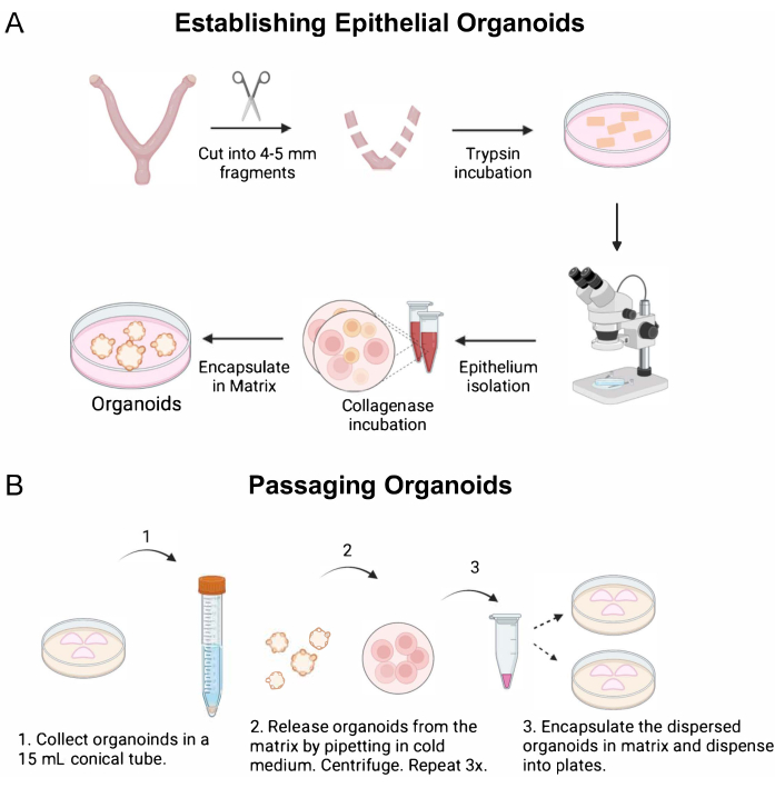
Figure 1: Procedures used to establish endometrial organoids. Diagram outlines the key steps that were followed to (A) establish and (B) passage epithelial organoids from the mouse endometrium. (A) Methods include the enzymatic and mechanical dissociation of endometrial epithelium from the mouse uterus, followed by single-cell dispersion and encapsulation of the epithelium into a gel matrix. To obtain the uterine tissues for enzymatic dissociation, the ovaries and oviducts are removed from the uterine horns with fine scissors, cut laterally into 4-5 mm fragments, and then placed in the enzyme solution. After enzymatic incubation, the endometrial epithelium is mechanically separated from the underlying stroma under a microscope using forceps and a pipette to "squeeze" out the epithelium from the uterine tube. The epithelium is further digested into epithelial cells using a short incubation in collagenase, followed by encapsulation into a gel matrix. (B) Passaging the endometrial organoids requires mechanical dissociation in cold medium and centrifugation to release the organoids from the matrix and to generate smaller organoid fragments. Once the organoids have been released from the matrix and mechanically dissociated into smaller fragments, they are encapsulated into matrix and replated. Please click here to view a larger version of this figure.
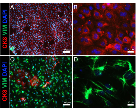
Figure 2: Immunostaining of isolated endometrial epithelium and stromal cell populations. Immunofluorescence images show epithelial and stromal cell populations of the (A,B) epithelial cell and (C,D) stromal cell fractions following enzymatic and mechanical separation of the uterus. Cytokeratin 8 (an epithelial cell marker) is shown in red, vimentin (a stromal cell marker) is shown in green, and DAPI (a nuclear marker) is shown in blue. Scale bars = 100 µm (A,C), 20 µm (B,D). Abbreviations: CK8 = cytokeratin 8; VIM = vimentin; DAPI = 4',6-diamidino-2-phenylindole. Please click here to view a larger version of this figure.
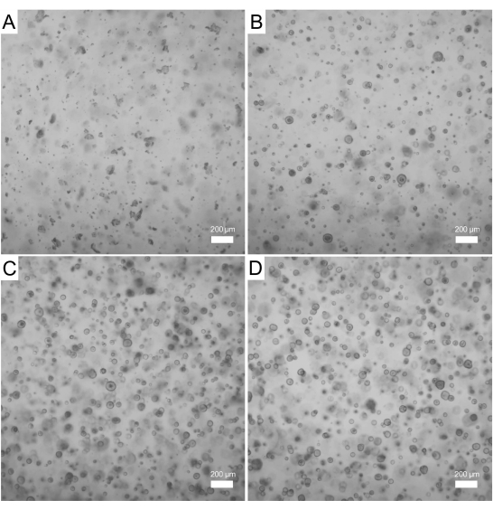
Figure 3: Formation of endometrial organoids from WT mice over the course of 4 days. Endometrial epithelium was isolated from a WT mouse and used to generate organoids. (A) Phase contrast image of the epithelial cells encapsulated in the gel matrix immediately after digestion (day 0). (B,C) A few small organoids can be observed on day 1 (B) and day 2 (C) of culture. (D) Larger and mature epithelial organoids can be observed on day 3 of culture. Images were captured with a 5x objective; scale bars = 200 µm. Abbreviation: WT = wild type. Please click here to view a larger version of this figure.
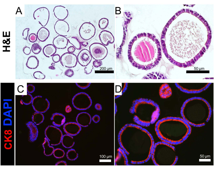
Figure 4: Histological analysis of endometrial organoids. (A,B) FFPE endometrial epithelial organoids were sectioned and stained with H&E. (C,D) FFPE epithelial organoids were immunostained with the epithelial cell marker cytokeratin 8 (red). Cell nuclei were stained with DAPI (blue). Scale bars = 200 µm (A), 100 µm (C), 50 µm (B,D). Abbreviations: FFPE = formalin-fixed paraffin embedded; H&E = hematoxylin & eosin; CK8 = cytokeratin 8; DAPI = 4',6-diamidino-2-phenylindole. Please click here to view a larger version of this figure.
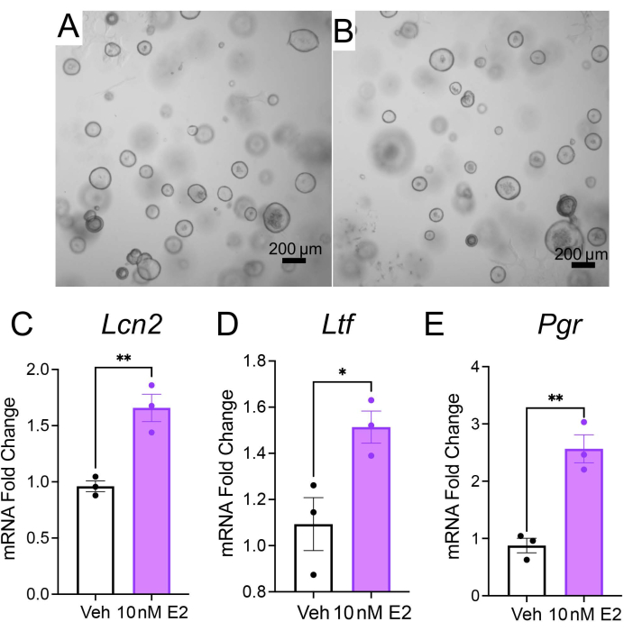
Figure 5: Gene expression analysis of mouse endometrial organoids. Mouse endometrial epithelial organoids from WT mice were cultured for 4 days in organoid growth medium, followed by treatment with vehicle or 10 nM E2 for 48 h in DMEM/F12 supplemented with 2% charcoal-stripped FBS. (A,B) Phase contrast images of the WT mouse organoids after treatment with vehicle (A) or 10 nM E2 (B). (C–E) Real time qPCR analysis of epithelial endometrial organoids treated with vehicle or 10 nM E2. Expression of E2-regulated genes, lipocalin 2 (Lnc2), lactoferrin (Ltf), and progesterone receptor (Pgr). Bars represent mean ± SEM, analyzed by a two-tailed t-test; *p < 0.033, **p < 0.002, ***p < 0.001. Repeated with organoids derived from n = 3 WT mice per condition. Scale bars = 200 µm (A,B). Abbreviations: WT = wild type; E2 = estradiol; qPCR = quantitative PCR. Please click here to view a larger version of this figure.
Primer Sequence | Forward (5'-3') | Reverse (5'-3') |
Lipocalin 2 (Lcn2) | GCAGGTGGTACGTTGTGGG | CTCTTGTAGCTCATAGATGGTGC |
Lactoferrin (Ltf) | TGAGGCCCTTGGACTCTGT | ACCCACTTTTCTCATCTCGTTC |
Progesterone (Pgr) | CCCACAGGAGTTTGTCAAGCTC | TAACTTCAGACATCATTTCCGG |
Glyceraldehyde 3 phosphate dehydrogenase (Gapdh) | CAATGTGTCCGTCGTGGATCT | GCCTGCTTCACCACCTTCTT |
PCR mixture contains: |
2x SYBR Green |
0.5 µM Fwd Primer |
0.5 µM Rev Primer |
cDNA |
RNAse/DNAse-Free H2O |
Cycler Conditions: | |||
Temperature | Time | Cycles | |
Hold | 95 °C | 10 min | 1 |
Denature | 95 °C | 15 s | 40 |
Extend | 60 °C | 60 s |
Table 1: Primer sequences and PCR conditions.
Here, we describe methods to generate endometrial epithelial organoids from mouse endometrium and the protocols routinely used for their downstream analysis. Endometrial organoids are a powerful tool to study the mechanisms that control endometrial-related diseases, such as endometriosis, endometrial cancer, and implantation failure. Landmark studies published in 2017 reported the conditions to culture long-term and renewable cultures of endometrial organoids from mouse and human epithelium4,5. Although organoids have been widely used to study biological pathways in other systems, the study of endometrial tissue in vitro has been limited by the absence of a suitable model to study the non-transformed primary epithelium in culture26. Endometrial organoids derived from human and mouse endometrium were successfully established using conditions typically used to culture organoids from other organ tissues, such as gut, kidney, and lung, and by embedding them in an extracellular matrix scaffold composed of gel matrix4. Since these initial reports, the use of endometrial organoids to study the biology of the endometrium has increased significantly in the scientific community.
By modifying a previously published method of epithelial isolation from the mouse uterus27,28, this protocol uniquely highlights key steps to isolate mouse uterine by directly placing the tissue into an enzyme solution, bypassing the need to transect the uterus, or use filtration. Using this method, we obtained epithelium with ~85%-90% purity, as determined by cytokeratin 8 and vimentin immunostaining (Figure 2). This protocol also clearly outlines the key details for encapsulation of the isolated epithelial cells into the growth matrix for generation, maintenance, and passaging of the organoids. The images in Figure 4 and our unpublished studies have identified the presence of mucins and glandular cells (i.e., FOXA2-positive) in our organoid cultures29. These indicate that the method presented here is effective for the isolation of both glandular and luminal-epithelial cell populations from the mouse endometrium.
Key limitations for the growth of these organoids include the use of the commercial matrix (i.e., Matrigel), which can result in batch-to-batch variations and contains growth factors that can affect organoid growth. Furthermore, while these organoids contain pure uterine epithelium, the use of additional endometrial cell types (i.e., immune, stromal) have been recently described in human organoid systems of the endometrium8,30. Other groups have shown the ability to isolate both luminal and glandular epithelial cells for organoid cultures from the mouse uterus14.
This approach uses tissues from a small animal model that is relatively common and available to most researchers; generating endometrial organoids from larger animal models, or humans, may pose ethical or technical challenges, which can be overcome by using induced pluripotent stem cell (iPSC)-based techniques. Such technologies have been previously described for endometrial tissues31, other tissues of the reproductive tract32 and have been effectively used to study endometrial/placental interactions in vitro33. Hence, using iPSCs as a source of endometrial stromal and epithelial cells of the endometrium from humans and other large-animal models poses a renewable and accessible source for generating endometrial organoids.
While the mouse is a widely used model for the study of implantation, there are key evolutional differences in the mechanism of implantation between mice and humans that should be noted. For example, while interstitial implantation is characterized by deep trophoblastic invasion through the luminal epithelium into the underlying stroma, the mechanism of the invasion process differs between mice and primates34. In mice, trophoblast invasion through the luminal epithelium involves apoptosis or entosis35,36, whereas trophoblasts migrate between luminal epithelium into the underlying stroma in primates34,35.
Providing endometrial organoids with the necessary growth factors to support self-assembly and long-term renewal is essential to obtain endometrial organoid cultures that recapitulate the native tissue. The complex media composition for the endometrial organoids indicates the multiple signaling pathways that contribute to epithelial cell characteristics, and would arise from both paracrine and autocrine sources. The stromal compartment of the endometrium is a complex assembly of numerous cell types, including fibroblast-like stromal cells, endothelium, macrophages, and other specialized immune cells that are constantly changing in response to estrogen and progesterone37.
For example, the organoid culture medium contains factors that activate WNT/β-catenin signaling in the organoids, such as WNT3a and R-Spondin. Studies in mice have demonstrated that WNT ligands are critical for uterine development and glandular function; therefore, activation of this signaling pathway is critical for organoid development and maintenance38,39. R-Spondin amplifies WNT signaling and is the ligand of the leucine-rich repeat-containing G-protein coupled receptor (LGR5)40. Endometrial organoid cultures also require inhibition of the transforming growth factor β (TGFβ) and bone morphogenetic protein (BMP) signaling pathways. For example, endometrial organoid culture medium contains the BMP inhibitor, Noggin, a secreted protein that binds to and inactivates BMP2, BMP4, and BMP741. The pharmacological inhibitor, A83-01, is also present in endometrial organoid medium. A83-01 is a small molecule inhibitor with high specificity to the receptors ALK4, ALK5, and ALK742. ALK4/5/7 are type 1 receptors of the TGFβ signaling family that bind to and transmit signals elicited by ligands such as TGFβ, activin, or nodal. BMP signals are critical for stem cell maintenance in tissues such as the gut43 and hair follicles44. Inhibition of TGFβ signaling contributes to the proliferative capacity and colony-formation efficiency of endometrial mesenchymal stem cells45, which occurs by remodeling key regions in the chromatin46. Thus, modulation of these two key signaling pathways, BMP and TGFβ, is necessary to maintain organoid cultures with self-renewal capacity.
Although not displayed here, we found that long-term culture of the mouse organoids with the ALK4/5/7 inhibitor, A83-01, led to morphological changes in the epithelial organoids after three or four continous passages (Kriseman et al., under review). The morphological changes in the epithelial organoids were reminiscent of the "dense" epithelial organoids described by Boretto et al.4, where the development of more secretory-like cells occurred due to decreased paracrine secretion of WNT ligands. Thus, it is likely that TGFβ and WNT signaling pathways intersect to control endometrial epithelial organoid regeneration and differentiation.
Recent studies have generated 3D endometrial epithelial/stromal co-culture systems from human tissues7,8,16. One method of co-culturing utilizes a scaffold-free system, in which endometrial epithelial and stromal cells are combined and allowed to assemble into 3D structures in a scaffold-free well using a defined culture medium. These 3D endometrial epithelial/stromal organoids not only express receptors for estrogen, progesterone, and androgen, but also faithfully recapitulate the response to ovarian hormones – estrogen and progesterone6,7. In another co-culturing method, used in a study by Rawlings et al., endometrial epithelial organoids are combined with stromal cells in a collagen matrix and grown in a modified organoid medium8. These epithelial/stromal co-cultured organoids, or assembloids, organize into a suspended 3D-system, where stromal cells surround epithelial gland-like structures.
The majority of endometrial organoids are cultured in growth-factor reduced gel matrix; however, the presence of active compounds in the matrix may exert undesired cellular responses within the organoids. To overcome this limitation, organoids have been established in gel matrix-free scaffolds, such as 3D printed agarose molds in which individual organoids are allowed to self-assemble6. Other groups have developed synthetic hydrogels to serve as 3D matrices for the culture of endometrial organoids47. These organoids assemble into 3D structures with apicobasal polarity and may also be combined with stromal cells of the endometrium to form complex organoids48. Since organoids can be successfully used for culturing non-transformed primary human and mouse endometrial tissues in ways that 2D cell cultures cannot, there is no doubt that endometrial organoids will advance the field of women's reproductive health and be an incredible tool for studying reproductive pathologies such as recurrent pregnancy loss, endometriosis, and endometrial cancer16,49,50.
The authors have nothing to disclose.
We thank Dr. Stephanie Pangas and Dr. Martin M. Matzuk (M.M.M.) for critical reading and editing of our manuscript. Studies were supported by Eunice Kennedy Shriver National Institute of Child Health and Human Development grants R00-HD096057 (D.M.), R01-HD105800 (D.M.), R01-HD032067 (M.M.M.), and R01-HD110038 (M.M.M.), and by NCI- P30 Cancer Center Support Grant (NCI-CA125123). Diana Monsivais, Ph.D. holds a Next Gen Pregnancy Award from the Burroughs Wellcome Fund.
Organoid Media Formulation | |||
Name | Company | Catalog Number | Final concentration |
Corning Matrigel Growth Factor Reduced (GFR) Basement Membrane Matrix, *LDEV-free | Corning | 354230 | 100% |
Trypsin from Bovine Pancreas | Sigma Aldrich | T1426-1G | 1% |
Advanced DMEM/F12 | Life Technologies | 12634010 | 1X |
N2 supplement | Life Technologies | 17502048 | 1X |
B-27™ Supplement (50X), minus vitamin A | Life Technologies | 12587010 | 1X |
Primocin | Invivogen | ant-pm-1 | 100 µg/mL |
N-Acetyl-L-cysteine | Sigma Aldrich | A9165-5G | 1.25 mM |
L-glutamine | Life Technologies | 25030024 | 2 mM |
Nicotinamide | Sigma Aldrich | N0636-100G | 10 nM |
ALK-4, -5, -7 inhibitor, A83-01 | Tocris | 2939 | 500 nM |
Recombinant human EGF | Peprotech | AF-100-15 | 50 ng/mL |
Recombinant human Noggin | Peprotech | 120-10C | 100 ng/mL |
Recombinant human Rspondin-1 | Peprotech | 120-38 | 500 ng/mL |
Recombinant human FGF-10 | Peprotech | 100-26 | 100 ng/mL |
Recombinant human HGF | Peprotech | 100-39 | 50 ng/mL |
WNT3a | R&D systems | 5036-WN | 200 ng/mL |
Other supplies and reagents | |||
Name | Company | Catalog Number | Final concentration |
Collagenase from Clostridium histolyticum | Sigma Aldrich | C0130-1G | 5 mg/mL |
Deoxyribonuclease I from bovine pancreas | Sigma Aldrich | DN25-100MG | 2 mg/mL |
DPBS, no calcium, no magnesium | ThermoFisher | 14190-250 | 1X |
HBSS, no calcium, no magnesium | ThermoFisher | 14170112 | 1X |
Falcon Polystyrene Microplates (24-Well) | Fisher Scientific | #08-772-51 | |
Falcon Polystyrene Microplates (12-Well) | Fisher Scientific | #0877229 | |
Falcon Cell Strainers, 40 µm | Fisher Scientific | #08-771-1 | |
Direct-zol RNA MiniPrep (50 µg) | Genesee Scientific | 11-331 | |
Trizol reagent | Invitrogen | 15596026 | |
DMEM/F-12, HEPES, no phenol red | ThermoFisher | 11039021 | |
Fetal Bovine Serum, Charcoal stripped | Sigma Aldrich | F6765-500ML | 2% |
Estratiol (E2) | Sigma Aldrich | E1024-1G | 10 nM |
Formaldehyde 16% in aqueous solution, EM Grade | VWR | 15710 | 4% |
Epredia Cassette 1 Slotted Tissue Cassettes | Fisher Scientific | 1000961 | |
Epredia Stainless-Steel Embedding Base Molds | Fisher Scientific | 64-010-15 | |
Ethanol, 200 proof (100%) | Fisher Scientific | 22-032-601 | |
Histoclear | Fisher Scientific | 50-899-90147 | |
Permount Mounting Medium | Fisher Scientific | 50-277-97 | |
Epredia Nylon Biopsy Bags | Fisher Scientific | 6774010 | |
HistoGel Specimen Processing Gel | VWR | 83009-992 | |
Hematoxylin solution Premium | VWR | 95057-844 | |
Eosin Y (yellowish) solution Premium | VWR | 95057-848 | |
TBS Buffer, 20X, pH 7.4 | GenDEPORT | T8054 | 1X |
TBST (10X), pH 7.4 | GenDEPORT | T8056 | 1X |
Citric acid | Sigma Aldrich | C0759-1KG | |
Sodium citrate tribasic dihydrate | Sigma Aldrich | S4641-500G | |
Tween20 | Fisher Scientific | BP337-500 | |
Bovine Serum Albumin (BSA) | Sigma Aldrich | A2153-100G | 3% |
DAPI Solution (1 mg/mL) | ThermoFisher | 62248 | 1:1000 dilution |
VECTASHIELD Antifade Mounting Medium | Vector Labs | H-1000-10 | |
Clear Nail Polish | Fisher Scientific | NC1849418 | |
Fisherbrand Superfrost Plus Microscope Slides | Fisher Scientific | 22037246 | |
VWR Micro Cover Glasses | VWR | 48393-106 | |
SuperScript VILO Master Mix | ThermoFisher | 11755050 | |
SYBR Green PCR Master Mix | ThermoFisher | 4364346 | |
Krt8 Antibody (TROMA-I) | DSHB | TROMA-I | 1:50 dilution |
Vimentin Antobody | Cell Signaling | 5741S | 1:200 dilution |
Donkey anti-Rat IgG (H+L) Highly Cross-Adsorbed Secondary Antibody, Alexa Fluor 594 |
ThermoFisher | A-21209 | 1:250 dilution |
Donkey anti-Rabbin IgG (H+L) Highly Cross-Adsorbed Secondary Antibody, Alexa Fluor 488 |
ThermoFisher | A-21206 | 1:250 dilution |
ZEISS Stemi 508 Stereo Microscope | ZEISS | ||
ZEISS Axio Vert.A1 Inverted Routine Microscope with digital camera | ZEISS | ||
Primer Sequence | Forward (5'-3') | Reverse (5'-3') | _ |
Lipocalin 2 (Lcn2) | GCAGGTGGTACGTTGTGGG | CTCTTGTAGCTCATAGATGGTGC | |
Lactoferrin (Ltf) | TGAGGCCCTTGGACTCTGT | ACCCACTTTTCTCATCTCGTTC | |
Progesterone (Pgr) | CCCACAGGAGTTTGTCAAGCTC | TAACTTCAGACATCATTTCCGG | |
Glyceraldehyde 3 phosphate dehydrogenase (Gapdh) | CAATGTGTCCGTCGTGGATCT | GCCTGCTTCACCACCTTCTT |