Bone therapy via endochondral ossification by implanting artificial cartilage tissue produced from mesenchymal stem cells has the potential to circumvent the drawbacks of conventional therapies. Hyaluronic acid hydrogels are effective in scaling up uniformly differentiated cartilage grafts as well as creating integrated bone with vascularization between fused grafts in vivo.
Conventional bone regeneration therapy using mesenchymal stem cells (MSCs) is difficult to apply to bone defects larger than the critical size because it does not have a mechanism to induce angiogenesis. Implanting artificial cartilage tissue fabricated from MSCs induces angiogenesis and bone formation in vivo via endochondral ossification (ECO). Therefore, this ECO-mediated approach may be a promising bone regeneration therapy in the future. An important aspect of the clinical application of this ECO-mediated approach is establishing a protocol for preparing enough cartilage to be implanted to repair the bone defect. It is especially not practical to design a single mass of grafted cartilage of a size that conforms to the shape of the actual bone defect. Therefore, the cartilage to be transplanted must have the property of forming bone integrally when multiple pieces are implanted. Hydrogels may be an attractive tool for scaling up tissue-engineered grafts for endochondral ossification to meet clinical requirements. Although many naturally derived hydrogels support MSC cartilage formation in vitro and ECO in vivo, the optimal scaffold material to meet the needs of clinical applications has yet to be determined. Hyaluronic acid (HA) is a crucial component of the cartilage extracellular matrix and is a biodegradable and biocompatible polysaccharide. Here, we show that HA hydrogels have excellent properties to support in vitro differentiation of MSC-based cartilage tissue and promote endochondral bone formation in vivo.
Autologous bone is still the gold standard for repairing bone defects due to trauma, congenital defects, and surgical resection. However, autogenous bone grafting has significant limitations, including donor pain, risk of infection, and limited bone volume that can be isolated from the patients1,2,3,4. Numerous biomaterials have been developed as bone substitutes, combining natural or synthetic polymers with mineralized materials such as calcium phosphate or hydroxyapatite5,6. Bone formation in these engineered materials is usually achieved using the mineralized material as a priming material to allow stem cells to differentiate directly into osteoblasts through the intramembrane ossification (IMO) process7. This process lacks the angiogenic step, resulting in insufficient in vivo vascularization of the graft after implantation8,9,10, and therefore, approaches using such a process may not be optimal for treating large bone defects11.
Strategies applied to recapitulate the endochondral ossification (ECO) process, an innate mechanism in skeletogenesis during development, have been shown to overcome significant problems associated with traditional IMO-based approaches. In ECO, chondrocytes in the cartilage template release vascular endothelial growth factor (VEGF), which promotes vascular infiltration and remodeling of the cartilage template into bone12. The ECO-mediated approach to osteogenesis via cartilage remodeling and angiogenesis, which is also activated during fracture repair, uses artificially created cartilage tissue derived from MSCs as a priming material. Chondrocytes can tolerate hypoxia in bone defects, induce angiogenesis, and convert a vascular-free cartilage graft into angiogenic tissue. Numerous studies have reported that MSC-based cartilage grafts generate bone in vivo by implementing such an ECO program13,14,15,16,17,18,19,20,21.
An essential requirement for the clinical application of this ECO-mediated approach is how to prepare the desired amount of cartilage graft in a clinical setting. Preparing clinical cartilage of a size that fits the actual bone defect is not practical. Therefore, graft cartilage must form bone integrally when multiple fragments are implanted22. Hydrogels may be an attractive tool for scaling up tissue-engineered grafts for endochondral ossification. Many naturally derived hydrogels support MSC cartilage formation in vitro and ECO in vivo23,24,25,26,27,28,29,30,31,32; however, the optimal support material to meet the clinical application requirements has remained undetermined. Hyaluronic acid (HA) is a biodegradable and biocompatible polysaccharide present in the extracellular matrix of cartilage33. HA interacts with MSCs via surface receptors such as CD44 to support chondrogenic differentiation25,26,28,30,31,32,34. In addition, HA scaffolds promote IMO-mediated osteogenic differentiation of human dental pulp stem cells35, and scaffolds combined with collagen promote ECO-mediated osteogenesis36,37.
Here, we present a method for preparing HA hydrogels using bone marrow-derived adult human MSCs and their use for hypertrophic chondrogenesis in vitro and subsequent endochondral ossification in vivo38. We compared the characteristics of HA with those of collagen, a material widely applied in bone tissue engineering with MSCs and a useful material for scaling up artificial grafts for endochondral ossification17. In an immunocompromised mouse model, HA and collagen constructs seeded with human MSCs were evaluated for in vivo ECO potential by subcutaneous implantation. The results show that HA hydrogels are excellent as a scaffold for MSCs to create artificial cartilage grafts that allow bone formation through ECO.
The protocol is divided into two steps. First, constructs of human MSCs seeded on hyaluronan hydrogel are prepared and differentiated into hypertrophic cartilage in vitro. Next, the differentiated constructs are implanted subcutaneously in a nude model to induce endochondral ossification in vivo (Figure 1).
This protocol uses 4-week-old male nude mice. House four mice in a cage under a 12 h light/dark cycle at 22−24 °C and 50%−70% relative humidity. All animal experiments were conducted in accordance with the guidelines approved by the Institutional Animal Care and Use Committee of Tokyo Medical and Dental University (approval ID: A2019-204C, A2020-116A, and A2021-121A).
1. Preparation of buffers and reagents
2. Expansion of human MSCs
NOTE: Prior to starting the experiments, MSCs with a high potential for MSC chondrogenesis should be selected in micro mass culture as described in17.
3. Preparation of MSC-encapsulated hydrogels
4. In vitro differentiation conditions
5. In vivo implantation of MSCs
6. Statistical analysis
MSC-encapsulated HA hydrogels were cultured in chondrogenic medium supplemented with TGFβ3, an inducer of chondrogenesis41 (step 4.1). We compared the properties of HA with those of collagen, which has been shown to be effective in creating MSC-based artificial cartilage grafts for endochondral ossification, as described previously38. Undifferentiated MSCs were not included as negative controls in this study because it has been demonstrated that undifferentiated MSCs require mineralized surfaces as a priming substrate to generate bone by osteogenic differentiation (i.e., intramembrane ossification)7.
The size of the hyaluronan hydrogels did not change throughout the in vitro culture period, as judged based on diameters measured under an inverted microscope, whereas the collagen hydrogels used for comparison began to shrink soon after the culture started. To reduce the size difference between the HA and collagen constructs after in vitro culture, the number of MSCs and the volume of the gel used to create the HA hydrogels were halved compared to those used for the collagen hydrogels. However, the wet weight of the HA constructs after 3 weeks of cartilage culture was 4x heavier than that of the collagen constructs.
Chondrogenic and hypertrophic differentiation in vitro.
After chondrogenic differentiation (at week 3 of in vitro culture), sulfated glycosaminoglycans (sGAG)-positive (Safranin O/fast green staining; Figure 3A) and type II collagen-positive extracellular matrix was observed in both constructs, indicating that both HA and collagen hydrogels supported chondrogenesis. However, compared to the collagen construct, the distribution of sGAG, type II collagen (Col II), and type X collagen (Col X) were more homogeneous throughout the tissue in the HA constructs. Differences between the two constructs were also apparent in cell morphology: cells with a chondrocyte-like rounded morphology were distributed throughout the tissue in the HA constructs. In the collagen constructs, however, cells in the periphery showed a heterogeneous morphology, ranging from irregular/stretched to rounded morphology. Consistent with this, the average roundness of cells in the HA constructs was higher than in the collagen constructs, both in the periphery and central regions (34% versus. 77.6% (p<0.01) and 78.3% vs. 100% (p<0.05), respectively; Figure 3B). At 5 weeks of culture, calcium deposition was detected at the outer edges in both constructs (alizarin red; Figure 3C). Furthermore, expression of chondrogenic (Sox-9, aggrecan (ACAN), Col II), hypertrophic (Col X, matrix metallopeptidase 13 (MMP-13)), and osteogenic (type I collagen (Col I), bone sialoprotein (BSP), osteocalcin (OCN)) markers14 was detected by quantitative real-time RT-PCR, confirming the progression of chondrogenic and hypertrophic differentiation during in vitro culture (Figure 4).
Endochondral ossification of subcutaneously implanted constructs in vivo.
Subcutaneous pockets were created on the back of nude mice, and two to three constructs were implanted in each pocket after hypertrophic differentiation (at week 5 of in vitro culture). At 4 or 8 weeks after implantation, all HA constructs were attached to each other in the implanted pockets (Table 1). In the collagen constructs, adhesion was observed in 60% of the pockets, while in the remaining pockets, the grafts were independent of each other. Hematoxylin and eosin (H&E) staining revealed that in both constructs at 8 weeks post-implantation, osteoid tissue with lamellar morphology was formed in the outer regions of the constructs (Figure 5Aa,f). sGAG staining disappeared in the HA constructs, indicating the loss of their cartilage phenotype (Figure 5Ah). In both constructs, a bone marrow component containing hematopoietic cells and adipose tissue was formed between the inner cartilage and the outer osteoid tissues. In all HA constructs, the bone tissues of the fused constructs were connected and surrounded by joint fibrous tissue and bone marrow developed along the space between the two adherent constructs, indicating that multiple HA constructs tend to form integrated bone tissue (Figure 5Ag). The adhered collagen constructs were similarly integrated in two of the three fused cases, but in the third case, the bone tissue of the two constructs was not connected (Table 1 and Figure 5Ab). In both constructs, vessels identified by CD31-positive endothelial cells42 were observed in the bone marrow (Figures 5Ad,i), and the inner cartilage regions were surrounded by TRAP-positive multinucleated cells of the osteoclast lineage, indicating that cartilage tissue was subject to remodeling (Figures 5Ae,j).
Mineral was deposited in the outer osteoid region in both HA and collagen constructs (Figure 5B). While the total mineral density of the new bone was similar between HA and collagen constructs, the mineral volume was significantly higher in the HA constructs than that in the collagen constructs, which may reflect the fact that HA hydrogels did not shrink during in vitro culture, resulting in larger constructs than collagen hydrogels (Figure 5C,D).
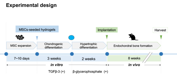
Figure 1: Experimental design. The first step of this protocol is to encapsulate expanded MSCs in HA hydrogels to promote chondrogenic and hypertrophic differentiation in vitro. Constructs are cultured in chondrogenic differentiation conditions for 3 weeks, followed by an additional 2 weeks in hypertrophic differentiation conditions. The second step of this protocol is to subcutaneously implant the constructs into nude mice at week 5 of in vitro culture to undergo endochondral ossification in vivo for 8 weeks. Please click here to view a larger version of this figure.
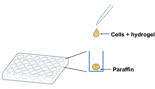
Figure 2: Preparation of MSCs-seeded hydrogels. Drop modified HA/crosslinker solution containing MSCs onto a paraffin-coated 24-well plate and allow to solidify at 37 °C for 30 min. Please click here to view a larger version of this figure.
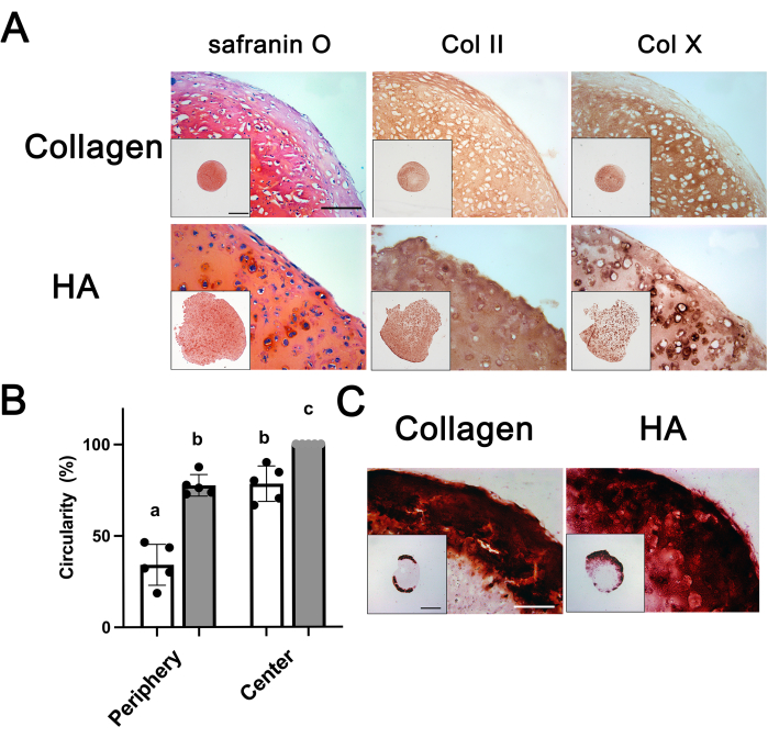
Figure 3: Histological and immunohistochemical analysis of HA and collagen constructs after in vitro culture22. (A) Constructs at 3 weeks (3W) of in vitro culture were stained for sGAG (safranin O/fast green), type II collagen (Col II), and type X collagen (Col X). (B) The average circularity values of cells at 3 weeks of in vitro culture (n = 5). Open and grey bars indicate collagen and HA constructs, respectively. Groups without a common letter are statistically different (a versus b, p<0.01; b versus c, p<0.05). (C) Constructs at 5 weeks (5W) of in vitro culture were stained for calcium (alizarin red). All pictures were captured with the same magnification (Scale bar: 200 µm). A low-magnification overview of the entire tissues is shown in the insets (Scale bar: 1 mm). Error bars represent mean ± standard deviation (SD). The one-way ANOVA followed by Tukey's multiple comparisons test was used to determine significant differences between groups. This figure has been modified from38. Please click here to view a larger version of this figure.
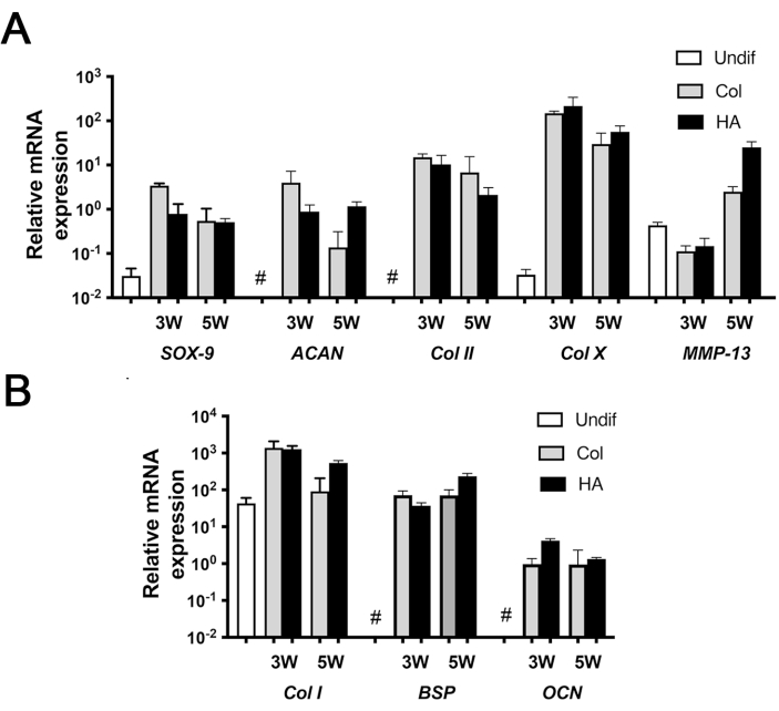
Figure 4: Gene expression analysis of HA and collagen constructs at 3 and 5 weeks of in vitro culture. (A) Chondrogenic and hypertrophic markers. (B) Osteogenic markers. Values are presented as mean ± standard deviation (SD) (n = 3). Undifferentiated MSCs expressed high levels of Col I and MMP-13 and low levels of Sox-9 and Col X; they did not express (#) ACAN, Col II, BSP, and OCN. Error bars represent mean ± standard deviation (SD). This figure has been modified from38. Please click here to view a larger version of this figure.
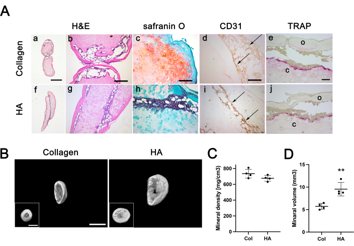
Figure 5: Immunohistochemical analysis and calcification of HA and collagen constructs post-implantation at 8 weeks. (A) Constructs were stained for Hematoxylin and Eosin (H&E, a, b, f, and g), safranin O/Fast Green (c and h), endothelial cells (Cluster of Differentiation 31, CD31) (d and i), and Tartrate-resistant acid phosphatase (TRAP, e and j). (a and f) Overview of the entire tissues (scale bar = 500 µm). H&E, safranin O, CD31: scale bar = 200 µm; TRAP: scale bar= 50 µm. Arrows indicate CD31-positive endothelial cells. Abbreviations: c = inner cartilage tissue; o = outer osteoid tissue. (B) MicroCT (µCT) imaging of collagen and HA constructs (main and inset image scale bars = 2 mm). (C,D) The total mineral density (C) and volume (D) of HA and collagen constructs (n = 4). ** indicates significant differences; p <0.01. Four constructs were analyzed per group using µCT. Error bars represent mean ± standard deviation (SD). Student's t-test was used to determine significant differences between groups. This figure has been modified from38. Please click here to view a larger version of this figure.
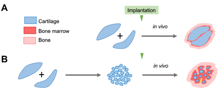
Figure 6: Strategy for facilitating ECOs using HA-based artificial cartilage. Schematic diagram of ossification and bone marrow development without and with fragmentation (µ-pellets) of implanted HA constructs. (A) HA hydrogels have an excellent fusion tendency to form an integrated bone with vascularization and marrow development between fused grafts in vivo. The resulting implanted tissue, however, remained predominantly in a cartilage state even 8 weeks post-implantation. (B) Given the tendency of HA constructs to fuse to form integrated bone, processing HA constructs into µ-pellets may accelerate the remodeling rate of transplanted tissue and promote bone formation. Micronizing the constructs increases surface area, which may promote the resorption of cartilage tissue and the formation of bone tissue and may also promote angiogenesis and bone marrow development because of the increased space for host cell infiltration. Please click here to view a larger version of this figure.
Hydrogel | Experiments | Adhered | United | % United |
Collagen | 5 | 3 | 2 | 40 |
HA | 5 | 5 | 5 | 100 |
Table 1: Unification rate when multiple constructs were implanted in a single pocket. Two or three constructs were subcutaneously implanted in a single pocket. Constructs were harvested 4- or 8-weeks post-implantation and histologically examined.
Using appropriate scaffold materials that promote the transition from hypertrophic cartilage to bone is a promising approach to scale up MSC-based engineered hypertrophic cartilage grafts and treat bone defects of clinically significant size. Here, we show that HA is an excellent scaffold material to support the differentiation of MSC-based hypertrophic cartilage tissue in vitro and to promote endochondral bone formation in vivo38. Furthermore, in vivo, HA constructs were shown to facilitate the fusion of multiple constructs implanted in the same pocket to form a single integrated bone. Because the interaction of MSCs with scaffold materials significantly affects cartilage and hypertrophic chondrogenesis, selecting suitable scaffold material for ECOs is important for generating clinically effective tissue-engineered cartilage grafts. Cells embedded in HA hydrogels uniformly exhibit a rounded morphology characteristic of chondrocytes from the center to the periphery, indicating that hyaluronan provides an environment conducive to uniform cartilage formation throughout the hydrogel21,30. Unlike HA, collagen interacts with MSCs via integrin binding sequences (RGDs); however, the persistence of this interaction prevents MSCs from further differentiating in the chondrogenic pathway43, which may account for the non-typical chondrocyte cell morphology that is prominent in the periphery of collagen constructs.
If multiple grafts unite to form a single bone, this ECO-mediated approach can be extended to more clinically relevant bone defects. When multiple MSC-based and scaffold-free hypertrophic cartilage were applied to a single bone defect, it was reported that the cartilage grafts integrated with the bone defect. However, adjacent grafts did not integrate with each other22. The approach presented here may offer one possible solution to overcome this limitation. Compared to collagen constructs, multiple grafts of HA constructs had a greater tendency to fuse to form a single bone. Furthermore, bone marrow was formed between fused HA constructs, which may be related to the fact that HA supports uniform cartilage differentiation throughout the hydrogel because hypertrophic cartilage secretes VEGF and provides an environment for the hematopoietic stem-cell niche in bone marrow development 44.
The key to a successful experiment using this protocol is the quality of MSCs. Confirming that the MSCs to be used have sufficiently high chondrogenic differentiation potential in advance is recommended. Second, the size of each construct should not be too large. In our experience, the volume of a single construct is limited to 10-15 µL. Larger constructs may result in insufficient oxygen and nutrient penetration into the constructs, leading to necrosis and poor differentiation in the center of the constructs. Therefore, gel thickness needs to be reduced to improve oxygen and nutrient penetration. In order to reduce gel thickness, it may be helpful to create thin gels on the porous membrane of Transwell inserts14,45. Alternatively, because HA constructs tend to fuse and form integrated bone, it may be adequate to implant many smaller constructs rather than a single large construct, as discussed below.
Limitations of this study include the need to accelerate the remodeling rate of the implanted tissues to facilitate bone formation.The implanted engineered tissue remained primarily in a hypertrophic calcified cartilage state even 8 weeks post-implantation. In bone formation via the ECO pathway, host-derived cells play a critical role in promoting ECO16,46. Providing additional channels in hypertrophic cartilage grafts has been reported to promote vascular invasion and graft mineralization in vivo20. Such channels serve as conduits for the infiltration of host cells, including osteoblasts, osteoclasts, and hematopoietic precursors, to form new tissue within the construct. Alternatively, it has been reported that multiple small fibrin-encapsulated pellets (µ-pellets) composed of chondrogenic differentiated MSCs can be implanted to form integrated bone47. Processing HA constructs into µ-pellets could increase the surface area surrounded by TRAP-positive osteoclasts, thus promoting the remodeling rate of cartilage tissue and enhancing osteogenesis (Figure 6)21,48. Furthermore, implantation of µ-pellets would increase the infiltration space of host cells, thereby facilitating angiogenesis and bone marrow development and generating integral bone formation with abundant vascularization. Thus, considering the tendency of HA constructs to fuse and create integrated bone, this µ-constructed bone regeneration strategy may accelerate the ECO process of hypertrophic cartilage grafts using HA hydrogels.
In conclusion, HA hydrogels are effective in scaling up tissue-engineered grafts with fewer cells and gels than collagen hydrogels. In addition, HA hydrogels have excellent fusion sensitivity and produce integrated bone with vascularization and marrow development between fused grafts. Therefore, modification of HA constructs to promote host cell infiltration and graft remodeling may lead to the development of implantable materials that can further promote vascularization and bone marrow development. With further optimization, this approach could be a promising alternative to current bone therapies in maxillofacial and orthopedic surgery.
The authors have nothing to disclose.
This work was supported by a Grant-in-Aid for Scientific Research (KAKENHI) from the Japan Society for the Promotion of Science (JSPS) (grant nos. JP19K10259 and 22K10032 to MAI).
0.25w/v% Trypsin-1mmol/L EDTA.4Na Solution | FUJIFILM Wako Pure Chemical | 209-16941 | |
Antisedan | Nippon Zenyaku Kogyo | ||
ascorbate-2-phosphate | Nacalai Tesque | 13571-14 | |
Bambanker | GC Lymphotec | CS-02-001 | |
basic fibroblastic growth factor | Reprocell | RCHEOT002 | |
bovine serum albumin | FUJIFILM Wako Pure Chemical | 012-23881 | 7.5 w/v% |
Countess Automated Cell Counter with cell counting chamber slides and Trypan Blue stain 0.4% | Invitrogen | C10283 | |
dexamethasone | Merck | D8893 | |
Domitor | Nippon Zenyaku Kogyo | ||
Dormicum | Astellas Pharma | ||
Dulbecco's Modified Eagle Medium | Merck | D6429 | high glucose |
Dulbecco's Modified Eagle's Medium/Nutrient Mixture F-12 Ham | Merck | D6421 | |
Fetal bovine serum | Hyclone | SH30396.03 | |
Gentamicin sulfate | FUJIFILM Wako Pure Chemical | 1676045 | 10 mg/mL |
Haccpper Generator | TechnoMax | CH-400-5QB | 50 ppm hypochlorous acid water |
Human Mesenchymal Stem Cells | Lonza | PT-2501 | |
HyStem Cell Culture Scaffold Kit | Merck | HYS020 | |
IL-1ß | PeproTech | AF-200-01B | |
ITS-G supplement | FUJIFILM Wako Pure Chemical | 090-06741 | ×100 |
L-Alanyl-L-Glutamine | FUJIFILM Wako Pure Chemical | 016-21841 | 200mmol/L (×100) |
L-proline | Nacalai Tesque | 29001-42 | |
L-Thyroxine | Merck | T1775 | |
MSCGM Mesenchymal Stem Cell Growth Medium BulletKit |
Lonza | PT-3001 | |
paraffin | FUJIFILM Wako Pure Chemical | 165-13375 | |
PBS / pH7.4 100ml | Medicago | 09-2051-100 | |
TGF-β3 | Proteintech | HZ-1090 | |
Vetorphale | Meiji Seika Kaisha | ||
Visiocare Ointment | SAVAVET/SAVA Healthcare | ||
β-glycerophosphate | FUJIFILM Wako Pure Chemical | 048-34332 |