The protocol presents a new tool to simplify intravital imaging using inverted confocal microscopy.
Understanding normal and aberrant in vivo cell behaviors is necessary to develop clinical interventions to thwart disease initiation and progression. It is therefore critical to optimize imaging approaches that facilitate the observation of cell dynamics in situ, where tissue structure and composition remain unperturbed. The epidermis is the body's outermost barrier, as well as the source of the most prevalent human cancers, namely cutaneous skin carcinomas. The accessibility of skin tissue presents a unique opportunity to monitor epithelial and dermal cell behaviors in intact animals using noninvasive intravital microscopy. Nevertheless, this sophisticated imaging approach has primarily been achieved using upright multiphoton microscopes, which represent a significant barrier for entry for most investigators. This study presents a custom-designed, 3D-printed microscope stage insert suitable for use with inverted confocal microscopes, streamlining the long-term intravital imaging of ear skin in live transgenic mice. We believe this versatile invention, which may be customized to fit the inverted microscope brand and model of choice and adapted to image additional organ systems, will prove invaluable to the greater scientific research community by significantly enhancing the accessibility of intravital microscopy. This technological advancement is critical for bolstering our understanding of live cell dynamics in normal and disease contexts.
Intravital microscopy is a powerful tool that allows the monitoring of cell behaviors in their unperturbed in vivo environments. This unique method has provided key insights into the inner workings of complex mammalian organ systems, including the lung1, brain2, liver3, mammary gland4, intestine5, and skin6. Furthermore, this approach has revealed cell behavioral alterations during tumor development7, wound healing8,9, inflammation10, and other diverse pathologies in situ. In this study, we focus on enhancing the accessibility of intravital microscopy to image live epithelial and stromal dynamics in intact mouse skin. Understanding cell behaviors in mammalian skin is of high clinical importance due to the remarkable regenerative and tumorigenic capacity of this tissue.
Intravital imaging in mice has been primarily performed using upright multiphoton microscopes due to their ability to provide high-resolution imaging at tissue depths >100 µm11,12. Nevertheless, these instruments lack the workhorse versatility and more general accessibility of inverted confocal microscopes, which are more user-friendly and cost-effective, provide the ability to image cultured cells, do not require complete darkness during image acquisition, and are generally safer, among other notable advantages13,14. In this study, we present a new tool that significantly enhances intravital imaging accessibility by adapting this approach for inverted confocal microscopes.
Here, we present a 3D-printed custom stage insert design that incorporates several key features to facilitate stable, long-term intravital imaging of mouse ear skin on an inverted confocal microscope (Figure 1, Figure 2, Figure 3, Figure 4, and Figure 5). These specialized features include an offset objective hole that allows the full body of an adult mouse to lay entirely flat during imaging. This minimizes the vibrational interference of mouse body movements on imaging and eliminates the need to administer ketamine and xylazine to dampen breathing, a practice often coupled with intravital imaging6. In addition, corner brackets on the insert correctly position an isoflurane nose cone to align with the face of the mouse, a metal ear clip immobilizes the mouse ear to a custom-built coverslip disk, and an optional detachable closed-loop biofeedback heat plate lies flush within the insert to support the mouse body temperature during long imaging sessions. The custom coverslip disk, which provides a flat surface essential for the mouse head and ear to lay flat, was generated in a machine shop by removing the walls of a generic coverslip-containing cell culture dish. The use of a 40x silicone oil immersion lens (1.25 numerical aperture [N.A.], 0.3 mm working distance) in conjunction with the coverslip disk and custom stage insert provides high resolution images >50 µm deep into the ear dermis.
To test the functionality of this new inverted microscope stage insert, we captured z-stacks spanning all epidermal epithelial layers over a 3 h time course in the ear of a live transgenic K14-H2B-mCherry15 adult mouse (epithelial nuclei in this mouse line contain a red fluorescent label) (Figure 6A–A'). We also captured z-stacks spanning several fibroblast layers within the skin dermis over a 3 h time course in the ear of a live transgenic Pdgfra-rtTA16; pTRE-H2B-GFP17 adult mouse (fibroblast nuclei in this mouse line contain a green fluorescent label following doxycycline induction) (Figure 6B–D'). Our high-resolution data demonstrate consistent stability by lack of drift in the x-, y-, and z-planes, thus proving the effectiveness of this new intravital imaging tool for use on inverted microscopes. Importantly, the dimensions of this 3D-printed stage insert can be adjusted, as described in Supplementary File 1, Supplementary File 2, and Supplementary File 3, to fit any inverted microscope, and the positioning of the objective opening can be moved to alternative locations within the insert to better suit imaging a particular tissue and/or animal model of interest. This invention can thus empower individual laboratories, or investigators with core facility confocal access, to adapt this tool for their unique intravital imaging needs, thereby streamlining evaluation of diverse in vivo cell biology.
This research was performed in compliance with Emory University and Atlanta Veterans Affairs Medical Center animal care and use guidelines and has been approved by the Institutional Animal Care and Use Committee (IACUC).
1. Installing the live imaging insert on the inverted microscope stage
2. Isoflurane configuration and mouse prep
3. Mouse placement on the insert for intravital imaging
4. Termination of imaging
Proper assembly of the live imaging insert on an inverted confocal microscope and appropriate orientation of a transgenic mouse atop the insert is validated by acquiring z-stacks of fluorescently-labeled, live ear tissue over a time course ≥1 h with minimal evidence of drift in the x-, y-, and z-axes. Images should be captured at consistent intervals (interval time will depend on the biological question, strength of fluorescence signal, etc.) so that cell dynamics and image drift can be tracked over time. Throughout the time course, monitoring individual z-planes to ensure they remain in focus reveals whether animal movement interferes with imaging stability. An example of single z-planes remaining in focus over an extended time course using the live imaging insert is depicted in Figure 6.
Images from four 60 min time points displayed in Figure 6A' were selected from a 3 h time-lapse movie of mCherry+ epidermal cells in the ear of a 3-month-old adult male K14-H2B-mCherry mouse (~30 g) captured at 2 min intervals using a z-step of 0.246 µm to achieve Nyquist sampling across a total z-depth of 24 µm (99 z-stack images acquired per time point).
Images from four 60 min time points displayed in Figure 6D' were selected from a 3 h time-lapse movie of GFP+ dermal fibroblasts in the ear of an 8-month-old adult female Pdgfra:rtTA; pTRE:H2B-GFP mouse (~30 g; Figure 6B). These were captured at 5 min intervals using a z-step of 2 µm to achieve Nyquist sampling across a total z-depth of 54 µm (28 z-stack images acquired per time point). This mouse was treated with 2 mg of doxycycline every other day for 6 days (four treatments, 8 mg total) prior to imaging (Figure 6C).
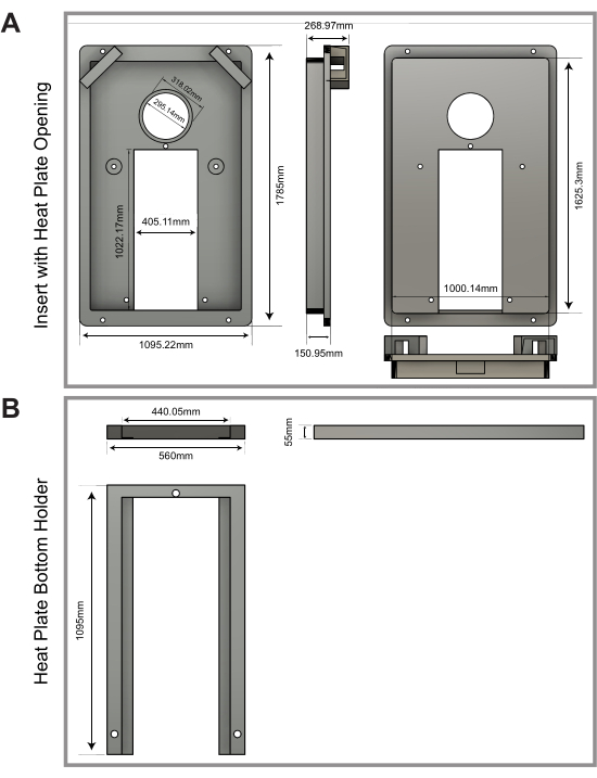
Figure 1: Customized 3D-printed stage insert design. (A,B) Design and dimensions of a custom, 3D-printed insert with a heat plate opening (A), as well as a heat plate bottom holder (B), which is printed separately and then screwed into the insert (see corresponding Supplementary File 1, Supplementary File 2, and Supplementary File 3). Please click here to view a larger version of this figure.
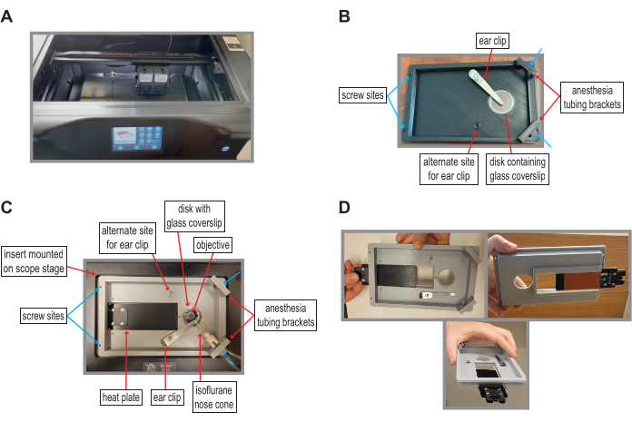
Figure 2: New stage insert streamlines intravital imaging on inverted confocal microscopes. (A) Insert being constructed using a 3D printer. (B) Simple insert model without a heating device; the live imaging insert contains four screw sites (blue arrows) for microscope stage attachment. The metal ear clip flattens and immobilizes the ear onto a 35 mm wide plastic disk containing a 20 mm wide glass coverslip. The insert contains two options for ear clip placement to provide flexibility with mouse orientation. Asymmetric placement of the objective hole allows the adult mouse to lay flat. Side brackets align and immobilize the isoflurane nose cone to facilitate mouse attachment. The simplified model requires the placement of a small heating pad (or alternative heat source) under the mouse to help regulate body temperature. (C) Advanced insert model with a built-in heat plate. (D) The heat plate is installed by sliding into a grooved opening of the insert. Please click here to view a larger version of this figure.
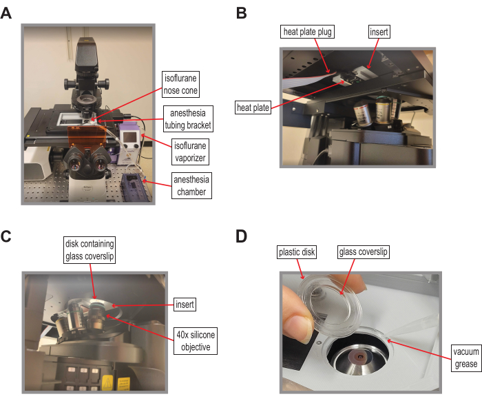
Figure 3: Intravital imaging insert installed on an inverted microscope stage. (A) Insert mounted on the stage of a laser scanning inverted confocal microscope. Proximity of the isoflurane vaporizer and chamber allows threading of the nose cone tubing through the insert bracket. (B) The heat plate plug extends below the microscope stage to connect to the controller. (C) The insert hole aligns with a 40x silicone objective. (D) A plastic disk (35 mm diameter) containing a glass coverslip (20 mm diameter) is laid atop the grooved opening of the stage insert and sealed in place with vacuum grease. The coverslip disk was created by removing the walls of a 35 mm x 10 mm glass-bottom cell culture dish. Please click here to view a larger version of this figure.
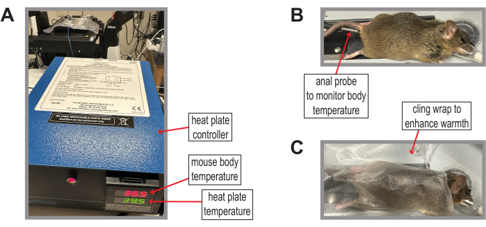
Figure 4: Monitoring animal body temperature using a heat plate controller. (A) Heat plate controller, which can be adjusted to stabilize mouse body temperature at the optimal 36 °C throughout the intravital imaging session. (B) An anal probe is used to monitor mouse body temperature once the mouse is laid atop the heat plate. (C) Plastic cling wrap can be used to trap heat to further elevate mouse body temperature. Please click here to view a larger version of this figure.
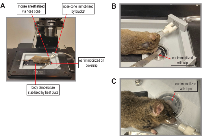
Figure 5: Mouse positioning on the intravital imaging insert. (A) The mouse body is spread along the top of the heat plate, with the ear centered onto the glass coverslip and immobilized with a metal ear clip. The bracket positions the isoflurane nose cone for mouse attachment. (B) Zoomed-in region of (A) showing mouse ear immobilization with a metal ear clip on the glass coverslip and the isoflurane nose cone attachment to the mouse. (C) Tape can be used as an alternative method of ear immobilization onto the glass coverslip. Please click here to view a larger version of this figure.
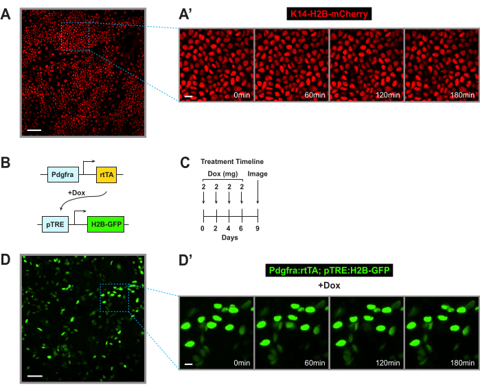
Figure 6: Custom insert facilitates stable long-term intravital imaging of mouse ear epidermis and fibroblasts. (A) A single z-plane captured from performing intravital imaging on the ear epidermal epithelium of a 3-month-old adult male K14-H2B-mCherry transgenic mouse. The dotted box indicates the zoomed-in region shown in (A'). Scale bar = 50 µm. (A') Zoomed-in region of (A) showing images every hour over a 3 h movie. Scale bar = 10 µm. (B) Schematic of the doxycycline-inducible transgenic system used to promote in vivo GFP labeling of dermal fibroblast nuclei. (C) Timeline of doxycycline injections. (D) Maximum intensity projection (representing a 54 µm total z-depth) of dermal fibroblasts captured by performing intravital imaging on the ear of a dox-injected 8-month-old female Pdgfra:rtTA; pTRE: H2B-GFP transgenic mouse. The dotted box indicates the zoomed-in region shown in (D'). Scale bar = 50 µm. (D') Zoomed-in region of (D) showing images every hour over a 3 h movie. Scale bar = 10 µm. These time courses demonstrate the stability of long-term intravital imaging using the 3D-printed custom insert. Please click here to view a larger version of this figure.
Supplementary File 1: Design file for the 3D-printed insert with a heat plate opening. Please click here to download of this File.
Supplementary File 2: Design file for the 3D-printed heat plate bottom holder. Please click here to download of this File.
Supplementary File 3: Design file for the 3D-printed insert without a heat plate opening. Please click here to download of this File.
In this study, we present a new tool that facilitates stable, long-term intravital imaging of intact mouse skin epithelia on inverted confocal microscopes. This invention is made of PLA, which is the most common and inexpensive 3D-printable material; all in-house 3D-printing costs for this insert amount to <$5. The two separate insert pieces (Figure 1, Supplementary File 1, and Supplementary File 2) can be easily assembled using set screws (see Table of Materials). Notably, the provided .stl files can also be used to order this insert by commercial means. An additional option is to use a computer numerical control (CNC) machine to generate the insert out of anodized aluminum, although this is significantly more costly.
To ensure reliable and efficient imaging over a duration of time using this new tool, it is critical to properly size the live imaging insert according to the user's microscope stage. The provided Supplementary File 1, Supplementary File 2, and Supplementary File 3 can be adapted to reflect the appropriate insert dimensions that are compatible with the microscope of choice prior to 3D-printing. Accurate insert dimensions with an immobilized coverslip disk minimize positional (x/y) and focal (z) drift throughout each imaging session. It is also important to ensure that the insert depth is sufficient to pass the heat plate plug port underneath the stage.
Prior to imaging, it is essential to confirm that the mouse is fully anesthetized, its body temperature remains stable at ~36 °C as measured with the anal thermometer probe, and the ear is firmly immobilized to avoid movement due to breathing. When using the metal ear clip to secure the ear to the coverslip, one should prevent cutting off normal blood circulation by ensuring that the ear clamp is not screwed down too tightly. It is also crucial to monitor and replenish eye ointment on the mouse when necessary to maintain ocular moisture during long imaging sessions.
While this unique insert design provides a new approach for intravital imaging, it has a few notable limitations. Due to the insert's low positioning in the microscope stage, stage movement in the x- and y-planes should be very limited after the objective is positioned and the coverslip disk is sealed into place. This limited movement is critical to avoid damage to the objective. Furthermore, it should be noted that the glass coverslip disk is not currently commercially available. To recreate the coverslip disk used in this study, collaboration with a machine shop may be required.
While we demonstrate that this confocal-based method can achieve stable long-term imaging deep into intact tissue, it should be noted that multiphoton microscopes cause less photodamage and penetrate to greater depths compared with confocal instruments19. Therefore, the utility of this tool relies on the strength of the fluorescence signal in the transgenic animal model of choice, as well as the tissue depth required to achieve the experimental goals. We therefore recommend using the lowest laser power possible to minimize photobleaching while achieving the desired resolution. It is also important that the objective lens used with this insert has a high N.A. for optimal resolution, as well as a long working distance so it can reach the coverslip disk. It should be noted that this approach is not meant to replace the well-validated upright multiphoton-based intravital imaging method described in Pineda et al.6. Instead, this new tool is intended to provide an effective alternative to labs that do not have access to multiphoton equipment, prefer a less cumbersome system, and/or own an inverted microscope.
Our new tool is remarkably customizable to individualized experimental requirements and preferences. This insert can be used with or without a heat plate, as some alternative options include the placement of a thin heating pad underneath the mouse, wrapping an objective lens warmer around its torso, and/or trapping heat by covering the mouse with plastic cling wrap (Figure 4C). Furthermore, tape may be used in conjunction with or in place of the ear clip to provide optimal tissue immobilization (Figure 5C). The orientation of the stage insert is reversible, so the user can decide whether it's optimal to orient the objective hole on the right or left side. Furthermore, the insert has built-in alternative placement options for the ear clip and isoflurane tubing to provide maximal flexibility based on the desired mouse orientation (imaging the right vs. left ear), location of isoflurane setup, and specific microscope configurations (Figure 2C). The insert is easily installable and removable, with a simplified design intended to be highly user-friendly so that intravital imaging can be made approachable to even novice microscopists. Additionally, the asymmetrically-localized objective hole provides the capacity to image diverse animal models as well as organs of varying sizes.
We intend for this invention to enhance the application of intravital microscopy within individual laboratories as well as microcopy cores that contain inverted confocal instruments. This highly accessible and customizable tool will provide investigators the freedom to visualize live cell dynamics across diverse organ systems to reveal significant cell biological insights.
The authors have nothing to disclose.
We thank Valentina Greco for the K14-mCherry-H2B mice. We are grateful to the Emory University Physics Department Machine Shop for generating the glass coverslip disks. This work was funded by Career Development Award #IK2 BX005370 from the US Department of Veterans Affairs BLRD Service to LS, NIH Awards RF1-AG079269 and R56-AG072473 to MJMR, and I3 Emory SOM/GT Computational and Data Analysis Award to MJMR.
3D Printer | Qudi Tech | i-Fast | 3D prints using PLA material |
40x 1.25NA silicone objective lens | Nikon | ||
AxR Laser Scanning Confocal Microscope | Nikon | ||
Cotton Tipped Swab | VWR | 76337-046 | Cream/ointment application |
Doxycycline hyclate | Sigma-Aldrich | D9891 | Induces GFP labeling of fibroblast nuclei in Pdgfra-rtTA; pTRE-H2B-GFP mice |
Flathead Screwdriver (2.5 mm) | Affiix insert to microscope stage | ||
Flathead Screws x 4 (#6-32) | Nikon | Screw insert into microscope stage | |
Glass Bottom Culture Dish | chemglass Life Sciences | CLS-1811-002 | Modified by removing walls of dish for use as coverslip disk compatible with live insert; 35 mm wide disk contains 20 mm wide glass coverslip; dish walls were removed by machine shop |
Heat Plate controller | Physitemp | TCAT-2LV | Animal Temperature Controller – Low Voltage; anal prob attachment for mouse body temperature monitoring |
Hex Wrench (1.5 mm) | For M3 setscrew adjustments | ||
Hex Wrench (2.5 mm) | Adjust tension on metal ear clip | ||
Intravital Imaging Insert | |||
Isoflurane | Med-Vet International | HPA030782-100uL | Mouse anesthesia |
Labeling Tape (or Scotch Tape) | VWR | 10127-458 | Alternative to metal ear clip to immobilize ear to coverslip |
Metal fastener | used as ear clip | ||
Mouse: C57BL/6-Pdgfraem1(rtTA)Xsun/J | The Jackson Laboratory | RRID: IMSR_JAX:034459 | Fibrroblast-specific promoter driving doxycycline-inducible rtTA expression |
Mouse: K14-H2BPAmCherry | Courtesy of Dr. Valentina Greco at Yale University | Labels epidermal epithelial cell nuclei with mCherry; referred to in text as "K14-H2B-mCherry" | |
Mouse: pTRE-H2B-GFP: STOCK Tg(tetO-HIST1H2BJ/GFP)47Efu/J |
The Jackson Laboratory | RRID: IMSR_JAX:005104 | Labels fibroblast nuclei with GFP when combined with Pdgfra-rtTA and induced with doxycycline |
Multipurpose Sealing Wrap | Glad | Enhance mouse warmth | |
Optixcare | VWR | MSPP-078932779 | Eye lubricant |
Set screws x 3 (M3; 6 mm) | Thorlabs | SS3M6 | Attachment for heatplate module |
Silicone Immersion Oil | Applied to 40x silicone objective | ||
Small Animal Heating Plate | Physitemp | HP-4M | Provides heat to animal |
Somnoflow Low-Flow Electronic Vaporizer | Kent Scientific | SF-01 | Mouse anesthesia |
Vacuum Grease | Flinn Scientific | AP1095 | Seals coverslip disk to insert |
Veet | hair removal | ||
Water circulating heat pad | Stryker Medical | TP700 | for mouse revival post-imaging |