Based on the safety and feasibility, this article presents an early weight-bearing rehabilitation protocol after anterior cruciate ligament reconstruction. The protocol is clear and easy to operate, which is helpful to promote its use in clinical practice and accelerate the functional recovery of patients.
Anterior cruciate ligament (ACL) injury is one of the common sports injuries. Anterior cruciate ligament reconstruction (ACLR) is the mainstream treatment for ACL injury, aiming to regain normal anatomical structure and stability of the knee joint and promote the patient’s return to sports. Under the guidance of the concept of enhanced recovery after surgery, early weight-bearing rehabilitation (EWB) is an important factor affecting patient function and quality of life. However, there is no consensus on whether EWB rehabilitation can be performed after ACL surgery.
This study aims to explore the safety and feasibility of EWB after ACL surgery. The study implemented a gradual EWB rehabilitation program in the experimental group, including weight-shifting training, balance training, and gait training on the affected lower limb, and assessed wound healing and stability of the knee joint. The study found that EWB after ACLR is safe and feasible. EWB rehabilitation not only does not pose a negative effect on the patient’s knee pain, swelling, wound healing, and stability, but also helps to improve knee active flexion and quality of life faster and better. The EWB program in this study is simple, safe, and effective, and it provides strong theoretical guidance and practical demonstration for accelerated rehabilitation after ACLR.
Enhanced Recovery after Surgery (ERAS) is a concept that promotes the early initiation of postsurgery rehabilitation care units, particularly after orthopedic operations, when conditions are appropriate1. This approach seeks to shift the clinical focus from treating the disease to functional rehabilitation promptly with the goal of reducing hospital stays, minimizing postoperative complications, improving patient prognosis and satisfaction, and enhancing overall rehabilitation outcomes. Since the introduction of ERAS, Chinese orthopedic rehabilitation specialist, Zhou Mouwang, has advocated for the active application of accelerated recovery protocols to further progress perioperative rehabilitation practices within the realm of Chinese orthopedics2. Internationally recognized consensus guidelines have also been established to facilitate enhanced surgical recovery for orthopedic conditions such as joint replacement and spine surgery3. Nonetheless, while considerable advancements have been made in implementing ERAS in recent years, the primary application has been in major operations, such as joint replacement surgeries4. As a result, there is a pressing need to further investigate and expand the application of ERAS within the rehabilitation of other orthopedic conditions.
The anterior cruciate ligament (ACL) plays a pivotal role in maintaining knee joint stability. ACL injuries are among the most prevalent sports-related injuries, with approximately 2 million cases occurring worldwide annually5. Anterior cruciate ligament reconstruction (ACLR) is the mainstream treatment approach, aiming to restore normal knee anatomy and stability, prevent secondary injuries, and enable patients’ return to sport. Rehabilitation following ACL injury has been an active research area. It is widely accepted that post ACLR rehabilitation is integral for optimizing surgical outcomes and facilitating patients’ recovery of sports-related function6,7. However, early weight-bearing (EWB) rehabilitation protocols after ACLR remain understudied, leading to a lack of consensus among clinicians.
The latest research in 2021 supports allowing immediate weight-bearing and full knee range of motion within tolerance after ACL autograft reconstruction surgery7. However, a consensus has not yet been reached in clinical practice. The authoritative British Journal of Sports Medicine published in 2016 also advocates for immediate weight-bearing post ACLR, finding it does not increase knee joint laxity. If gait is proper without increased pain or effusion during/after walking, immediate weight-bearing is considered acceptable6. In contrast, a review in 2022 summarizes that accelerated weight-bearing rehabilitation following ACLR may increase risks of knee joint laxity and bone tunnel widening compared to delayed weight-bearing protocols, advising cautious selection of postoperative regimens8. Additional concerns with EWB include knee pain, swelling, effusion, wound healing, and fall risks after ACLR. Although substantial literature endorses EWB after ACLR, clinical practices still vary. A study in 20209 found that surgeon experience influences post ACLR weight-bearing timelines in France; more experienced surgeons were more likely to favor EWB rehabilitation.
Under the guidance of the ERAS concept, EWB is an important factor affecting patient function and quality of life. However, whether the EWB program could impair wound healing, increase pain and swelling, or impact knee stability remains a key concern for orthopedic and rehabilitation clinicians. In view of the current dilemma of clinicians and inconsistent research results, this study aims to explore the safety and feasibility of EWB after ACLR.
This protocol has been approved by the Ethics Committee of Shanghai First People's Hospital (project number: 2022SQ470).
1. Study design
2. Participant recruitment (see Figure 1)
NOTE: The research team selected 38 patients who underwent ACLR at Shanghai First People's Hospital from July 2022 to November 2022 based on the inclusion and exclusion criteria. They were divided into experimental and control groups with 19 participants. General demographic information and clinical indicators, including gender, age, graft type, degree of meniscal repair, pain intensity, and wound healing status, were collected.
3. Rehabilitation treatment plan
4. Outcome assessments
NOTE: The evaluations are completed by rehabilitation therapists on the 2nd, 7th, and 14th day after surgery. The primary outcome measures include wound healing, stability of knee joint, pain, and edema of the knee.
This study included 38 patients, all of whom completed the 2-week study (CG = 19, PG = 19). There were no significant differences in sex, age, surgical site, graft type, or degree of meniscus repair between the two groups (P > 0.05; see Table 1).
The intensity of pain of patients in EG on postoperative days 2, 7, and 14 was (2.2 ± 1.3), (1.1 ± 0.6), and (0.6 ± 0.5), respectively, indicating a gradual decrease in the intensity of pain. The intensity of pain was (2.5 ± 1.2), (1.5 ± 0.8), and (0.8 ± 0.5) in CG on postoperative days 2, 7, and 14, respectively, also indicating a gradual decrease in pain. DKE in both groups gradually decreased on postoperative days 2, 7, and 14 but there were no significant differences in pain intensity or DKE between the two groups (P > 0.05; see Table 2). All patients had good knee stability during rehabilitation, with good wound healing. None had positive anterior drawer tests, knee giving way, or falls.
AKROM in EG at 2, 7, and 14 days after surgery were (70.5 ± 17.0), (83.4 ± 13.5), and (98.1 ± 13.0), respectively, indicating gradual improvement in AKROM. AKROM in CG was (56.8 ± 13.1), (74.7 ± 12.9), and (87.0 ± 10.6), respectively, also indicating gradual improvement in AKROM. The ADL in EG on postoperative days 2, 7, and 14 were (72.8 ± 8.9), (85.7 ± 5.8), and (94.2 ± 1.9), respectively, indicating that the patients' ability to perform ADL was gradually improving. The ADL in CG were (64.5 ± 9.1), (78.7 ± 6.6), and (89.7 ± 4.6), respectively, indicating the same improvement effect. There were significant differences between the two groups at different time points in AKROM and ADL scores (P < 0.05). The EG had greater flexion angles and ADL scores compared to CG at the same time points (see Table 3).
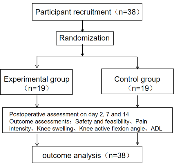
Figure 1: Schematic diagram of the protocol. The schematic diagram of the protocol includes the sample size, grouping, evaluation time, and outcome indicators of the research subjects. Please click here to view a larger version of this figure.
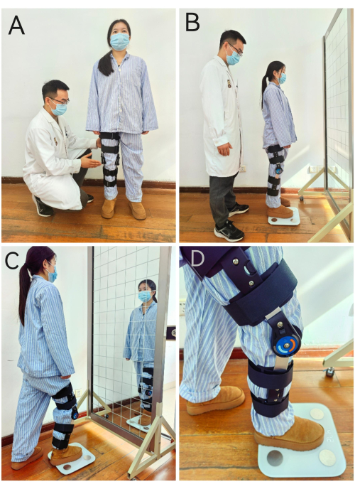
Figure 2: Early weight-bearing standing training. (A–D) The methods and steps of weight-bearing standing. Please click here to view a larger version of this figure.
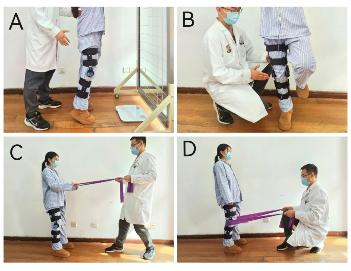
Figure 3: Early weight-bearing balance training. (A–D) Four different methods of balance training. Please click here to view a larger version of this figure.
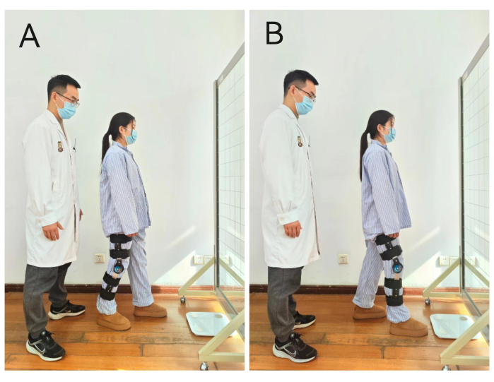
Figure 4: Early weight-bearing gait training. (A,B) Gait training methods with different foot support. Please click here to view a larger version of this figure.
EG (n=19) | CG (n=19) | P-value | |
Sex (M/F) | 16/3 | 14/5 | 0.426a |
Age (years) | 29.8 ± 9.4 | 33.5 ± 7.3 | 0.224b |
Affected limb (L/R) | 9/10 | 11/8 | 0.516a |
Graft type (ALL/ARL) | 9/10 | 4/15 | 0.087a |
Meniscus repair (N/M) | 11/8 | 10/9 | 0.744a |
Table 1: Basic characteristics. This table shows the sample size, gender, age, surgical site, graft type, and meniscus repair degree of the two groups. Data are presented as mean ± SD. Abbreviations: EG = Experimental group; CG = Control group; M = male; F = female; L = left; R = right; ALL =autologous ligament; ARL =artificial ligament; N = no repair; M = mild repair; Superscript a = χ2 test; "b" = t-test.
G | NRS | DKE | ||||||||||
Day 2 | Day 7 | Day 14 | Day 2 | Day 7 | Day 14 | |||||||
Mil | Mod | Sev | Mil | Mod | Sev | Mil | Mod | Sev | ||||
EG | 2.2±1.3 | 1.1±0.6 | 0.6±0.5 | 4 | 11 | 4 | 9 | 9 | 1 | 12 | 6 | 1 |
CG | 2.5±1.2 | 1.5±0.8 | 0.8±0.5 | 3 | 10 | 6 | 8 | 10 | 1 | 15 | 3 | 1 |
P-value | 0.850b | 0.059b | 0.198b | 0.744b | 0.946b | 0.513b |
Table 2: Pain intensity and degree of knee edema in two groups. This table records the intensity of pain and knee joint edema degree of the two groups at different time points. Data are presented as mean ± SD. Abbreviations: DKE = Degree of knee edema; EG = Experimental group; CG = Control group; Mil = Mild; Mod = Moderate; Sev = Severe; "b" = t-test.
G | AKROM (degree) | BI | ||||
Day 2 | Day 7 | Day 14 | Day 2 | Day 7 | Day 14 | |
EG | 70.5±17.0 | 83.4±13.5 | 98.1±13.0 | 72.8±8.9 | 85.7±5.8 | 94.2±1.9 |
CG | 56.8±13.1 | 74.7±12.9 | 87.0±10.6 | 64.5±9.1 | 78.7±6.6 | 89.7±4.6 |
P-value | 0.009b | 0.049b | 0.006b | 0.008b | 0.001b | 0.001b |
Table 3: Active knee flexion range of motion and daily living ability score of two groups after surgery. This table shows the angle of active knee flexion and daily living ability scores of the two groups at different time points. Data are presented as mean ± SD. Abbreviations: AKROM = Active knee flexion range of motion; BI = Barthel index; EG = Experimental group; CG = Control group.
This study protocol contains details of the EWB rehabilitation after ACLR. Its process is clear and not complicated, and because only simple rehabilitation equipment is needed for the implementation of the EWB rehabilitation program, it is clinically feasible and convenient. In addition, safety is an important consideration in this study protocol. The EWB rehabilitation program not only adds protective measures to prevent iatrogenic sports injury but also follows the step-by-step principle. Therefore, the results of this study show that the EWB rehabilitation program within 2 weeks after ACLR does not increase knee pain and edema and helps improve the active knee flexion angle and the ability for daily living of patients, demonstrating its safety and effectiveness.
In this study, there were no significant differences in pain intensity, knee swelling, or wound healing at 2, 7, and 14 days after surgery between EG and CG (Table 2). The pain intensity was mild in both groups, indicating that EWB after ACLR did not cause additional pain for the patients, exacerbate wound swelling, or affect wound healing. On the one hand, this may be related to postoperative analgesia measures. On the other hand, it is also related to scientific and standardized weight-bearing rehabilitation regimens. The EWB rehabilitation regimen in this study emphasizes full knee extension during weight-bearing standing and walking as much as possible within 2 weeks after surgery and allows activity within 30° of knee flexion when sensation is good. During walking, patients were required to concentrate attention, enhance overall exertion of the affected lower limb, activate the quadriceps as much as possible, improve knee stability, reduce traction and excessive stimulation on the wound caused by instability, and avoid aggravating the inflammatory response. Moreover, ACLR is a minimally invasive surgery with little trauma and good wound suturing. As patients are generally young with strong healing abilities, theoretically, EWB after surgery will not cause more trauma.
This study comprehensively assessed knee stability after ACLR through the anterior drawer test and sudden knee flexion during rehabilitation. No patients had positive anterior drawer tests or reported sudden knee flexion, suggesting that EWB rehabilitation after surgery is safe and feasible. Knee stability after ACLR is critical for graft safety. Classic previous studies show that the integrity and stability of the knee joint are maintained by the passive and active structures surrounding it. The ACL is considered the primary restraint to anterior tibial translation (ATT). Excessive ATT may place the ACL graft at risk of injury12.
Some studies13 show that partial weight bearing starts 1-2 weeks after ACLR, with full weight bearing even later. However, other studies believe that allowing immediate or even full weight bearing after ACLR does not affect knee stability and helps prevent complications14,15. Therefore, whether immediate weight-bearing rehabilitation after surgery leads to excessive anterior tibial translation, increasing graft tension and causing graft elongation, poor tendon-bone healing, and enlarged bone tunnels, has become a focus of debate. One study on healthy subjects found that in non-weight bearing conditions, knee flexion of 20° leads to increased ATT with increased quadriceps load, especially with sudden load increases, which may cause sudden rises in ACL tension16. Another important study on ACL-deficient subjects found that in 20° of knee flexion during weight-bearing, the degree of ATT was unrelated to the amount of weight borne, and ATT was minimal and significantly less than during the Lachman test and non-weight bearing17. EWB in this study emphasizes isometric contractions and weight-shifting exercises in full knee extension after surgery. One study18 also found that ATT during side-to-side and anterior-posterior weight-shifting exercises 2 weeks after ACLR did not differ from healthy controls. Although weight-shifting training started 2 weeks after surgery in that study compared to 1-2 days in this study, considering the incorporation process of autografts and allografts, graft strength is generally greatest at initial implantation19. Thus, there are reasons to believe that EWB in this study is less likely to cause excessive anterior tibial translation and graft elongation.
This study also found that EWB after ACLR increased patients’ AKROM and improved the quality of life (Table 3). The increased postoperative AKROM may be related to earlier activation of lower limb flexors and inhibition of antagonist muscle tension and spasms with EWB20. Weight-bearing activities also increased fluid flow, promoting joint fluid secretion and absorption, facilitating sliding between joint surfaces, and reducing blocking sensation during knee flexion21. The increased AKROM allows patients to choose more positions, improving convenience in daily life. Earlier out-of-bed weight-bearing rehabilitation increased patients’ activity area, enhancing recovery confidence, and reducing depression or anxiety caused by prolonged bed rest. This is also helpful in improving patients’ enthusiasm for participating in rehabilitation.
However, this study lacked a quantitative assessment of knee stability and long-term follow-up. Future studies should aim to increase sample size, lengthen follow-up duration, objectively quantify knee stability through instrumentation, and explore the effects of EWB rehabilitation on patients’ mid-to-long term knee joint stability. In summary, under ERAS guidance, EWB rehabilitation protocol after ACLR appears safe and feasible, which can help accelerate the functional recovery of patients and improve rehabilitation efficiency.
The authors have nothing to disclose.
This research was supported by two grants from the research project grant from Shanghai First People's Hospital (grant number: SYYG20221007) and the Medical Innovation Research Special Project in the Science and Technology Innovation Action Plan of the Shanghai Science and Technology Committee (grant number: 22Y11912100).
Elastic band | JOINFIT | 10722038422 | |
Electronic body weightometer | China's Xiaomi Technology (W0LONOW) | 100021480693 | |
Movable mirror | Guangzhou Compaq Medical Equipment Co. LTD | 10073735389717 | |
SPSS 21.0 | statistical analysis |