In this video, we perform the isolation of mononuclear cells from a mouse’s brain using density gradient centrifugation. The isolated mononuclear cell suspension can be used for further downstream analysis.
Protocol
All procedures involving animal models have been reviewed by the local institutional animal care committee and the JoVE veterinary review board.
1. Single-Cell Suspension Preparation from Brain
Dilute density gradient medium in 9:1 ratio with PBS in a 15 mL conical tube to yield a final 100% solution.
Anesthetize mice at the peak of EAE with an intraperitoneal injection of 1% sodium pentobarbital (50 mg/kg) and perfuse intracardially with 20 mL of sterile ice-cold PBS. Achieve this by slowly and steadily injecting PBS into the left ventricle of the heart using a 20 mL syringe and opening the right atrium.
Cut the cranium carefully from the nose to the neck, then remove the brain from the cranial box into 10 mL of RPMI in 50 mL conical tubes. Mix well to remove adherent red blood cells. Then, remove the medium by aspiration and add 10 mL of RPMI.
Place the brains and medium in a 100 mm dish. Finely chop with a razor blade.
Transfer 6 mL from the dish to an ice-cold 7 mL sintered glass homogenizer with a clean pipette. Avoid leaving large quantities of tissue in the pipette. A small amount is unavoidable.
Grind the brain using the "loose" plunger of the pestle first, then use the "tight" plunger until the suspension is homogeneous and pour into a prechilled 15 mL conical tube and keep on ice.
After all the samples are homogenized, estimate the volume. Adjust the volume with RPMI to 7 mL. Then, place 3 mL of ice-cold 100% basement membrane matrix in a chilled 15 mL conical centrifuge tube and add 7 mL of the brain homogenate to yield a final 30% density gradient medium. Mix by inversion a couple of times. Do not vortex.
To ensure a sharp interface, carefully and slowly add 1 mL of 70% underlay density gradient medium in RPMI with a 3 mL pipette.
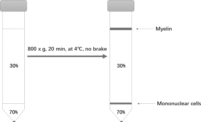
Centrifuge at 800 x g for only 20 min at 4 °C. Set the acceleration to 1 and deceleration to 0. After centrifugation, aspirate almost all of the top phase, being careful to completely remove the myelin at the top (Figure 1).
Remove the interface into a new 15 centrifuge tube. Adjust the volume to 10 mL with RPMI.
Centrifuge at 500 x g for 10 min. After centrifugation, aspirate the supernatant. Resuspend the pellet in ~200 μL of flow cytometry staining (FSC) buffer.
Representative Results
Figure 1: Schematic of the Percoll gradient setup for isolation of mononuclear cells.
Mononuclear Cell Isolation from Mouse Brain: A Density Gradient Centrifugation Technique to Isolate Brain Resident Mononuclear Cells. J. Vis. Exp. (Pending Publication), e20621, doi: (2023).