TTC Staining of Rat Brain Tissue Slices: A Staining Procedure to Differentiate Viable and Necrotic Tissue in Tissue Slices After Ischemia-Reperfusion Injury
PREPARAÇÃO DO INSTRUTOR
CONCEITOS
PROTOCOLO DO ALUNO
All procedures involving animal models have been reviewed by the local institutional animal care committee and the JoVE veterinary review board.
1. Brain staining and slicing
- After the middle cerebral artery occlusion experiment, remove the brain, including the brainstem, from the skull, and wash it in ice-cold PBS.
- Choose the correct size of the brain stainless-steel matrix (see the Table of Materials) depending on the weight of the animals (Figure 1B). Place the brain with its ventral side up in the brain matrix.
NOTE: When seated in the matrix, the brain's ventral surface must be parallel to the top surface of the mold. - Using blades, restrict the frontal and caudal parts (2 blades from both sides) of the brain.
NOTE: Slicing matrix-compatible razor blades must be used. In general, a compatible, single-edge razor blade (thickness of up to 0.01 inch (0.254 mm)) can be used for rat brain slicing. - Put the blades partially (not fully cutting the brain) into the channels between the first and the last blades. When all the blades are inserted and arranged in parallel, press all the blades down with the palm at the same time to cut the brain into 2 mm coronal slices.
- Grasp the blades firmly along the sides with two fingers and remove them together with the sliced brain from the matrix.
- Arrange the brain slices one by one in a tray (70 mL, 72 x 72 mm). When arranging the slices, ensure that the anterior surface of each slice is always facing up.
- Pour warm (+37 °C) 1% TTC solution in PBS onto the brain slices and incubate them for 8 min at 37 °C in the dark.
NOTE: The brain slices must be fully immersed in TTC solution during the incubation. - After incubation in 1% TTC solution, transfer the brain slices to the blue plastic tray to capture images. Arrange the brain slices in sequential order from the frontal to the caudal part and use a scalpel to separate the hemispheres in the sagittal plane.
TTC Staining of Rat Brain Tissue Slices: A Staining Procedure to Differentiate Viable and Necrotic Tissue in Tissue Slices After Ischemia-Reperfusion Injury
Learning Objectives
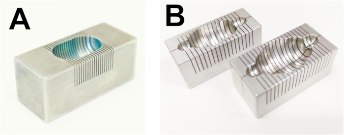
Figure 1: Matrices for the rat heart and brain slicing. (A) Rat heart, (B) rat brain.
List of Materials
| 2,3,5-Triphenyltetrazolium chloride (TTC) | Sigma-Aldrich | 298-96-4 | |
| Adult Rat Brain Slicer Matrix | Zivic Instruments | BSRAS001-1 | |
| Single Edge Razor Blades | Zivic Instruments | BLADE012.1 | |
| Sony Alpha a6000 Mirrorless Digital Camera | Sony | ILCE6000 | Can be repalaced by any up-to-date digiatal camera |
| Surgical blade | Heinz Herenz Hamburg Germany | BS2982 | |
| Weigh tray, 70 mL | Sarsted | 71,99,23,212 | |
| Thermo-Shaker | BioSan | PST-60HL-4 | |
| Toothed tissue forceps | Agnthos | 11021-12 | |
| Sodium chloride | Fisher bioreagents | BP358-10 | |
| Cover Glass Forceps, Angled | Fine Science Tools | 11073-10 |
Preparação do Laboratório
Source: Liepinsh, E., et al. Rodent Heart and Brain Tissue Preparation for Digital Macro Photography after Ischemia-reperfusion. J. Vis. Exp. (2022)
This video describes the 2,3,5-triphenyl-2H-tetrazolium chloride staining procedure to visualize rat brain tissue slices and differentiate viable and damaged tissue following middle cerebral artery occlusion and reperfusion.
Procedimento
Source: Liepinsh, E., et al. Rodent Heart and Brain Tissue Preparation for Digital Macro Photography after Ischemia-reperfusion. J. Vis. Exp. (2022)
This video describes the 2,3,5-triphenyl-2H-tetrazolium chloride staining procedure to visualize rat brain tissue slices and differentiate viable and damaged tissue following middle cerebral artery occlusion and reperfusion.