A subscription to JoVE is required to view this content. Sign in or start your free trial.

Method Article

Label-Free Neutrophil Separation from Tracheal Secretion Using Spiral Microfluidics Channel

854 views

⸱

July 8th, 2025

In This Article

Abstract

Source: Ryu, H., et al., Label-free Neutrophil Enrichment from Patient-derived Airway Secretion Using Closed-loop Inertial Microfluidics. J. Vis. Exp. (2018).

This video demonstrates separating neutrophils from tracheal secretions using a microfluidic device with spiral channels. The device achieves efficient separation based on cell size by utilizing varying thicknesses and trapezoidal-shaped channels. The interplay between forces results in large neutrophils accumulating near the inner wall while erythrocytes and mucin aggregates remain near the outer wall, ensuring effective neutrophil separation.
Protocol

All procedures involving human participants have been performed in compliance with the institutional, national, and international guidelines for human welfare and have been reviewed by the local institutional review board.

1. Device Fabrication and Soft Lithography

NOTE: Standard soft lithography techniques were used to create the polydimethylsiloxane (PDMS) microchannel.

  1. Mix the PDMS precursor in a 10:1 ratio of base and curing agent.
  2. Pour 30 g of the PDMS precursor mixture into the micro-machined aluminum mold (see Figure 1 for dimensions)....

Access restricted. Please log in or start a trial to view this content.

Results

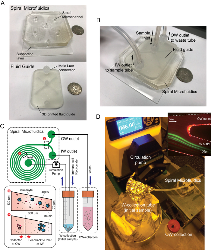
Spiral microfluidics diagram and setup for immune-cell separation; includes 3D fluid guide and circulation pump.
Figure 1: Spiral microfluidic device and experimental settings. Photograph images of (A) spiral microfluidics and (B) the device assembled with the fluidic adaptor. (C) Schematic diagram of closed-loop separation using spiral inertial microfluidics. By recirculating the par.......

Access restricted. Please log in or start a trial to view this content.

Disclosures

No conflicts of interest declared.

Materials

List of materials used in this article
NameCompanyCatalog NumberComments
PDMS precursorDow corning184 SIL ELAST KIT 3.9KG10:1 ratio of base and curing agent
VWR gravity convection ovenVWR414005-128PDMS precursor to be cured in 90 deg.
100mm petri dishVWR89000-324Fabrication of PDMS Supporting layer
Harris Uni-core puncherSigma-aldrichWHAWB1000762mm diameter or other depending on the tubing size
Air plasma machineFemto ScienceCuteSurface plasma treatment for PDMS device to bottom base.
2" x 3" glass slideTED PELLA, INC.2195To support PDMS device
Masterflex spooled platinum-cured silicone tubing, L/S 14Cole-ParmerEW-96410-14Tubing for microfluidics and peristlatic pump
1/16 inch Luer connector, maleHarvard apparatusPC2 72-1443Connector for fluid guide
50mL Falcon tubeCorning21008-940Sample collection & preparation
Phosphate-Buffered Saline, 1X Without Calcium and MagnesiumCorning45000-446 Buffer solution to dilute sample
Halyard Closed suction Catheter, Elbow, 14F/ channel 4.67mmHALYARD HEALTH22113Tracheal seceation suction catheter
0.9% Sterile Normal saline, 10mL pre-filled syringeBD PosiFlushNHRIC: 8290-306547For tracheal seceation collection from the patients
SecurTainer™ III Specimen Containers, 20mLSimport1176R36Sterile sputum (airway secretion) collection container
Syringe with Luer-Lok Tip, 10mLBDBD309604To pipette homogenize the mucus sample and reach the bottom of sample tube
BD  Blunt Fill Needle, with BD Luer-Lok  TipBDTo pipette homogenize the mucus sample and reach the bottom of sample tube
40µm nylon cell strainer Falcon21008-949To remove large chunk or blood clots, which can block the microfluidics access hole or the channel.
Peristaltic pump (Masterflex L/S Digital Drive)Cole-ParmerHV-07522-30 Operation of microfluidics

Explore More Articles

Inertial MicrofluidicsDean Drag ForceInertial Lift ForceCell Size SeparationMicrofluidic Channel DesignPeristaltic PumpPhosphate Buffered Saline