Introduction
Mitochondria from rat heart is one of the most common preparations for past and current studies of cellular metabolism. They can be obtained quickly and reliably in great quantity from wild type or knock-out animals. The general procedure consists of tissue digestion by trypsin, fractionation and differential centrifugation.
There are several conditions that have to be kept during Isolation of mitochondria from various tissues, including the heart (Fig. 1).
- The tissue has to be thoroughly minced, since it directly affects the yield of obtained mitochondria. Heart tissue is best to mince with small curved scissors and can be followed by use a Latapie tissue grinder or, if a tissue grinder is not available, a garlic press with outlet holes diameter of around 1.0 mm diameter.
- It is necessary to maintain the temperature of solutions at each step. Therefore the entire procedure has to be carried out in a cold room and all instruments and solutions have to be prepared and cooled in advance. If the temperature conditions are not kept stable several properties of the preparation can be lost. Mitochondrial structure is very sensitive to freezing so overfreezing of the suspension (for example during centrifugation) is not permitted, especially if studies of active transport are important.
- An important parameter is the time-length of the procedure; the isolation should be performed as quickly as possible and the obtained preparation has to be used immediately. Therefore, surgical instruments, solutions and appliances have to be prepared in advance.
Materials and instruments
Instruments
- Straight operating scissors, sharp point
- Curved scissors
- Forceps
- Petri dish
- Small funnel + piece of nylon filter (prewashed)
- 6 beakers 50 ml
- Two magnetic stirrers and two ice baths
- One big ice bath
- 2 small magnetic bars
- Spatulas for pellet scraping
- Centrifuge tubes
- Latapie tissue press (can be substituted with garlic press)
- Teflon-glass homogenisers 20ml and 1-2ml
- Waste beaker
- Eppendorf tubes for final mitochondrial suspension and samples for protein determination
- Ice baths
Solutions (calculated per 2 rats):
- Washing buffer: 0.3 M Sucrose, 10 mM HEPES (pH=7.2), 0.2 mM EDTA (1000 ml)
- Isolation buffer: 0.3 M Sucrose, 10 mM HEPES (pH=7.4), 0.2 mM EDTA, 1 mg/ml BSA (Sigma A6003) (100ml prepared from the Washing buffer). Bovine serum albumin can be substituted by less expensive fatty acid-free analogues. The same buffer or its isotonic variations can be used as Resuspending buffer.
- Trypsin (Sigma T8003) solution: 2.5mg/ml in 1mM HCl (0.5 ml)
- Trypsin inhibitor from soy bean (Sigma T9128) 6.5 mg/10 ml of Isolation buffer
Protocol
The general scheme of the procedure is shown on Fig. 1. Hearts should be cooled and washed in Washing buffer, ventricular tissue excised, minced and homogenised during photolytic treatment with trypsin. The suspension is centrifuged at low speed and the obtained supernatant is centrifuged again at higher speed to collect mitochondria. Depending on the planned experiments the resuspending buffer can be replaced by any other isotonic solution.
Animals were terminated in accordance with the U.K. “Animals (Scientific Procedures) Act.” by cervical dislocation followed by decapitation. It is necessary to extract the heart immediately after decapitation of the animal, so that there is no delay between animal termination and heart extraction. If parallel isolation of liver mitochondria is expected the rats should be fasted overnight prior to the experiment.
- Prepare three beakers containing 40 ml of Washing Buffer. Cool them in the ice-salt bath for 3 to 5 min before proceeding to the next step. Make sure not to overfreeze them and use immediately just after clouds of ice crystals start to appear.
- Open the thorax of the decapitated rat and extract the heart. Make small incisions and then immediately transfer the heart to the ice-cold solution in the first beaker. Squeeze the heart to remove blood and put it into the second beaker. Repeat this operation for each heart.
- Dry the hearts on the filter paper. Remove all fat, clotted blood, auricles and fasciae and pool the ventricular tissue.
- Mince the pooled tissue with small curved scissors on the Petri dish on ice for around 5 min until the size of the particles is about 1-2 mm.
- Put the minced tissue into the tissue press and pass it through. Be aware that a substantial amount of the material can remain in the press so collect all the heart tissue from the walls and bottom of the press.
- Transfer the material into the beaker with 50 ml Washing Buffer and stir. Then filter the suspension through a nylon filter. Wash the minced tissue on the filter 3 times with the Washing Buffer and discard the filtrate.
- Transfer the washed tissue into 20 ml of Washing Buffer and place it in the ice bath on the magnetic stirrer. Add 0.5 ml of trypsin solution under constant stirring and start the timer. Prepare another magnetic stirrer, ice bath and a second 50 ml beaker.
- On the 4th minute briefly homogenise the suspension portion by portion using a small loosely fitted glass-Teflon homogeniser (10-15ml) with several up and down strokes to disperse the suspension. After homogenisation transfer the suspension into the second beaker. Repeat the procedure on the 9th minute, so that you perform the homogenisation of the suspension two times in total. After 15 min of incubation, add 10 ml of isolating buffer containing trypsin inhibitor and incubate for 1 min.
- Filter the suspension again through a nylon filter and save the filtrate. Collect the particulate fraction left on a filter and homogenise for 1 min in 15-20 ml of Washing Buffer.
- Combine the homogenate with the filtrate and centrifuge for 15 min at 600g.
- Carefully filter the resulting supernatant into a new centrifuge tube. Avoid contamination by the pellet and centrifuge again for 15 min at 8500g.
- After high speed centrifugation discard the supernatant and rinse the pellet 2-3 times with 1 ml of the buffer discarding the fluffy white outer rim layer. Break up the pellet with the spatula but avoid the red spot of erythrocytes at the bottom. If the blood cells got loos, remove the red bits from the suspension before homogenisation.
- Resuspend the pellet with a small homogeniser or alternatively by gentle pipetting. Dilute the suspension with more Isolationbuffer and centrifuge again for 15 min at 8500g.
- Discard the supernatant, resuspend the pellet in Resuspending Buffer (an isotonic buffer of choice) and centrifuge again.
- The resulting brown pellet contains washed intact mitochondria. Resuspend the pellet in a very small volume of isotonic buffer of your choice.
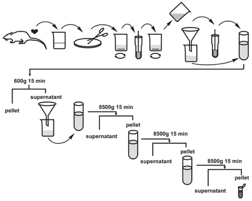
Figure 1. Scheme demonstrating the step by step procedure of isolation of rat heart mitochondria. Top part, Stages 1-9: tissue disruption, trypsin treatment and homogenisation; bottom part, stages 10-15: differential centrifugation.
