Isolating Tumor-Infiltrating Immune Cells from a Murine Brain

Published: July 31, 2024

Abstract

Source: Baker, G. J., et al., Lowenstein, P. R. Isolation and Flow Cytometric Analysis of Glioma-infiltrating Peripheral Blood Mononuclear Cells. J. Vis. Exp. (2015)

The video demonstrates the isolation of tumor-infiltrating peripheral blood mononuclear cells from a mouse brain using mechanical dissociation and enzymatic digestion. This is followed by density gradient centrifugation to collect the immune cells for further analysis.

Protocol

All procedures involving sample collection have been performed in accordance with the institute's IRB guidelines.

Prepare density centrifugation media mix solution by placing 90 ml of 10x PBS into 264 ml of ultrapure deionized water (3.93x) in a sterile plastic container. Titrate to pH 7.0-7.2 with HCl and filter sterilize through a 0.22 µm filter. Store at 4 °C.

Prepare 70% density centrifugation media by combining 18 ml of density centrifugation media mix solution with 30 ml of density centrifugation media (see materials list for details) in a 50 ml polypropylene centrifuge tube. Store at 4 °C, but bring to RT before use.

Prepare 37% density centrifugation media by combining 9.6 ml of DPBS with 10.4 ml 70% density centrifugation media in a 50 ml centrifuge tube. Store at 4 °C, but bring to RT before use.

1. Isolation of Glioma-infiltrating PBMCs

  1. Using a clean, single-edged razor blade, isolate the area of the brain containing the tumor by making a sagittal cut down the center of the brain to bisect the two hemispheres. Turn the ipsilateral hemisphere medial side down and make two coronal cuts at the levels of the cerebellum and the olfactory bulb to isolate the target tissue containing the tumor implant (Figure 1A). An equivalent portion of the contralateral hemisphere may also be isolated and used as a negative control.
  2. Place the target tissue into the respectively labeled glass Dounce tissue grinder containing 1 ml of DPBS, push the plunger all the way down, and twist 7 times to initially disrupt the tissue. Lift the plunger to allow the liquid to settle back to the bottom of the tissue grinder. Repeat this step thrice; however, only twist the plunger 4 times during the next two repeats and 3 times during the last repeat to avoid over-triturating the tissue.
  3. Using a P-1000 micropipette, sequentially apply three 1 ml volumes of ice-cold DPBS along the sides of the plunger to rinse into the tissue grinder.
  4. Resuspend the triturated brain matter by pipetting up and down and placing it into a labeled 15 ml centrifuge tube on ice. Rinse the sides of the Dounce tissue grinder with 1 additional ml of ice-cold DPBS and add to the same 15 ml centrifuge tube. Keep all tubes containing triturated brain matter on ice until all samples have been processed.
  5. Once all samples have been processed, spin down the triturated brain matter in the 15 ml centrifuge tubes at 740 x g (max RCF) for 20 min at 4 °C.
  6. Remove the supernatant with a 10 ml serological pipette and pipette gun and resuspend the pelleted brain matter in 1 ml of the previously prepared collagenase/DNase-I digestive enzymes using a P-1000. Place the 15 ml centrifuge tubes into a test tube rack and place in a 37 °C water bath for 15 min.
  7. Gently agitate the samples twice throughout the incubation period by flicking the tubes to facilitate tissue disaggregation.
  8. Add 6 ml of ice-cold DPBS using a 10 ml serological pipette to each tube to dilute the digestive enzymes. Pipette up and down to resuspend, and filter the total volumes through sterile 70 µm nylon mesh filters into new labeled 15 ml centrifuge tubes on ice.
  9. Spin the brain cell suspension down at 740 x g (max RCF) for 20 min at 4 °C to achieve a single-cell pellet (Figure 1B). After removing the 15 ml tubes from the centrifuge, place them on ice and increase the temperature setting on the centrifuge to about 21 °C in preparation for the next step.
  10. Fully remove the supernatant from each tube containing brain cells and initially resuspend the pellets in 1 ml of 70% density centrifugation media using a P-1000 micropipette. Then add 4 additional milliliters of 70% density centrifugation media to each tube using a 10 ml serological pipette. Screw the caps on securely and homogenize the cell suspension by gently inverting to tubes several times.
  11. Remove the 15 ml centrifuge tubes containing brain cells resuspended in 70% density centrifugation media from ice, and place in a test tube rack at RT. One at a time, carefully overlay 2 ml of 37% density centrifugation media solution onto the 5 ml of 70% density centrifugation media with a P-1000 micropipette to form a clean interface between the two density centrifugation media layers (Figure1C).
    1. Use a fine-tipped marker to indicate the position of the interface so it can be easily identified after centrifugation when the distinction becomes less apparent.
  12. Spin down the 15 ml centrifuge tubes at 740 x g (max RCF) for 20 min at RT with no break to avoid disrupting the interface.
  13. After centrifugation, collect the PBMCs that have accumulated at the interface between the two density centrifugation media layers (Figure 1D) by introducing a P-200 micropipette into the tube along its side, carefully bypassing the lipid layer.
    1. Once at the level of the PBMC band, slowly extract 200 µl from the surface of the 70% density centrifugation media layer and place it into a respectively labeled polypropylene FACS tube on ice. Repeat once for a total volume of 400 µl per sample.
  14. Add 3 ml of flow buffer to each FACS tube containing 400 µl of PBMCs to sufficiently dilute the density centrifugation media, then spin the cells down at 660 x g (max RCF) for 20 min at 4 °C.

Representative Results

Figure 1
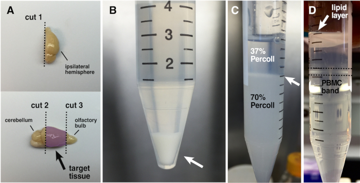
Figure 1: Isolation of Glioma-infiltrating PBMCs. (A) Strategy for the dissection of mouse brain tissue containing early-stage orthotopic glioma implants. The top panel demonstrates the sagittal bisection of the ipsilateral hemisphere away from the contralateral hemisphere. The bottom panel demonstrates the two additional coronal cuts necessary to isolate the target tissue containing the tumor implant (highlighted in purple). (B) Single-cell brain tissue pellet (white arrow) after enzymatic digestion, filtration through a 70 μm nylon mesh, and centrifugation. (C) Properly poured density centrifugation media gradient prior to centrifugation. The white arrow indicates the clean interface formed between the two density centrifugation media layers. (D) Density centrifugation media gradient after centrifugation demonstrating the lipid layer that forms at the top of the 37% density centrifugation media layer white arrow) and the PBMC band (outlined by the dashed black lines) at the interface between the two layers.

Declarações

The authors have nothing to disclose.

Materials

Dulbecco's Modification of Eagles Medium Gibco 12430-054 With 4.5g/L D-glucose, L-glutamine, 25mM HEPES and phenol red
Dulbecco's Phosphate-buffered Saline Gibco 14190-144 Without calcium chloride or Magnesium chloride
Fetal bovine serum Omega Scientific Inc. FB-11
Penicillin Streptomycin Gibco 15140-122 10,000U/ml Penicillin; 10,000μg/ml Streptomycin
0.6ml conical polypropylenemi microtubes Genesee Scientific 22-272
Polyurethane ice bucket Fisherbrand 02-591-45 Capacity: 0.152 oz. (4.5L)
Collagenase (Type I-S) Sigma Aldrich C1639 De Clostridium histolyticum
Deoxyribonuclease I Worthington Biochemical Corp. LS002007 >2,000 Kunitz units per mg dry weight
Large dissection scissors Ted Pella Inc. 1316
Small dissection scissors FST 14094-11
Blunt end forceps Ted Pella Inc. 13250
7ml glass Dounce tissue grinder Kontes KT885300-0007
Percoll Density Centrifugation Media GE Healthcare GE17-0891-01
10 mL serological pipettes Genesee Scientific 12-104 Single use; sterile
70μm sterile nylon mesh cell strainers Fisherbrand 22363548
15ml polypropylene centrifuge tubes Genesee Scientific 21-103 Conical bottom
50ml polypropylene centrifuge tubes Genesee Scientific 21-108 Conical bottom
12x75mm round-bottom polypropylene FACS tubes Fisherbrand 14-956-1B
check_url/pt/22387?article_type=t

Play Video

Citar este artigo
Isolating Tumor-Infiltrating Immune Cells from a Murine Brain. J. Vis. Exp. (Pending Publication), e22387, doi: (2024).

View Video