Cell manipulation in microfluidic devices is desirable for the sorting or selective placement of single cells or for population studies.2 Laminar flow is used in conjunction with valves and pumps to manipulate cells within microfluidic devices. However, these methods alone are challenging and require detailed fabrication processes and skills.3 Centrifugation can simplify the demands for cell placement but simultaneous imaging is a challenge; furthermore, the channel architecture for centrifugation must be considering carefully when designing the desired manipulations and considering effects of centripetal forces.4 Laser tweezers can be used for cell placement but the method is expensive and not amenable for high-throughput cell sorting.5 Yet, DEP has been demonstrated as an effective system of “electrical tweezers” for effective cell placement, characterization and manipulation.6,7
Specifically, DEP has been used to selectively capture and sort cells for on-chip processing of living and dead cells,7-10 and for collecting cells on resonant sensors for the measurement of cell mass.11 We have previously demonstrated that by increasing the DEP force on bacteria or beads above the fluidic drag force, the on-chip concentration and trapping of polystyrene beads and Listeria monocytogenes V7 can be accomplished.12 Mixed populations of E. coli and L. monocytogenes bacteria can also be directed and released through DEP pulses. Furthermore, larger particles can be differentially captured and concentrated on the electrodes based on the large particle size, whereas smaller particles are not capture but are removed with the fluidic flow.13 When DEP forces do not overcome the fluidic dragging forces on the particles, the bead or cell is not captured, but rather moved within the fluidic stream. As shown in Figure 2C, beads can be focused into the central region of the fluid stream sufficient to retain the beads in the central channel. This could be due to the combined effect of particle-to-particle influences within the electrical field, fluid velocities greater than DEP dragging forces, the combination of these, or some other undefined effect.
Newer advances in contactless DEP allow for maximizing cell capture and manipulation with the minimum required field, thus protecting cell types, to the greatest extent, during DEP manipulation.1,9 Contactless DEP offers promise to the microfluidics community for sorting, collecting and positioning cells within microfluidic devices. We anticipate that with the increased demand for, and implementation of, DEP for manipulation within microfluidic devices, further discoveries and innovations will expand the understanding and influence of DEP forces.
PCBs can be manufactured through high-volume processes at an affordable price with a rapid fabrication turnaround time, making them good platforms for commercialization. In addition, PCBs are easy to use and accessible for scientists in a range of disciplines for on-chip cell manipulations and sorting.
When designing the layout of the PCB electrodes, consideration should be given to the desired particle/cell trajectory. Key factors for consideration include particle size and type (bead and/or cell), cell type, microchannel size, flow velocity, electrode spacing (which determines the electrical field strength), coverslip thickness, and the fluid conductivity. These factors influence the force required and available to manipulate the particle or cell, and ultimately the separation efficiency. Our protocol presented here demonstrates an effective setup for initiating DEP for microsphere and cell separation. For potential applications, users should match the architecture of the microfluidic channel design with the desired flow paths for cells and beads, as well as the electrode patterns to optimize efficiency for each application. Electrode spacing and coverslip thickness can be used according to the previously reported guidelines when designing the microfluidic channel layout.1
The conductivity and permittivity of the cell/particle and the surrounding media must be different enough to facilitate positive or negative DEP, while keeping the cells intact. The polarity of DEP for a spherical particle can be determined from the real part of a complex value of the following Clausius-Mosotti factor at the frequency ω of DEP voltage.
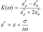
Equation 1
In this equation ε, is permittivity, σ is conductivity, and ε* is complex permittivity. The subscripts p and m denote the particle and media, respectively. When a cell has a higher permittivity than the media, or the real part of the Clausius-Mosotti factor becomes positive, the particle becomes more polarized than the surrounding media. Due to a non-uniform electric field, the particle’s polarization becomes non-uniform and this creates a positive DEP (+DEP) force that draws the cell toward the region with higher electric field intensity. If a cell has a lower permittivity than the surrounding media, or the real part of the Clausius-Mosotti factor becomes negative, it will undergo negative DEP (-DEP), and the cell is forced toward the minimal field region. If the cell and media have approximately the same complex permittivity, no force can be generated to manipulate the cell. For this reason, pure water is a preferred media for the DEP particle manipulation. However, to avoid the osmotic stress on the cell from pure water, low conductivity media was formulated to keep the conductivity unchanged, but to increase osmolarity to decrease osmotic stress to the cells. Conventional cell culture media or physiological buffers, such as DMEM or PBS, have high conductivity, which is not suitable for the DEP manipulation.
We also have previously demonstrated that cells can be captured on sensors with DEP using low-conductivity media. After a brief period for cell attachment, the low conductivity media can be replaced for the required cell culture media to support cell growth for days.11
From our experience, fluorescent beads are very bright with respect to the live cell fluorescence, thus it can be a challenge to match the bead intensity to the intensity of a living and fluorescing cell. To improve the visualization of both the cells and the beads, we used DIC microscopy on an upright microscope for imaging both. To display the cells and beads, we presented the data in an intensity glow-scale image, which retains the data in a wide color spectrum for easier viewing. Thus, when designing the study of interest, the imaging parameters and resources must be considered.
In summary, we demonstrate the ability to selectively actuate beads and cells into separate channels using DEP. With the increasing utility of microfluidic channels for cell biology, biochemistry and bioengineering applications, DEP is a desirable option for cell collection, placement and sorting. The PCB electrodes fabrication is inexpensive and convenient, the electrodes are easy to use, and the rapid fabrication times are ideal for the implementation of DEP.
