1. Extracting Collagen from Rat Tails
- Place frozen fresh rat tails in 70% ethanol.
- Deglove the overlying skin with a scalpel exposing tendons.
- Strip tendons away from tail and place in 70% ethanol to sterilise.
- Weigh the collected tendons and transfer to acetic acid (1g tendon to 250ml 0.5M acetic acid). Mix for 48hr at 4°C.
- Centrifuge the tendon/acetic acid mix at 10,000g for 30min and discard the pellet.
- Add an equal volume of 10% (w/v) NaCl to the supernatant and allow the mix to stand overnight at 4°C.
- Collect the collagen-rich, insoluble bottom layer and centrifugate for a further 30min at 10,000g.
- Resuspend the collagen-rich material in 0.25M acetic acid at 4°C and dialyse against 1:1000 acetic acid at 4°C for 3 days, changing the dialysis buffer twice daily.
- Further sterilise the collagen solution by centrifugation (20,000g for 2hr) and store at 4°C.
- Dilute collagen as required with the addition of sterile 1:1000 acetic acid to a stock concentration of 1mg/ml.
2. 3D Assay Creation
The 3D assay is based on a cell line assay, published previously2.
- Multiple core biopsies are harvested from consenting patients at the time of curative surgical resection for invasive breast cancer.
- By eye, divide the cores using a scalpel into 1mm3 fragments. Trim and discard macroscopically distinct fat.
- Immerse the fragments in MEGM Complete media. The tissue can be stored in this state for up to 1hr.
- To create the collagen gel, mix on ice 1mg/ml rat tail collagen, 1:1000 filtered acetic acid, 10x DMEM/F12 and 0.22M NaOH in a ratio of 3:5:1:1 to create a final collagen concentration of 0.3mg/ml.
- Inject 1.2ml of collagen mixture into individual wells of a 24 well plate and place into an incubator at 37 °C for 10min to initiate gelling.
- After 10min, remove the 24 well plate from the incubator. Place the tumour pieces on to the gelling collagen and gently push them in to the centre of the gel using a 10μl pipette tip. Return the 24 well plate to the incubator for 1hr.
- For ER+ tumours, supplement MEGM Complete media with β-oestradiol to a concentration of 3.2×10⁻¹⁰ M. Once the gel assays have gelled for 1hr, carefully introduce 1.2ml of the supplemented media into each well. The final β-oestradiol concentration within the assay will equilibriate to 1.6×10⁻¹⁰ M to recapitulate physiological oestrogen levels found in breast tissue3.
- Run a 10μl pipette tip around the inner edge of each well, releasing each gel circumferentially, thus allowing each gel to float freely within the media. Return the 24 well plate to the incubator.
- Exchange supplemented media twice weekly until termination of the experiment. Terminate the experiment by removing media and adding a formalin fixative.
3. Antibody Staining of Assay in preparation for Optical Projection Tomography
We have adopted a previously published method for whole sample staining4.
- Wash formalin fixed specimens for 30min in PBS. Dehydrate the gels stepwise in methanol diluted in PBS containing 0.05% Tween 20 (0.05% PBST): 33%, 66%, and 100% for ≥15min at each step.
- Incubate the tissue in freshly prepared MeOH: DMSO: 30%H2O2 at a ratio of 2:1:3 (15% v/v H2O2) at room temperature for 24hr to quench the autofluorescence.
- Wash samples twice for 30min in methanol. Freeze the samples to -80°C five times for at least 1hr each time and back to room temperature (RT) to ensure that antigens in the deeper parts of the tissue are rendered accessible.
- Rehydrate the tissue back to 0.05% PBST with methanol as diluent (33%, 66% and 100% for 15min at each step).
- Block the tissue in blocking solution (10% serum in 0.05% PBST) for 24hr.
- Incubate the tissue with primary antibody to identify the epithelial content, using rabbit anti-cytokeratin at a concentration of 1:500 diluted in 1.5ml of blocking solution containing 5% DMSO for 48hr.
- Wash samples four times with 0.05% PBST for 30min each.
- Incubate the tissue with fluorescent secondary antibody (1:100 Goat anti-rabbit Alexa555), diluted in 1.5ml blocking solution containing 5% DMSO for 48hr.
- Wash the samples again with 0.05% PBST four times for 30min each.
4. Optical Projection Tomography
Optical projection tomography is a microscopy technique previously developed to produce high-resolution 3D images of fluorescent biological specimens with a thickness of up to 15 millimeters5.
- Mount the stained gel in 1% low melting point agarose and dehydrate in 100% methanol for 24hr. Clear the specimen in BABB solution (1:2 Benzyl alcohol, Benzyl benzoate) for 48hr and scan using the OPT 3001M Scanner (Bioptonics).
- Both target and autofluorescence are captured using the Cy3 filter set (exciter 545nm/30 nm, emitter 610/75 nm) and GFP1 filter set (exciter 425nm/40 nm, emitter LP475 nm) respectively.
5. Reconstruction and 3D quantification
- The set of angular projections acquired by OPT may be reconstructed using NRecon software (SkyScan) to create a series of cross section slices through the invasion assay. This software is available to download for free at http://www.skyscan.be/products/downloads.htm.
- Analyse the reconstructed data using Volocity software (PerkinElmer). Free demonstration software is available to download at http://www.cellularimaging.com/products/Volocity/demo/.
- In order to quantify epithelial volume by cytokeratin staining, set a minimum voxel intensity threshold to reduce interference from background fluorescence in the target channel. We defined this threshold empirically at 9000 a.u. Using a list of available parameters in the measurement application, we quantified the volume of epithelium in continuity with the original core (demarcated in the autofluorescent channel).This allowed us to quantify volumes of epithelial outgrowth between different tumour samples.
6. Representative Results:
During the development of the assay, a total of 52 breast cancer biopsy specimens with a wide range of histopathology have been used. Of these <10% (5/52) failed to provide at least one viable invasion assay. Assay failure was due to either bacterial supra-infection or insufficient tumour biopsy material. Therefore application of the assay is not restricted to a specific subtype of breast cancer.
Typical assays (figure 1) fixed after 20 days in collagen culture and subsequently stained for cytokeratin. Volocity software is able to recognise both the epithelial bulk continuous with the original core and detached groups of invading cells and quantify each volume separately.
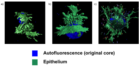
Figure 1. Invasion assays following fixation and quantification. The assay supports samples obtained from multiple breast cancer subtypes: a) ER+ b) HER2+ and c) ER- .
