Zebrafish were raised using standard protocols13 in the Queens College Animal Facility at 28°C. Individual fish pairs or group matings should be preformed and the resulting embryos cleaned and stored in a 28°C incubator. Fish health should be examined daily. At 5-day post fertilization (dpf), fish should be separated into tanks at a 10-15 fish density and entered into the fish facility nursery. For the protocol presented here zebrafish were collected at larva through adult stages: 15, 30, 45, 60, 90, 180 dpf.
1. Zebrafish subject collection and fixation
- Remove fish from habitat tank using net and place in 0.2% Tricaine to anesthetize.
- Place anesthetized fish in ice water for 15 minutes to euthanize.
- Make fresh 4% paraformaldehyde (PFA) in 1x Phosphate-buffered saline (PBS).
- Incubate collected fish in 4% PFA for 1-2 hours at RT then overnight at 4°C in a petri dish wrapped in parafilm. This keeps the fish flat and makes later dissection much easier.
- After fixation, rinse twice in PBS with Tween (PBT). Fish can be keep in PBT at 4°C for up to 10 days.
2. Measuring fish and dissection
- Place single fish into petri dish half filled with fresh PBS and lay it on its right side.
- Measure length of fish from snout to base of tail (the caudal peduncle), not including tail fins, using a digital mm caliper (Figure 1). This is the standard length (SL).
- Each fish can be assigned a number/letter combination to allow an individual fish and the resulting dissected heart to be tracked for later analysis. Data, including parental stock number, age at fixation, SL measurements, were entered into a spreadsheet and organized based on these labels.
- Before dissection, label PCR tube strips with assigned tracking number and fill with PBT or other solution appropriate for further analysis.
- Orient the fish ventral side up in petri dish while stabilizing the body with forceps holding the head between the eyes and gills (Figure 2).
Smaller fish (smaller than 12mm SL):
- a) Using sharp forceps, or a micro-needle in a pin holder, remove the pectoral muscles and fins from the body to reveal the heart. The constant motion of the forceps during dissection can cause currents in the PBT that move the fish. This is particularly problematic for smaller fish, thus a micro-needle can be used to remove pectoral muscles and fins and open the body cavity.
- a) Gently use the forceps to scoop the heart out of the cavity from under the atrium. Alternately, in some fish you may be able to remove the heart by gently pulling the bulbous out and the rest of the heart will follow. Be careful of the location of the atrium if using this technique as it can easily be damaged. If there is extra tissue that comes with the heart, that is fine and can be cleared after.
- a) If the fish contains a fluorescent cardiac marker such as Tg(myl7:GFP)twu34, use fluorescent scope for dissection and verify removal of the heart by florescence (Figure 3).
- a) Once the heart is removed from the cavity, use the forceps to hold the heart and a micro-needle to remove extra non-cardiac tissue and the epicardial lining from the outside of the heart.
Larger fish (greater than 12mm SL):
- b) Using spring handled micro-scissors, make three incisions of less than 1mm deep: 1- transverse cut through the gills 2- transverse cut at anterior belly 3- sagittal cut on ventral side connecting both cuts (Figure 4).
- b) Using sharp forceps remove the pectoral muscles and fins from the body to open the body cavity and reveal the silvery tissue of the pericardium (Figure 5A). Remove this pericardial tissue and the heart will become visible (Figure 5B).
- b) Use the micro-scissors to cut the artery connected to the bulbous arteriosus, located superior to the heart. Once this artery is cut, put the forceps tips under the atrium, use the forceps to scoop the heart out of the cavity. If there is extra tissue that comes with the heart, that is fine and can be removed later.
- b) Once the heart is removed from the cavity, use the two forceps to hold the heart and remove extra tissue, or one pair of forceps to hold the heart and a micro-needle to remove extra, non-cardiac tissue. The epicardial lining, evident by its black pigmentation, can be removed from the outside of the heart if desired by gently scraping the tissue.
3. Photography of hearts
- Prepare an 23g/L nutrient agar petri dish and cover solidified agar with PBS.
- Using the forceps, make a well in the agar that allows the heart to rest in the desired orientation for photographing.
- Remove the heart from the PCR tube by holding the tip of the bulbous with the forceps firmly. Place the heart in the agar dish.
- Orient the heart in agar to photograph. The sample can be rotated after each picture to all possible orientations (Figure 6).
- Use overhead light and short exposure time to photograph the hearts. Make adjustments for light quality using the exposure as necessary to obtain the best photograph.
4. Representative Results:
An example of a well-dissected clean heart is shown in multiple orientations in Figure 6. Each heart will have some differences in overall morphology, but the heart should be complete, containing an intact ventricle, atrium and bulbous arteriosus. Some examples of this variation in overall morphology are shown in Figure 7. The black spotted epicardial tissue can also removed from the heart.
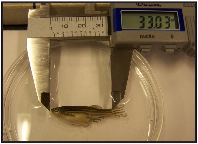
Figure 1. Measure the fish from snout to caudal peduncle using a digital mm caliper.
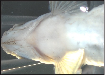
Figure 2. Hold the fish with forceps between the eyes and gills.
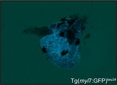
Figure 3. A cardiac specific fluorescent transgenic marker such as Tg(myl7:GFP)twu34, shown here, can aid in visualizing hearts dissected from smaller fish.
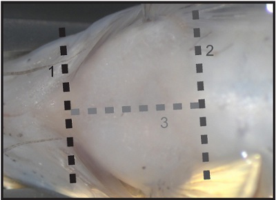
Figure 4. Three incisions to remove the pectoral muscles and skin: 1- transverse cut through the gills 2- transverse cut at anterior belly 3- sagittal cut on ventral side connecting both cuts.
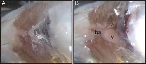
Figure 5. Black letters label the chambers of the heart: bulbous arteriosus (ba), ventricle (v), and atrium (a).
A- The upper body cavity is open to show the silvery tissue of the pericardium.
B- After removing the pericardium, the heart is visible and easily removed.
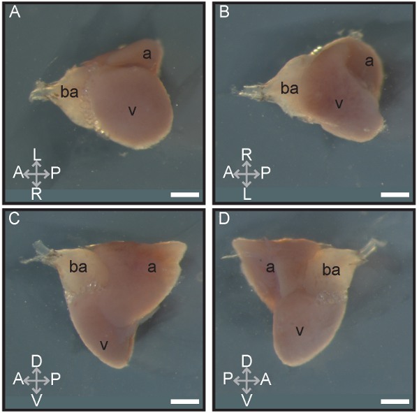
Figure 6. Different orientations of the heart are shown. Black letters label the chambers of the heart: bulbous arteriosus (ba), ventricle (v), and atrium (a). Grey arrows indicate location and orientation of the heart in reference to its normal location in the body: anterior (A), posterior (P), dorsal (D), ventral (V), left (L), and right (R). Scale bar indicates 400μm.
A- ventral view of the heart
B- dorsal view of the heart
C- lateral view from the left side
D- lateral view from the right side
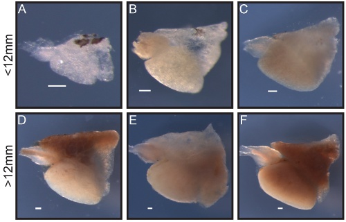
Figure 7. Morphology of hearts of different sizes is shown. Black letters label the chambers of the heart: bulbous arteriosus (ba), ventricle (v), and atrium (a). Scale bar indicated 400 μm.
A-C- Hearts form fish with SL less than 12mm
D-F- Hearts form fish with SL greater than 12mm
