1. Assay Set-up
- Thaw a small aliquot (<500 μl) of Matrigel on ice at 4 °C (approximately 2 hr).
- Prepare gel recipe (see example volumes in table below): 10x PBS (1x in total volume), 1N sodium hydroxide (equivalent to 0.023 volumes of added collagen, or per the collagen manufacturer’s recommendations, as appropriate), Matrigel and collagen type I to final concentrations of 1 mg/ml and 1.3 mg/ml respectively (other matrix formulations may be used depending on cell type and experiment).
Example gel recipe
| Components | Stock Concentration | Final Concentration | Add volume |
| 10X PBS | 10X | 1X | 0.090 ml |
| Sterile water | 0.346 ml | ||
| 1N NaOH | 0.008 ml | ||
| Matrigel | 9.90 mg/ml | 1 mg/ml | 0.101 ml |
| Rat tail type I collagen | 3.66 mg/ml | 1.3 mg/ml | 0.355 ml |
- Mix components of the gel on ice in the same sequence as above and incubate final solution for 1 hr at 4 °C. In our experience, a 1 hr incubation prior to cell seeding results in a more uniform gelation of the collagen. Make sure to keep Matrigel and collagen on ice at all times and to work as fast as possible to prevent gelation.
- Place 12 mm diameter 8 μm pore cell culture inserts into a 12-well plate using sterilized forceps.
- Count cells and resuspend in serum-free media at 5 x 10⁶ cells/ml (total volume should be 10% of the gel solution).
- Add 100 μl of cell suspension to 900 μl of gel solution (final cell concentration 5 x 10⁵ cells/ml) and mix thoroughly by pipetting gently up and down.
- Add 150 μl of the final mixture to each insert and transfer to a 37 °C, 5% CO2 incubator for 30 minutes until gel polymerizes.
- Using serum-free media,
- Add 100 μl on top of the gel and 1200 μl under the insert for the STATIC condition. The fluid levels inside the insert and outside in the well should be approximately equal, resulting in minimal pressure difference across the gel and no interstitial flow.
- Add 100 μl under the insert and 650 μl above gel for the FLOW condition. Be careful to avoid any air bubbles beneath the insert, as they will prevent cells from migrating through the membrane at these locations. The pressure difference generated by these volumes is approximately 1.3 cm H2O (or 1 mm Hg).
- Place plate in a 37 °C, 5% CO2 incubator for 24 hr.
2. Cell Staining and Counting
- Add 500 μl of 1X PBS per well into new 24-well plate; this will be used to wash the inserts.
- Remove the medium remaining in the upper portion of the flow transwells and determine the volume. Calculate total eluted volume by subtracting the remaining volume from the total volume originally added, 650 μl (this will be used for flow rate calculations, see below). Use cotton swabs to remove the gel from the inserts and to wipe the top surface of the membrane to remove non-invaded cells. Place the inserts into the 24-well plate containing 1X PBS for 15 s to wash inserts.
- Remove PBS and add 500 μl of 4% paraformaldehyde (PFA) underneath each insert and incubate for 30 minutes at room temperature to fix the transmigrated cells.
- Remove the PFA and rinse once with 500 μl 1X PBS to remove residual fixative.
- Add 500 μl of 0.5% Triton X-100 solution beneath the inserts and incubate for 10 minutes at room temperature to permeabilize the cells.
- Cut membranes out of the insert using a razor blade and place into 100 μl of 2 μg/ml DAPI in 1X PBS solution, being careful to place the underside of the membrane face down (transmigrated cells face down).
- Wrap plate in aluminum foil and place on a shaker at 150 rpm for 30 minutes at room temperature.
- Wash membranes in 500 μl 1X PBS on shaker (repeat 3 times for 10 minutes each) to remove free DAPI.
- Place membranes on glass slides with transmigrated cells facing up, add mounting solution and cover slip.
3. Data Analysis
- Count DAPI-stained nuclei on 5 randomly selected locations of each membrane (stay away from the edges and use a 10X or 20X objective lens).
- Calculate the average cell count and obtain the percent invasion by using the following formula:
Percent invasion = 100% x (average cell count x membrane surface area) / (surface area of the microscope image x number of cells seeded)
The percent invasion can be normalized to some control condition (typically the static condition) to allow for comparison between independent experiments. - Calculate the average flow rate: divide the total eluted volume in the flow condition by the incubation time (e.g., 24 hr).
- Calculate the average flow velocity: divide the average flow rate by the cross-sectional area of the gel/membrane (in this case 60 mm2).
4. Representative Results
To measure tumor cell invasion under interstitial flow, we performed our 3-D flow invasion assay using MDA-MB-435S metastatic melanoma cells. These cells have previously been shown to invade in response to interstitial fluid flow 13-14. The cells were embedded in a matrix composed of 1.3 mg/ml rat tail tendon collagen type I and 1 mg/ml Matrigel basement membrane matrix at a final cell concentration of 5 x 10⁵ cells/ml. Two different conditions were compared: (1) average interstitial flow = 0.1 μm s-1 and (2) static condition = no measurable flow rate (Figure 1).
After 24 hr, the cells that invaded through the pores of the membrane were stained with DAPI to facilitate cell counting. Figure 2 shows a representative image of the invaded cells. Under brightfield, only the pores of the membrane are visible. Using fluorescence, the DAPI-stained nuclei were used for cell counting and the phalloidin stained F-actin structures were used to visualize the cell body (optional). Using a Student’s t-test assuming equal variances, we showed that interstitial flow significantly increases MDA-MB-435S cell invasion by 2.3-fold over static conditions (p = 0.003) (Figure 3). This corroborates similar findings (but with different matrix conditions and therefore flow velocities) using this cell line 13-14.
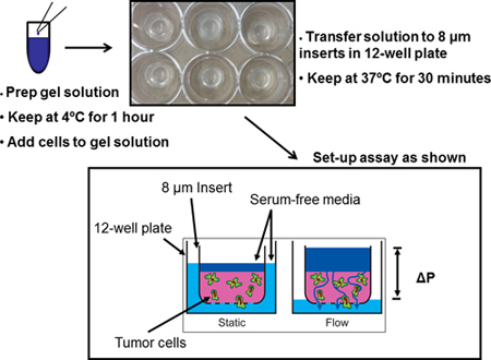
Figure 1. Schematic of the 3-D interstitial fluid flow invasion assay. First prepare gel solution using appropriate concentrations and volumes. Then add cells to gel solution and transfer to cell culture inserts. Finally add appropriate volume of media to each condition and incubate. Interstitial fluid flow is driven by a fluid pressure head.
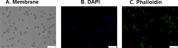
Figure 2. Transmigrated MDA-MB-435S cells on membrane. Invaded cells were fixed after our interstitial fluid flow invasion assay and stained with DAPI and Alexa Fluor 488-conjugated phalloidin to facilitate counting of invaded cells; A) Picture of the membrane under bright field; B) DAPI stained nuclei (in blue); C) Alexa Fluor 488-phalloidin stained F-actin (in green). Scale bar represents 50 μm.
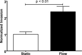
Figure 3. Increased invasion of MDA-MB-435S cells under flow. Cell invasion after 24 hr significantly enhanced by interstitial flow (p = 0.003). The results are normalized to the average static condition and values represent mean ± SEM of 6 cell culture inserts.
