1. Preparing Spleen Tissue
- Note- all human material should be treated as potentially infectious and all procedures should be conducted in a Class II Laminar Flow Cabinet. Using sterile scissors and forceps, remove fat and fibrous tissue from spleen sections (~1-2 cm in size) and trim off as much of the outer capsule material as possible.
- Cut a small piece (1-2 cm3) of spleen tissue and place each piece into a sterile 50 ml Falcon tube.
- Snap-freeze the pieces of tissue by immersion in liquid N2.
- Store at -80 °C. A similar protocol is suitable for other tissue(s).
2. Preparing Human Islet for Storage
- Culture islets in CMRL media. Collect islets in a 10 ml conical bottom tube and wash twice in PBS by centrifuging at 1,500 rpm for 5 min. Pour off the PBS and drain the residual buffer by placing the inverted tube briefly on a paper towel. Be careful not to dislodge the islets.
- Once drained, re-cap the tube and snap-freeze in liquid nitrogen and store at -80 °C.
3. Preparing Extract
- Prepare BAW mix (10:30:60 %v/v) and store at 4 °C.
- Remove the tube from -80 °C. Thaw at room temperature.
- Add sufficient ice-cold BAW to cover the piece of tissue. For islets use 3-5 ml. For spleen tissue use 10-20 ml depending upon the size of the piece of tissue.
- Assemble the tissue homogenizer. Clean by ‘homogenizing’ 10-20 ml of 70% ethanol/water.
- Homogenize the tissue in multiple bursts, after placing the homogenizer probe into the tube with the tissue and BAW solution. Keep the tube in an ice-bucket thereafter.
- Thoroughly clean the homogenizer between samples, by homogenizing 10-20 ml of 70% ethanol/water and then BAW buffer. This prevents cross-contamination of tissue between samples.Dismantleand clean with 70% ethanol/water after use.
- If an extract containing only soluble material is required, centrifuge homogenized tissue extract at 4,000 rpm at RT for 10 min. If a more crude extract is required, spin at 1,000 rpm at RT for 5 min. The most appropriate technique for extracting the BAW-insoluble proteins depends upon the downstream analysis of the proteins. For use in functional immunological assays we would suggest attempting to dissolve the insoluble fraction in 8 M urea as we have previously found this to be well tolerated6.
- Transfer supernatant to a clean tube and put it on ice.
- Determine the concentration of protein in the extract using a BCA assay or similar.
4. Freeze Drying Extracts
- Depending on the mass of protein required (i.e., 100 μg per tube), dilute the homogenate accordingly and dispense aliquots into labeled 5.0 ml sterile, Falcon (12×75 mm) tubes. We frequently use 100 μg/tube.
- Use an 18-20 gauge sterile syringe needle to make 3 holes in the cap of each tube.
- Freeze tubes either by placing them on dry ice for ~10 min or in a -80 °C freezer for >1 hr. Store at -80 °C until ready to put on the lyophilizer.
- Switch on freeze-drier and allow to equilibrate (-100 °C). This takes about 30 min.
- Depending upon the volume, freeze-drying can be completed within 3 hr but we routinely leave our samples overnight.
- After completion of the drying cycle, switch off vacuum pump and slowly allow pressure into the chamber. Remove rack.
- In a sterile hood, remove the perforated caps from the tubes and replace with new ones (Falcon 352032).
- Store tubes at -20 °C. Samples can be reconstituted in culture media, or other buffers, and used in function or biochemical assays.
5. Representative Results
Figure 1 shows the staining of a protein gel loaded with extract from spleen and islet depleted pancreatic tissue (labeled acinar) and purified human islets (labeled islets). The results show a good representation of proteins of different molecular weight for each tissue.
The capacity of tissue extracts to stimulate human T-cell proliferation was tested using a CFSE-based proliferation assay7 (Figure 2). The PBMC used in this assay were isolated from an individual with type 1 diabetes. The magnitude of the response is expressed as a ratio of the number of CFSEdim cells per 5,000 CD4+, CFSEbright cells without antigen: of CFSEdim cells per 5,000 CD4+, CFSEbright cells with antigen from triplicate samples7. The results show a weak, but detectable, proliferation in response to acinar (CD1=3.5) and a stronger response to islet extract (6.8). Inactivated influenza virus (CDI= 142.6) is included as a positive control.
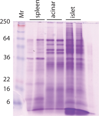
Figure 1. Protein gel.
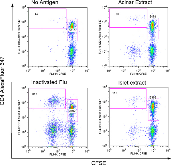
Figure 2. Results from a CFSE-based proliferation assay against acinar and islet extract. Click here to view larger figure.
