This protocol describes a rapid and low material consuming procedure for the integration of transgenic extrachromosomal arrays into the Caenorhabditis elegans genome using ultra violet (UV) irradiation. Furthermore, this protocol is particularly well suited for transgenic lines that transmit extrachromosomal arrays at a high rate.
Microinjecting DNA into the cytoplasm of the syncytial gonad of Caenorhabditis elegans is the main technique used to establish transgenic lines that exhibit partial and variable transmission rates of extrachromosomal arrays to the next generation. In addition, transgenic animals are mosaic and express the transgene in a variable number of cells. Extrachromosomal arrays can be integrated into the C. elegans genome using UV irradiation to establish nonmosaic transgenic strains with 100% transmission rate of the transgene. To that extent, F1 progenies of UV irradiated transgenic animals are screened for animals carrying a heterozygous integration of the transgene, which leads to a 75% Mendelian transmission rate to the F2 progeny. One of the challenges of this method is to distinguish between the percentage of transgene transmission in a population before (X% transgenic animals) and after integration (≥75% transgenic F2 animals). Thus, this method requires choosing a nonintegrated transgenic line with a percentage of transgenic animals that is significantly lower than the Mendelian segregation of 75%. Consequently, nonintegrated transgenic lines with an extrachromosomal array transmission rate to the next generation ≤60% are usually preferred for integration, and transgene integration in highly transmitting strains is difficult. Here we show that the efficiency of extrachromosomal arrays integration into the genome is increased when using highly transmitting transgenic lines (≥80%). The described protocol allows for easy selection of several independent lines with homozygous transgene integration into the genome after UV irradiation of transgenic worms exhibiting a high rate of extrachromosomal array transmission. Furthermore, this method is quite fast and low material consuming. The possibility of rapidly generating different lines that express a particular integrated transgene is of great interest for studies focusing on gene expression pattern and regulation, protein localization, and overexpression, as well as for the development of subcellular markers.
Transgenes are extensively used in C. elegans for a large range of applications. Transgenic strains are typically generated by DNA injection into the syncytial hermaphrodite gonad1,2. The DNA carrying the transgene of interest is coinjected along with plasmids encoding coinjection markers1,3,4. Coinjection markers are established transgenes leading either to the expression of a fluorescent protein or to a specific behavioral or morphological phenotype. The injected DNAs rearrange to form multi copy extrachromosomal arrays; thus the transgene of interest and the coinjection marker are transmitted together5,4. Transgenic lines are selected in the F2 generation of injected animals by following the phenotype induced by the expression of the coinjection marker (fluorescence or specific phenotype)1. Transgenic lines exhibit partial and variable transmission rates of the extrachromosomal arrays to the next generation. Depending on the strain, 10-100% (100% being a rare event) of the animals inherit extrachromosomal arrays. In addition, transgenic animals are mosaic and express the transgene in a variable number of cells. This mosaicism is likely to be correlated to the transmission rate to the next generation. Indeed, in both germ line and somatic cells the transmission rate depends on the segregation of the extrachromosomal arrays during cell divisions. One way to avoid this issue is to integrate the transgene into the genome. Classically, the integration of a transgene into a chromosome relies on irradiation (ultraviolet or gamma) of transgenic worms carrying extrachromosomal arrays3. Briefly, 50-100 hermaphrodite animals are irradiated at the L4 larval stage. In the F1 generation 200-800 transgenic animals are selected and individually cultured onto plates. According to Mendelian segregation, transgenic F1 animals carrying a heterozygous integration of the transgene into the genome would produce 1/2 of transgenic descendants that are heterozygous for the integrated array, 1/4 of transgenic descendants being homozygous for the integrated array, and 1/4 of descendants that have not integrated the array (Figure 1). Thus, plates containing the F2 generation are visually screened for ≥75% transgenic animals as observed by the expression of the coinjection marker. About 1/3 of the F2 animals selected for being integrant candidates are assumed to be homozygous for the integrated array and to produce 100% of homozygous F3 animals. Hence, from each selected plate, three to eight transgenic F2 animals are individually cultured and plates with 100% of transgenic descendants are selected. Next, eight F3 animals are individually cultured to confirm the 100% inheritance of the transgene.
The main disadvantages of this method are that
The irradiation of worms presumably induces double-strand breaks in the DNA and integration of extrachromosomal arrays into chromosomes occurs during DNA repair. Thus, it is likely that chances to obtain successful irradiation mediated integration are directly proportional to the number of germ line cells carrying an extrachromosomal array. Strains with a high transmission rate may contain more transgenic DNA arrays than strains with a low transmission rate as suggested in2, and exhibit a higher number of germ line cells carrying extrachromosomal arrays. Thus, we reasoned that in highly transmitting lines, transgenes might be easier to integrate than in lines with a low transmission rate. However, the standard protocol excludes the possibility of integrating transgenes with high transmission frequency as the percentage of transgenic animals with a nonintegrated extrachromosomal array can hardly be distinguished from that of the progeny of a F1 animal carrying a heterozygous integration of the transgene into the genome (Figure 1).
Here we show that for a particular transgene, the number of integrated lines recovered after irradiation with ultraviolet (UV) light is dependent on the initial percentage of transmission of the nonintegrated array. We present an improved protocol for UV irradiation-mediated transgene integration that is particularly relevant to lines exhibiting a high rate of transgene array transmission.
1. Culturing Transgenic Nematodes Prior to Integration
Transgenic lines can be generated as described in1. These lines usually contain a coinjection marker plasmid leading either to the expression of a fluorescent protein or to a specific behavioral or morphological phenotype at a given development stage1,3. This integration protocol can be applied to strains expressing fluorescent or nonfluorescent coinjection markers. In the example to follow, a strain with a fluorescent coinjection marker was used.
2. UV Irradiation and Recovery of Transgenic Worms
3. Isolation of Integrated Transgenic Lines
Integrations of transgenes in two different lines transmitting extrachromosomal arrays at about the same frequency (50-60%) were performed using the standard and the improved protocols (Table 1). The standard protocol allowed the recovery of three independent integrated lines after UV irradiation of 91 transgenic L4 animals, while the improved protocol allowed the recovery of one integrated line after UV irradiation of 30 transgenic L4 animals. This suggests that the efficiency of integration is similar for both protocols (one integrated line per 30 irradiated L4 animals). However, we cannot rule out the possibility that one protocol could be more efficient than the other when compared for the integration of the same transgene. The main difference between the protocols is that with the improved method all F1 plates exhibiting fluorescent progeny were selected regardless of the percentage of fluorescent F2 animals, while with the standard method a visual screen of F1 plates was performed and only plates with ≥75% transgenic F2 progeny were selected. This screening step is highly time consuming, in particular if the initial rate of transmission is close to 75%. In the improved protocol, it is sufficient to single four F2 worms from each F1 plate carrying transgenic F2 animals and to screen for plates with 100% transgenic progeny (F3), which can be performed quickly. In addition, the improved protocol allows for a decrease in the number of analyzed F2 and F3 animals when compared to the standard protocol. Altogether, the time and the number of plates necessary per integrated line are optimized in the improved protocol.
Next, we tested the hypothesis that a higher transgene transmission rate in a nonintegrated line renders easier the integration of the transgene into the genome. One particular transgene was integrated in lines transmitting at different rates using the improved protocol (Table 2). Gonads of wild type worms were injected with 10 ng/µl of a plasmid (pKG45) carrying the Pdyc-1::dyc-1::gfp transgene and with 30 ng/µl of the coinjection marker plasmid carrying Pmyo-2::dsRed10. Several transgenic strains were recovered and three of them that transmitted at different levels (10-15%, 50-60%, and >80%, respectively) were selected. The transgene was integrated in each strain using the improved protocol. The protocol was performed under the same condition for each strain, except that a higher number of transgenic L4 animals were irradiated for the line with the lowest transmitting rate (10-15%). For the highest transmitting line (>80%) three integrated lines were recovered from only 90 selected F1 animals, while for the transgenic line transmitting at 50-60%, one integrated line was recovered from 110 selected F1 animals. For the line with the lowest transmission rate, we could not recover a line carrying the integrated transgene, although 524 F1 animals were selected. Furthermore, the number of plates required per integrated line was three times lower with the highest transmitting line than with the line transmitting at 50-60% (Table 2).
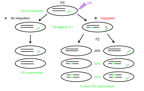
Figure 1. Transgene transmission in the progeny of transgenic worms with a nonintegrated or a heterozygous integrated transgene. A) Without integration, a transgenic animal will transmit the transgene at a frequency of X%, B) If integration of the transgene into a chromosome occurs; the F1 is most likely heterozygous for the integration and may carry or not the extrachromosomal array (as indicated by the question mark). The F2 progeny will be composed of at least 75% of fluorescent worms due to the Mendelian segregation. Worms without integrated transgene can be fluorescent, if they have retained the nonintegrated array (left column of Panel B).
Click here to view larger image.
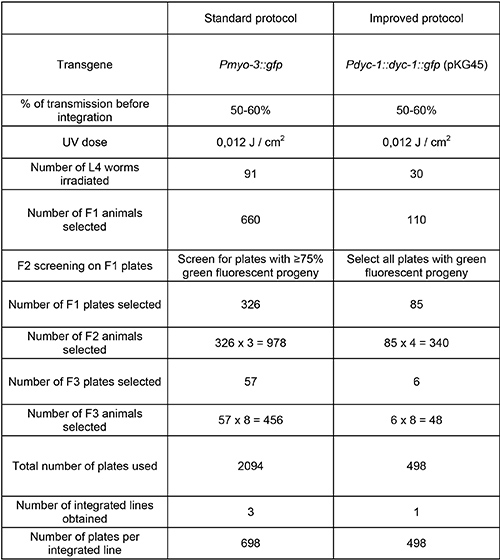
Table 1. Comparison of the standard and improved protocols for extrachromosomal arrays integration into the C. elegans genome. Two nonintegrated transgenes transmitting at about the same frequency were integrated using the standard or the improved protocol. The main difference lies in the fact that in the standard protocol a screen of the progeny on all F1 plates is performed to estimate the percentage of transgenic F2 worms, which should be ≥ 75%. This step takes a long time, particularly if the initial rate of transmission is close to 75%. In the improved protocol, it is sufficient to single four F2 worms from each F1 plate carrying transgenic F2 animals and to screen for 100% of transgenic worms in the progeny (F3), which can be performed quickly. The standard protocol is adapted from3.
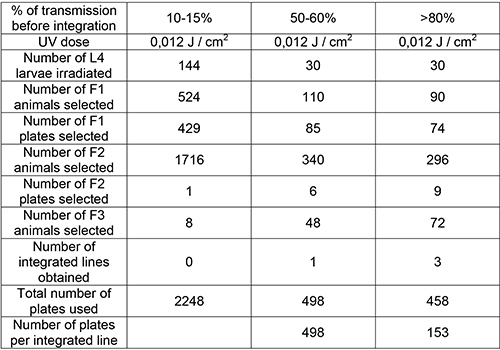
Table 2. Efficiency of transgene integration into the genome depends on the initial percentage of transgene transmission. Different nonintegrated lines, which transmitted the same transgene (Pdyc-1::dyc-1::gfp) at different rates (10-15%, 50-60%, and >80%, respectively) were used. Worms from each line were irradiated at the same UV dose (0.012 J/cm2) and the F3 generation screened for homozygous integration. The improved integration protocol applied to the highest transmitting line (>80%) recovered three integrated lines for only 74 selected F2 animals. The data presented with the line transmitting at 50-60% are those presented in Table 1 with the improved protocol.
The protocol described here provides an efficient method to integrate extrachromosomal transgenic arrays into the genome of C. elegans. Using different nonintegrated lines, which transmitted the same transgene Pdyc-1::dyc-1::gfp (pKG45) at different rates (10-15%, 50-60%, and >80%), we showed that the number of integrated strains recovered after UV irradiation is dependent of the initial level of transmission of the nonintegrated line (Table 2). Therefore, transgenes are easier to integrate in lines exhibiting high transmission rate.
Integrated transgenes are of interest for a wide range of purposes including studies related to gene regulation and gene expression pattern. Integrated transgenes are also useful tools to establish fluorescent subcellular markers. When integrating transgenes it can be crucial to isolate several independent lines. Several studies require introducing the transgene in different genetic backgrounds. However, the transgene could be inserted on a chromosome near to the gene of interest or create a mutation that may lead to an undesired phenotype in a given genetic background. It is therefore valuable to generate several independent integrated lines with an easy and rapid method.
Integration of extrachromosomal arrays, which are generally composed of multiple copies of the transgenes, allows for the overexpression of the respective transgenes. Strains that overexpress a given transgene are useful tools to study i) the subcellular localization of proteins, which exhibit a low endogenous expression level, ii) the effect of the overexpression of a protein on worm physiology, or iii) the effect of the overexpression of a protein in a particular tissue11. Several applications require however the use of low or single copy integrated transgenes that can be generated with alternative methods such as coinjection of the transgene of interest along with single stranded oligos2, bombardment12, Mos1 transposon mediated Single Copy gene Insertion (MosSCI)13 or Cas-9-triggered homologous recombination14. The irradiation of worms with UV induces chromosomal breaks and thereby mutations. To limit this undesirable effect, doses between 0.010 and 0.015 J/cm2 may be used, which allows recovering of about 80-90% of healthy and fertile animals. Effects on worm physiology of the dose delivered depend on the distance between the UV lamp and the animals, which may vary between different UV cross linkers. Similar doses may lead to experimental variations if the conditions of irradiation are not carefully controlled. Thus, it is important for each lab to determine the most suited UV dose for its particular settings and strains. Irradiated worms presenting fertility and/or survival rates close to that of nonirradiated worms indicate that the UV dose used was too low. Conversely, a high mortality rate (more than 40% of the animals) after UV irradiation indicates that the UV irradiation dose is potentially too high. Importantly, we noticed that worms placed at 15 °C for 24 hr after irradiation at 15° were healthier and gave greater progeny than worms directly grown at 23 °C. Thus, a recovery step at 15 °C after irradiation as presented here is critical for the survival and health of the animals. Alternatively, gamma irradiation can be used instead of UV irradiation to integrate extrachromosomal arrays in the C. elegans genome. This method requires however more sophisticated equipment3.
Several reasons could be considered in case the improved protocol presented here fails to recover integrated transgenic lines. The number of irradiated P0 animals or the number of selected F1 or F2 animals may be insufficient to recover integrated lines. It is also possible that nontransgenic eggs or larvae were carried along during transgenic F2 animals picking, thus leading to false negatives in the F3 progeny. Finally, some transgenes may be toxic, when expressed at high levels. Another pitfall of the integration procedure can come from toxicity of the transgene, especially when a strong promoter is used to express the gene of interest. The toxicity of a transgene can be masked by its mosaic expression in nonintegrated lines but revealed in integrated lines provoking the death of the animals. In this case aforementioned alternative methods could be used to produce lines with low copy integrated transgenes.
The presented protocol was optimized for the integration of extrachromosomal arrays in highly transmitting lines (>80%) that have higher chances to integrate a transgene into their genome. However, for particular transgenes, highly transmitting lines may not be available after worm transformation. When using lines transmitting the transgene at a rate of ≤50%, the standard method that includes an extra screening step of F1 plates may be more appropriate for successful transgene integration.
The authors have nothing to disclose.
Authors would like to acknowledge Claire Lecroisey and Laurent Ségalat for generating nonintegrated Pdyc-1::dyc-1::gfp; Pmyo-2::dsRed and Pmyo-3::gfp transgenic strains. We thank Andy Fire for the gift of the Pmyo-2::dsRed plasmid and Pamela Hoppe for the gift of the Pmyo-3::gfp plasmid. This work was founded by the French National Center of Scientific Research (CNRS) and the Claude Bernard University of Lyon.
Agar | Prolabo | 20768.292 | for NGM (nematode growth medium) preparation (6,7) |
NaCl | Chimie-Plus | 40045 | for NGM preparation |
Bacto-peptone | BD | 211820 | for NGM preparation |
KH2PO4 | Roth | 3904-1 | for NGM preparation |
K2HPO4 | Prolabo | 26930.293 | for NGM preparation |
MgSO4 | Roth | P027.2 | for NGM preparation |
CaCl2 | Prolabo | 22317.297 | for NGM preparation |
Cholesterol | Rectapur | for NGM preparation | |
LB-broth | Sigma | L3022 | for culture of OP50 bacteria |
EQUIPMENT | |||
Name of Equipment | Company | Catalogue number | Comments |
U.V. cross-linker | Fischer Scientific | Wavelength 254 nm | |
Microscope SteREO Discovery V8 | Carl Zeiss, Inc. | ||
Petri dishes, 60 mm | CML | BPS55E6 | |
Platinium wire 0.25 mm | Alfa Aesar | 7440-06-4 | To make a pick for worms (7) |