Contrast Enhanced Ultrasound imaging is a reliable in-vivo tool for quantifying spinal cord blood flow in an experimental rat spinal cord injury model. This paper contains a comprehensive protocol for application of this technique in association with a contusion model of thoracic spinal cord injury.
Reduced spinal cord blood flow (SCBF) (i.e., ischemia) plays a key role in traumatic spinal cord injury (SCI) pathophysiology and is accordingly an important target for neuroprotective therapies. Although several techniques have been described to assess SCBF, they all have significant limitations. To overcome the latter, we propose the use of real-time contrast enhanced ultrasound imaging (CEU). Here we describe the application of this technique in a rat contusion model of SCI. A jugular catheter is first implanted for the repeated injection of contrast agent, a sodium chloride solution of sulphur hexafluoride encapsulated microbubbles. The spine is then stabilized with a custom-made 3D-frame and the spinal cord dura mater is exposed by a laminectomy at ThIX-ThXII. The ultrasound probe is then positioned at the posterior aspect of the dura mater (coated with ultrasound gel). To assess baseline SCBF, a single intravenous injection (400 µl) of contrast agent is applied to record its passage through the intact spinal cord microvasculature. A weight-drop device is subsequently used to generate a reproducible experimental contusion model of SCI. Contrast agent is re-injected 15 min following the injury to assess post-SCI SCBF changes. CEU allows for real time and in-vivo assessment of SCBF changes following SCI. In the uninjured animal, ultrasound imaging showed uneven blood flow along the intact spinal cord. Furthermore, 15 min post-SCI, there was critical ischemia at the level of the epicenter while SCBF remained preserved in the more remote intact areas. In the regions adjacent to the epicenter (both rostral and caudal), SCBF was significantly reduced. This corresponds to the previously described “ischemic penumbra zone”. This tool is of major interest for assessing the effects of therapies aimed at limiting ischemia and the resulting tissue necrosis subsequent to SCI.
Traumatic spinal cord injury (SCI) is a devastating condition leading to significant impairment in motor, sensory and autonomous functions. To date, no therapy has demonstrated its efficiency in patients. For such reason, it is important to identify new techniques that will improve the assessment of potential treatments and can further elucidate injury pathiophysiology1.
SCI is divided into two sequential phases, referred to as primary and secondary injuries. The primary injury corresponds to the initial mechanical insult. Whereas the secondary injury groups a cascade of various biological events (such as inflammation, oxidative stress and hypoxia) that further contribute to the progressive expansion of the initial lesion, tissue damage and therefore neurological deficit2,3.
At the acute phase of SCI, neuroprotective therapies are aimed at reducing the secondary injury pathology and should accordingly improve neurological outcomes. Among the many secondary injury events, ischemia plays a crucial role 4,5. At the level of the SCI epicenter, the damaged parenchymal microvessels impede effective spinal cord blood flow (SCBF). Moreover, SCBF is also significantly reduced in the region surrounding the injury epicenter, an area specifically known as the “ischemic penumbra zone”. If SCBF cannot be quickly restored within these regions, ischemia can lead to supplementary parenchymal necrosis and further nervous tissue damage. As even the slightest tissue preservation can have substantial effects of function, it is of major interest to develop drugs and therapies that can reduce ischemia post-SCI. To highlight this phenomenon, previous work has shown that preservation of only 10% of myelinated axons was enough to permit walking in cats post-SCI 6.
Although several techniques have been described to assess SCBF, they all have significant limitations. For example, the use of radioactive microspheres7,8 and C14-iodopyrine autoradiography9 requires subsequent animal sacrifice and cannot be repeated at later time-points. The hydrogen clearance technique10 depends on the insertion of intraspinal electrodes, which may further damage the spinal cord. While laser Doppler imaging, photoplethysmography14,15 and in-vivo light microscopy16 have a very limited depth/area of measurement11-13.
Our team has previously shown that contrast enhanced ultrasound (CEU) imaging can be used to assess real time and in-vivo the SCBF changes in the rat spinal cord parenchyma17. It is important to note that a similar technique was applied by Huang et al. in a porcine model of SCI18. CEU applies a specific mode of ultrasound imaging which allows to associate grayscale morphological images (obtained by the conventional B-mode) with spatial distribution of blood flow 19. The SCBF imaging and quantification relies on intravascular injection of echo-contrast agents. The contrast agent is made up of sulphur hexafluoride microbubbles (mean diameter of about 2.5 μm and 90% having a diameter less than 6 μm) stabilized by phospholipids. The microbubbles reflect the ultrasound beam emitted by the probe thus enhancing blood echogenicity and increasing contrast of the tissues according to their blood flow. It is therefore possible to assess the blood flow in a given region of interest according to the intensity of the reflected signal. The microbubbles are also safe and they have been clinically applied in humans. The sulphur hexafluoride is quickly cleared (mean terminal half-life is 12 min) and more than 80% of the administered sulphur hexafluoride is recovered in exhaled air within 2 min after injection. This protocol provides a simple way to use CEU imaging to assess SCBF changes in rat.
NOTE: The methods described in this manuscript were approved by the bioethics committee of the Lariboisière School of Medicine, Paris, France (CEEALV/2011–08-01).
1. Instrument Preparation
2. Jugular Vein Catheterization (Figure 3)
3. Accessing the Spine, Laminectomy and Rat Positioning (in the 3D-frame)
4. CEU Probe Positioning
5. Preparation of Contrast Agent – Microbubble Reconstitution
6. Assessment of SCBF in the Intact Spinal Cord
7. Experimental SCI
8. Assessment of SCBF 5 min Post-SCI
9. Animal Sacrifice
10. Quantification of SCBF by Offline Analysis
With the protocol described above, it is possible to map the SCBF along a longitudinal spinal cord sagittal segment.
In the intact spinal cord, there appears to be SCBF irregularities within the parenchyma (Figure 12). This can be explained by the variable distribution of radiculo-medullary arteries (RMA) from one animal to another. RMA refers to segmental arteries that reach the anterior spinal artery (ASA) and therefore provide blood supply to the spinal cord parenchyma. In contrast, the radicular arteries correspond to segmental arteries, which do not reach the ASA and therefore do not provide spinal cord blood supply. Therefore, in spinal cord segments where the RMA anastomoses with the ASA, there is more blood flow (as shown in our results).
After SCI, real-time CEU imaging shows a deficiency in circulation at the injury epicenter. The epicenter remains dark (no contrast agent signal), as there is no active blood flow. More detailed analysis of the blood flow using several ROIs shows three unique blood flow territories. First, at the level of the epicenter, the blood flow is the lowest with a mean decrease of about -90%. Second, in the territories adjacent to the epicenter (both rostral and caudal), SCBF was also significantly decreased (ranging from -50% to -80%). Third, in the most remote areas corresponding to intact tissue, SCBF is preserved. The second region corresponds to the “ischemic penumbra zone”, which should be the target of potential neuroprotective therapies. Being able to readily visualize and quantify SCBF changes post-SCI is useful for assessing the efficiency of therapies aimed at reducing tissue ischemia, and therefore highlights the importance of this technique (Figure 13).
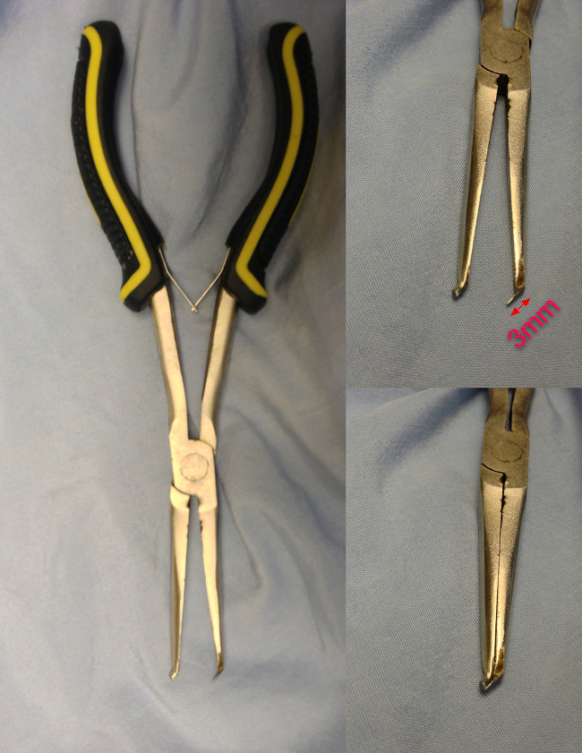
Figure 1. The custom-made bone cutter for laminectomies. The thin blade is designed to slide beneath the lamina. Please click here to view a larger version of this figure.
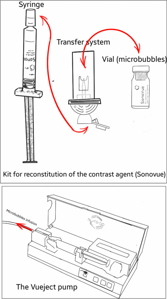
Figure 2. Schematic representation of the kit for microbubbles reconstitution and the Vueject° pump used for microbubbles infusion. The transfer system allows for the delivery of microbubbles and saline between the vial and the syringe. Please click here to view a larger version of this figure.
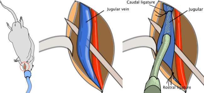
Figure 3. Jugular catheter. The catheter is to be inserted in the jugular vein, then pushed toward the heart and finally fasten with a knot. Please click here to view a larger version of this figure.
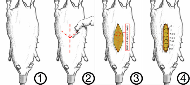
Figure 4. Method for correct identification of the vertebral levels. In the rat, the last rib is attached to the XIIIth vertebra. The latter can be palpated through the skin as a landmark for the last thoracic vertebra, the XIIIth. Muscles are detached on either side of the spinous processes. Please click here to view a larger version of this figure.
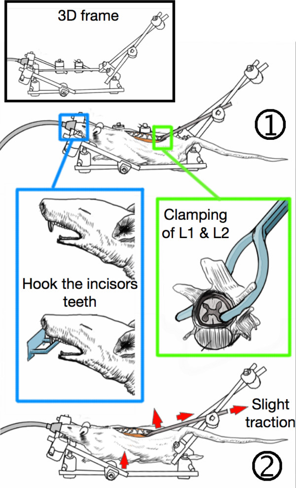
Figure 5. Stabilization of the animal in the 3D-frame. (1) The incisor teeth are hooked on the frame while the first and second lumbar vertebras (L1 & L2) are clamped with custom-made forceps. (2) The lumbar spine is slightly tightened which stabilizes the animal and elevates the thorax from the bench, thereby allowing free respiratory motions without spine movements. Please click here to view a larger version of this figure.
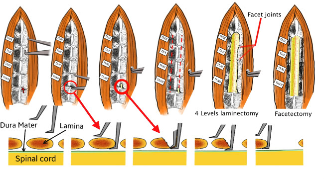
Figure 6. Technical details of the laminectomy. First, the thin blade of the custom-made bone cutter is passed beneath the lamina without damaging the spinal cord. Then the bone cutter is closed, which cuts and removes a part of the lamina. The procedure is repeated on both sides and from ThXII to TxIX in order to achieve a four-level laminectomy. Finally, the facet joints are also removed. Please click here to view a larger version of this figure.
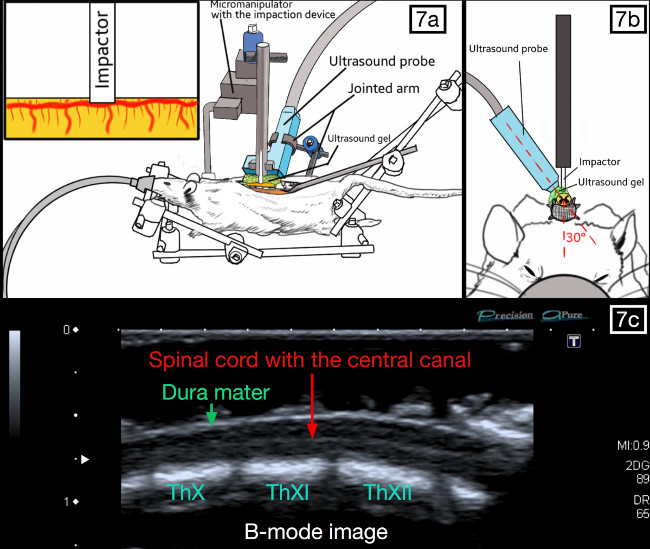
Figure 7. Positioning of the ultrasound probe and the impaction device. The probe is parallel to the spinal cord and slightly oblique (20-30°), so that the weight-drop impactor can be placed against the posterior aspect of the dura. The spinal cord should be visible with the central canal present throughout the middle segment on the ultrasound imaging “B-Mode”. Please click here to view a larger version of this figure.
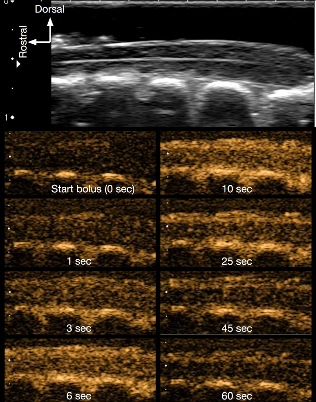
Figure 8. Contrast imaging of the intact spinal cord. The successive figures in contrast mode (orange coloured images) show how the contrast agent (microbubbles) progressively appears following the infusion, thereby enhancing the contrast of the spinal cord. Bolus infusion lasts about 10 sec and the contrast data was recorded for 1 min. Please click here to view a larger version of this figure.
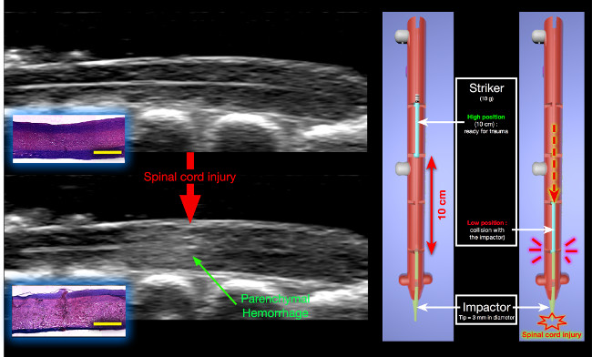
Figure 9. Changes in B-mode following experimental SCI. A hyperechoic lesion appears inside the parenchyma, corresponding to the initial parenchymal hemorrhage post-SCI. Histology (H&E staining) : The hemorrhage results from massive traumatic disruption of small blood vessels leading to blood extravasation in the parenchyma (yellow scale bar = 2,000 μm). The impaction device is shown on the right. The striker is released from a 10 cm height and collides with the impactor that subsequently generates the spinal cord injury. Please click here to view a larger version of this figure.
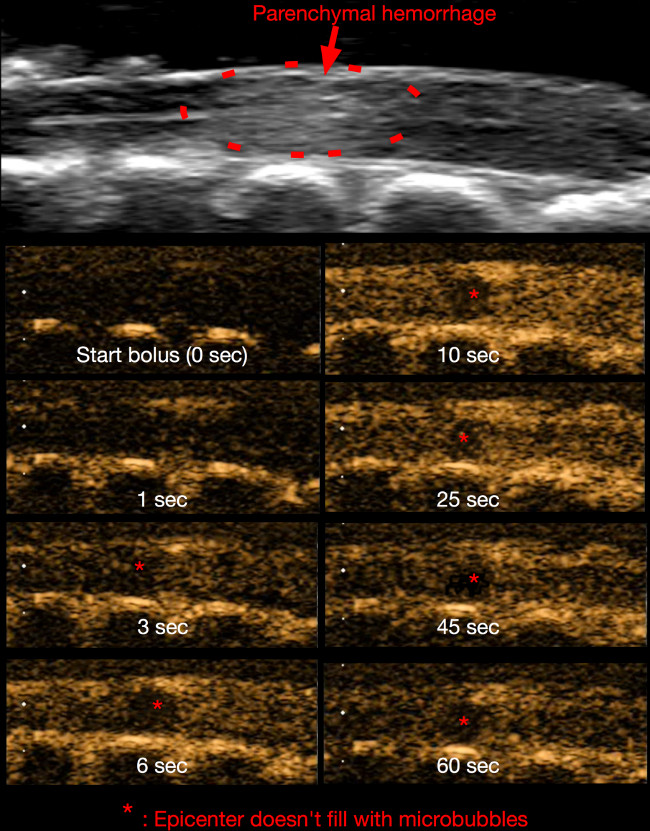
Figure 10. Contrast imaging 15 min post-SCI. Similar to Figure 8, the microbubbles are visible as they pass through the spinal cord microvasculature. At the epicenter (asterisk), the blood flow is obstructed by microvascular disruption. Please click here to view a larger version of this figure.
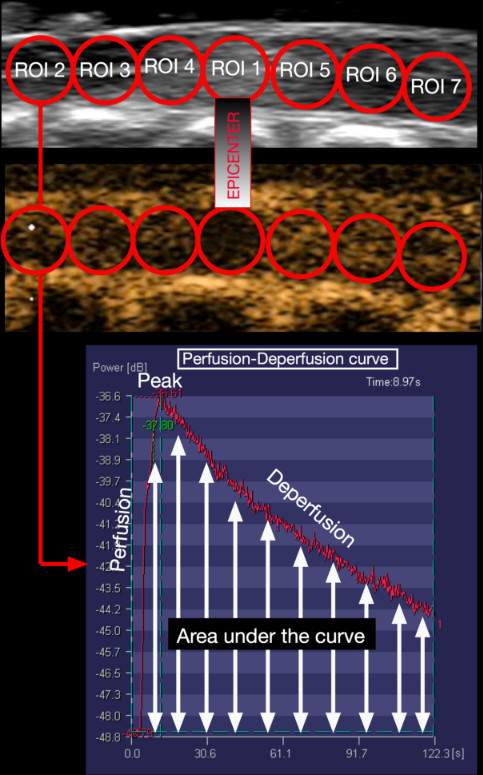
Figure 11. Protocol for SCBF quantification. With Ultra-Extend Software, seven circular and adjacent regions of interest (ROI) are drawn on the longitudinal spinal cord image. The first ROI is placed on the injury epicenter. In each ROI, the software generates a perfusion-deperfusion curve and calculates the area under this curve. This value correlates with the blood flow in this area. Please click here to view a larger version of this figure.
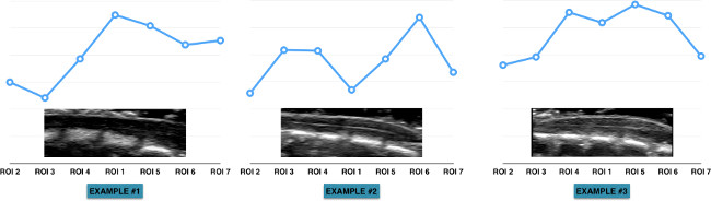
Figure 12. Heterogeneity of the blood flow along the spinal cord. These graphs display the heterogeneity of spinal cord blood flow as well as the variability between animals. This can be largely explained by the vascular anatomy of the spinal cord. However, due to the heterogeneity and variable vascular anatomy, one must use the blood flow measurements (from each ROI) prior to injury as the baseline. The measurements made at the following time-points (post-SCI) are expressed as the percentages change of the baseline. Please click here to view a larger version of this figure.
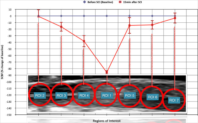
Figure 13. Changes in spinal cord blood flow (SCBF) induced by the experimental spinal cord injury (SCI). 15 min after SCI there is critical ischemia at the level of the epicenter while SCBF remained preserved in the more remote intact areas. In the regions adjacent to the epicenter (both rostral and caudal), SCBF is significantly reduced. This corresponds to the previously described “ischemic penumbra zone”. Please click here to view a larger version of this figure.
Although we have described how to use CEU in a rat SCI contusion model, this protocol can be modified to fit other experimental objectives or SCI models. We have chosen to measure SCBF at only two time points (before injury and 15 min post-SCI), however the number of time points and the delay between SCBF measurements can be adapted to fulfill the needs of other studies. For example, in our previous work 17, we have measured SCBF at five successive time points throughout the first hour post-SCI. It is important to note that in the sham group (no SCI), we were surprised to observe a progressive decrease in SCBF. While we initially feared that repeated microbubble infusion might harm the spinal cord vasculature, further experimentation (unpublished data) confirmed that these changes were caused by progressive alterations in local tissue physiological conditions (temperature, hydration) induced by the laminectomy, as well as the prolonged exposition of the dura and surrounding tissue to the ambient air and the ultrasound gel. These problems are common in all experiments dealing with microcirculation, as the circulation is extremely sensitive to many parameters and therefore prone to vascoconstriction or vasodilatation. Therefore, we recommend that the period during which the surgical wound remains open is as short as possible. If multiple SCBF measurements are needed over a prolonged period, it would be preferable to close the animal incision between the acquisitions in order to restore physiological conditions around and inside the spinal cord.
It is also possible to modify the shape, size, location and the number of ROIs for SCBF analysis. One of the major advantages of CEU is that the measurements can be made anytime following experimental completion by processing the recorded data offline. It is also possible to repeat the measurements or to modify the measurement settings/standards if necessary.
In the protocol described here, we applied a contusion model of SCI. However, there are other models, including clip compression or cord transection 21 that can be easily adapted to measure SCBF with this protocol. Once the spinal cord is injured, one simply needs to place the ultrasound gel on the dura mater and position the ultrasound probe. We also choose to measure SCBF at the lower thoracic level because it corresponds to the model that we currently use in our lab. However, the same technique can be used at other levels of the spinal cord. Since the whole spine is stabilized between the lumbar spine (clamping at L2) and the incisors teeth, one simply needs to make a laminectomy at the desired level and position the probe accordingly.
Spatial resolution of ultrasound imaging is proportional to the frequency of the ultrasound waves. The higher the ultrasound frequency, the better the spatial resolution. We have used a high-frequency (12-14 MHz) probe, which provides an image with a pixel resolution of about 100 µm. With very-high-resolution systems, the frequency increases up to 55 MHz and each pixel is about 20 µm 20. Such devices can also be used for CEU, which depict much more precisely the distribution of SCBF in the parenchyma. However, the very-high-resolution systems are much more expensive.
Several other techniques have been proposed to measure SCBF in SCI, but they all have unique limitations. Some, such as radioactive microspheres7,8 or the C14-iodo-antipyrine autoradiography9, require animal sacrifice. In these cases, the spinal cord must be harvested for analysis. On the other hand, the hydrogen clearance technique10, requires intraspinal electrode insertion which may actually modify the SCBF. Moreover, the measurement can only be made in a very restricted region of the spinal cord parenchyma. Light microscopy through a spinal window also provides a way to assess microcirculation, but this approach has a very restricted depth of observation. It only allows to observe the circulation in the superficial pia matter and not within the parenchyma 16.
In literature, real time in-vivo assessments of SCBF are usually performed by laser Doppler imaging 11-13. However, even this technique has several limitations. Firstly, since the laser is less than 1 mm in diameter, SCBF can only be assessed in a very restricted area corresponding to a semi-sphere of about 1 mm in diameter. Since the rat’s spinal cord is about 3 mm in diameter, the limited area of analysis is a major constraint. Moreover, as we have shown that SCBF in the intact spinal cord is not homogeneous, it is important to measure SCBF in a larger area for a proper representation of tissue microcirculation. Secondly, the laser has a limited depth of penetration and therefore detects superficial SCBF. As a result, it not only measures parenchymal SCBF but also that of the pia mater (that surrounds the parenchyma). Since the pia mater has a unique vascular system and is not subjected to the same auto-regulatory mechanisms as parenchymal vessels, this information may be misleading. Lastly, the laser-Doppler doesn’t provide any morphological information. CEU overcomes such limitations by displaying morphological images of the cord (B-mode), while uniquely presenting the contrast agent that can be clearly identified within the parenchyma.
Despite its many advantages to other approaches, CEU also has some distinct limitations. Since measurements are made on a bi-dimensional sagittal slice (usually parallel to the central canal), SCBF from other regions of the parenchyma are inaccessible. Further, the information generated by a single bi-dimensional sagittal spinal cord segment may not be representative of the entire cord. Nevertheless, this can be controlled by several precautions. First, by repeating measurements at the same location, the first measurement made (intact spinal cord) can be used as a baseline value. Second, by injuring at the spinal cord midline (bilateral injury), the SCBF changes should be symmetrical between left and right (unpublished data). These precautions help ensure that analysis of single sagittal slice is enough to reflect the global longitudinal distribution of SCBF.
The high cost of ultrasound machines is another limitation. However, several solutions exist to target this problem. First, some labs can negotiate a temporary loan by the manufacturer for their experiments. As ultrasound machines are transportable, temporary loans are possible. This has been the approach used by our lab. Alternatively, a group of labs can pool resources to buy the machine and split the costs. Otherwise, many university institutions have imaging facilities and ultrasound machines can be recommended as essential tools. Thus, animals can be transported the imaging facility for CEU assessment and then brought back for other experiments.
To assess vascular changes, contrast agent (microbubbles) must be injected intravenously. Although catheterization of the jugular or femoral vein is invasive and risky, the veins are easily accessible and clearly identifiable. In contrast, tail vein injection is much less invasive, but the vessel is poorly distinguished/visible for proper catheterization. Therefore, there is a risk that the needle tip will not be properly placed inside the vein or that it may move during injection, compromising the entire experiment. For such reason, we prefer to use the jugular vein and introduce a catheter for consistent microbubble infusion.
Vertebra bones surround the spinal cord. As ultrasound waves are reflected by bone and cannot pass through the spinal cord laminas, imaging requires bone removal (laminectomy) to open an acoustic window. The easiest way to open the vertebral canal is to remove the posterior arch of the vertebra through a laminectomy. In this protocol, we require a four-level laminectomy to visualize a long segment of spinal cord, including the epicenter, the penumbra zone and remote areas of intact spinal cord. Although a majority of experimental SCI models require a laminectomy (for clip application or impactor contusion), these usually consist of removing 1-2 lamina. The extensive 4-level laminectomy is another limitation of our study. However, if one only needs to study the epicenter and penumbra zone, a less extensive laminectomy can be made and is recommended.
In conclusion, despite the several limits described above, CEU is a useful tool for assessing SCBF changes and the effect of various therapies (research purposes). This reliable, real-time, in-vivo approach is ideal for looking at treatments to reduce ischemia and subsequent tissue necrosis post-SCI.
The authors have nothing to disclose.
We acknowledge Stephanie Gorgeard, Thierry Scheerlink (Toshiba France), and Christophe Lazare (Bracco France).
Name of Reagent/ Equipment | Company | Comments/Description | |
External Fixator Hoffman 3 | Stryker, Kalamazoo, USA | Modular system used to build the custom made 3D frame and the jointed arm holding the ultrasound probe | |
Toshiba Applio | Toshiba, Tokyo, Japan | Ultrasound machine | |
Sonovue | Bracco, Milan, Italy | Contrast agent : microbubbles | |
Vueject pump | Bracco, Milan, Italy | Electric pump for infusion of microbubbles bolus | |
Aquasonic Ultrasound Gel | Parker Laboratories, Fairfield, NJ, USA | Ultrasound gel used to transmit the ultrasound waves | |
Isovet | Piramal Healthcare, Mumbai, India | Isoflurane used for anesthesia | |
Ultra Extend | Toshiba, Tokyo, Japan | Software used for quantification of spinal cord blood flow | |
Mastercraft Five-piece Mini-pliers Set, Product #58-4788-6 | Canadian Tire, Toronto, Canada | Set of pliers for Do-it-yourself job |