Summary

Een endotheliale Planar Cell Model for Imaging Immunologische Synapse Dynamics

Published: December 24, 2015
doi:

Summary

Adaptive immunity is controlled by dynamic ‘immunological synapses’ formed between T cells and antigen presenting cells. This protocol describes methods for investigating endothelial cells both as understudied physiologic APCs and as a novel type of ‘planar cellular APC model’.

Abstract

Adaptieve immuniteit wordt geregeld door dynamische interacties tussen T-cellen en antigeen-presenterende cellen ("APC's") genoemd "immunologische synapsen. Binnen deze intieme cel-cel-interfaces discrete sub-cellulaire clusters van MHC / Ag-TCR, F-actine, hechting en signaalmoleculen vormen en snel te verbouwen. Deze dynamiek wordt gedacht dat ze kritische factoren voor zowel de efficiëntie en kwaliteit van de immuunresponsen die zich ontwikkelen en dus van beschermende immuniteit tegen pathologische. Huidige begrip van immunologische synapsen met fysiologische APC's wordt beperkt door de ontoereikendheid van de verkregen beeldresolutie. Hoewel kunstmatig substraat modellen (bijvoorbeeld vlakke lipidendubbellagen) bieden een uitstekende resolutie en zijn zeer waardevolle hulpmiddelen geweest, ze zijn inherent niet-fysiologische en ongenuanceerd. Vasculaire en lymfatische endotheelcellen naar voren zijn gekomen als een belangrijke perifere weefsels (of stromale) compartiment van 'semi-beroepal APCs. Deze APC's (die het merendeel van de moleculaire machinerie van professionele APC's tot expressie) hebben de unieke eigenschap van het vormen nagenoeg vlak celoppervlak en gemakkelijk te transfecteren (bijvoorbeeld met fluorescente proteïne reporters). Hierin een fundamentele benadering van endotheelcellen als nieuw en fysiologische "vlakke cellulaire APC model" voor verbeterde beeldvorming en ondervraging van fundamentele antigene signaleringsprocessen uitvoering worden beschreven.

Introduction

T lymfocyten een tak van het adaptieve immuunsysteem gekenmerkt door het vermogen om efficiënt te herkennen peptide-antigeen (Ag) gebonden aan major histocompatibility complex (MHC) moleculen door hun T-celreceptoren (TCR's) 1. Naïeve lymfocyten constitutief migreren en scannen 'professionele Ag presenterende cellen (APC's, bijvoorbeeld, dendritische cellen) in lymfeklieren, terwijl het geheugen / effector-T-cellen nodig hebben om effectief te overzien een zeer breed scala van APC's en potentieel doelwit cellen in perifere weefsels.

In de min na de eerste opname van verwante Ag op een APC, lymfocyten arresteren hun migratie en beginnen aan een gespecialiseerde intieme cel-cel-interface genaamd 'immunologische synaps' vormen (IS). Aanhoudende (dwz 30-60 min) IS contacten nodig zijn om te versterken en te ondersteunen signalering 2-7. Opkomende studies identificeren die binnen de IS, is de continue vorming en snelle remodeling van discrete sub-cellulaire signalering micro-clusters (dwz bevattende MHC / Ag-TCR, F-actine, adhesie en signaalmoleculen) de sterkte en de kwaliteit van de verkregen immuunreacties 07/02 bepalen. Echter, dynamische details en regulerend mechanisme van dit proces onvolledig begrepen 8,9. Dit vloeit grotendeels voort uit de technische uitdagingen in verband met onregelmatige topologieën van APC oppervlakken en slecht gecontroleerde oriëntatie van de cel-cel interactie vliegtuigen, kwesties die diep beperken de vereiste spatiotemporele imaging benaderingen 8-10 (Figure1A).

Figuur 1

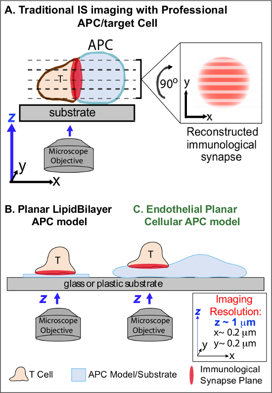
Figuur 1. Een Fysiologische Planar Cell APC Model for Imaging Immunologische Synapse Dynamics. Het schema illustreert traditionele beeldvorming van immunologische synaps tussen een T-cel en een professio nal APC (A) en T-cel en een traditionele vlakke lipide dubbellaag APC model (B) in relatie tot deze nieuwe endotheliale vlakke APC model (C). Professionele APCs bieden fysiologische immunologische synapsen maar bieden weinig georiënteerde cel-interface (dat wil zeggen ten opzichte van de optimale xy beeldvlak; resolutie ~ 0,2 pm), die dramatisch compromissen ruimtelijke (z beeldvlak resolutie ~ 1 micrometer) en tijd (dat wil zeggen, vanwege de noodzaak om herhaaldelijk gescand door alle z beeldvlakken) oplossing van beeldvorming. Bilaag modellen hebben een vlakke topologie die optimale spatiotemporele beeldvorming resolutie voorziet, maar ook sterk vereenvoudigd, niet-fysiologische en rigide. Deze endotheelcellen model combineert de vlakke topologie van lipidendubbellagen met de fysiologische substraat van een klassieke APC om een ​​optimale ruimtelijke en temporele beeldvorming resolutie leveren in een fysiologische omgeving.m / files / ftp_upload / 53.288 / 53288fig1large.jpg "target =" _ blank "> Klik hier om een ​​grotere versie van deze figuur te bekijken.

Vorige werk is gedeeltelijk omzeild deze obstakels door het ontwikkelen vlakke ondergrond modellen (dwz lipidendubbellagen en antilichaam-gecoate oppervlakken) die optimale spatiotemporele resolutie (dwz bieden, via de vaststelling van de T-cel activatie oppervlak in een enkel plan dat parallel aan de optimale xy beeldvorming vliegtuig) 11-15 (Figuur 1B). Deze modellen hebben belangrijke inzichten vergemakkelijkt in de subcellulaire / moleculaire dynamica dat antigene signalering in T-cellen, met inbegrip van de ontdekking van dynamische actine / TCR signalering micro-clusters 7,11-14 controleren. Echter, dergelijke modellen inherent oversimplified, evenals stijve (evenwel de ontwikkeling / studie van 3-dimensionale topologische kenmerken) (Figuur 1B). Daarom blijft het onzeker hoe dergelijke bevindingen aan phy betreffensiologic cel-cel immune surveillance.

Hoewel nog weinig bestudeerd, vasculaire en lymfatische endotheelcellen zijn in opkomst als een grote (dwz, groter in aantal dan alle professionele APC's, door ~ 1000-voudig) perifere compartiment van 'semi-professioneel "APC's 16-18. Deze cellen brengen MHC-I, MHC-II en een veelheid aan co-stimulator moleculen (bijvoorbeeld CD40, LFA3, ICOSL, 4-1BB, OX40L, TL1A, PD-L1, maar niet CD80 en CD86) en strategisch gepositioneerd aan de bloed-weefsel interface waar ze dienen gespecialiseerde sentinel functies 16-18. Vorige studies aangetoond dat endotheelcellen effectief opnieuw stimuleren effector / geheugen, maar niet naïeve T-cellen 19-25. Aldus endotheelcellen waarschijnlijk unieke APC rollen in effector fase van adaptieve immuunreacties in de perifere weefsels, zoals lokale invloed op T-celactivering, differentiatie, geheugen en tolerantie 16,17,26. Critisch, wanneer gekweekt in vitro endotheliale cellen vormen vrijwel vlakke celoppervlakken en gemakkelijk te transfecteren (bijvoorbeeld met fluorescente proteïne reporters). Deze functies zijn ideaal voor hoge spatiotemporele resolutie beeldvorming van topologische dynamiek in cel-cel interacties 19,27. Zo endotheelcellen kunnen dienen als een fysiologische "vlakke cellulaire APC-model duidelijk geschikt voor het onderzoek van de subcellulaire / moleculaire remodeling mechanismen antigeenherkenning drijven en te reguleren responsen (figuur 1C) 19,20.

Eerder vastgestelde complementaire beeldvormingstechnieken (waaronder transfectie van endotheel cellen met fluorescent eiwit makers van de plasmamembraan en cytosol) voor het bestuderen van de details van leukocyt-endotheel interactie tijdens hechting en transendotheliale migratie 27, bleek dat leukocyten actief sonde het oppervlak van het endotheel door dynamische inbrengen van eend terugtrekken van sub-micron-schaal, actine-rijke cilindrische uitsteeksels (~ 200-1000 nm in diameter en diepte) genoemd invadosome-achtige uitsteeksels (dat wil zeggen, 'POPs') 27,28. Deze beeldvormingstechnieken zijn verder uitgebreid met de creatie protocollen te profiteren van endotheliale APC functie om de eerste werkwijzen voor spatiotemporele hoge resolutie afbeelding van de T-cel-endotheliale immunologische synaps gerapporteerde 19,20 ontwikkelen en verder hierin beschreven. Een centrale bevinding afgeleid van deze nieuwe vlakke cellulaire APC model is dat T-cel POPs functioneren, zowel in het bevorderen van de initiële Ag detectie en bij het instandhouden van de daaropvolgende signalering. Inderdaad, arrays van meerdere POPs (die werden gestabiliseerd en opgebouwd in reactie op calciumflux initiële) tonen verrijking in TCR en moleculen die wijzen op actieve signalering zoals PKC-Q, ZAP-70, fosfotyrosine en HS1. Daarom POPs lijken een driedimensionale fysiologische equivalent aan het TCR-signalering micro vertegenwoordigenclusters gezien op vlakke dubbellaag modellen. Deze aanpak, dus onthult gevoelig / rapporten moleculaire en architecturale (en impliciete biomechanische) dynamiek anders niet aantoonbaar.

De hierin beschreven werkwijze moet nuttig zijn om de kloof tussen professionele APC en kunstmatige APC substraat modellen om ons vermogen om fundamentele mechanismen van adaptieve immuunreacties ondervragen verbeteren zijn. Hoewel hier de nadruk op de activatie van CD4 + Th1-type effector / geheugencel kan deze basisaanpak gemakkelijk worden aangepast aan een breed scala van T-cel soorten en Ags bestuderen, zoals hieronder besproken.

Protocol

Alle experimenten beschreven in dit protocol worden uitgevoerd met primaire menselijke T-cellen en in de handel verkrijgbaar primaire menselijke endotheelcellen (huid of longen microvasculaire EC) Je verder onderzoeksprotocol met mensen moet worden goedgekeurd door een institutionele review board en schriftelijke geïnformeerde toestemming moet worden verstrekt uit elke bloeddonor. Experimenten uitgevoerd met behulp van dit protocol werd goedgekeurd door de IRB van Beth Israel Deaconess Medical Center. 1. Voorbere…

Representative Results

Een nieuwe imaging benadering middels endotheliale cellen en het combineren van de resolutie voordelen van de vlakke lipide dubbellagen model met de fysiologische complexiteit en vervormbaarheid van professionele APC werd ontwikkeld (figuur 1). Figuur 2 geeft voorbeelden van normale migratie, calcium flux en topologische dynamiek waargenomen met dit aanpak. Aangezien SAg op endotheel, SAg-specifieke CD4 + Th1 lymfocyten snel verspreiden, polar…

Discussion

Kortom, dit protocol beschrijft methoden voor het onderzoeken van endotheelcellen als i) weinig bestudeerd fysiologische APC's en ii) als een nieuw type 'vlakke cellulaire APC model'. Wat het eerstgenoemde is geworden steeds duidelijk zijn dat niet-hematopoietische perifere (of "stromale") APCs spelen cruciale, niet-redundante functies (bijv tegenover hematopoietische APCs) de vormgeving adaptieve immuunreacties 16-18. Onder dergelijke 'semi-professioneel "APC's, va…

Declarações

The authors have nothing to disclose.

Acknowledgements

We thank Dr. Peter T. Sage for his assistance in generating some of the representative images. This work was supported by an NIH R01 grant to C.V.C. (HL104006).

Materials

BD Vacutainer stretch latex free tourniquet BD Biosciences 367203
BD alcohol swabs BD Biosciences 326895
BD Vacutainer Safety-Lok BD Biosciences 367861 K2 EDTA
BD Vacutainer Push Button Blood Collection Set BD Biosciences 367335
RPMI-1640 Sigma-Aldrich R8758-1L
Ficoll-Paque  Sigma-Aldrich GE17-1440-02 Bring to RT before use
FCS-Optima Atlanta Biologics s12450 Heat inactivated
Penicillin-Streptomycin  Sigma-Aldrich  P4458-100ML  
Trypan blue Sigma-Aldrich T8154-20ML
staphylococcal enterotoxin B  Toxin Technology BT202RED Stock solution 1mg/ml in PBS
toxic shock syndrome toxin 1  Toxin Technology TT606RED Stock solution 1mg/ml in PBS
human IL-15 R&D Systems 247-IL-025 Stock solution 50ug/ml in PBS
PBS Life Technologies 10010-049
Fibronectin Life Technologies 33016-015 Stock solution 1mg/ml in H20
HMVEC-d Ad-Dermal MV Endo Cells Lonza CC-2543 Other Human Microvascular ECs can be used, i.e. HLMVECs
EGM-2 MV bullet kit Lonza CC-3202
Trypsin-EDTA Sigma-Aldrich T-4174 Stock solution 10x, dilute in PBS
amaxa-HMVEC-L Nucleofector Kit Lonza vpb1003 Required Kit for step 4
IFN-g Sigma-Aldrich I3265 Stock solution 1mg/ml in H20
TNF-alpha 10ug, human Life Technologies PHC3015 Stock solution 1mg/ml in H20
phenol Red-free HBSS  Life Technologies 14175-103
Hepes Fisher Scientific BP299-100
Calcium Chloride Sigma-Aldrich C1016-100G Stock solution 1M in H20
Magnesium chloride Sigma-Aldrich 208337 Stock solution 1M in H20
Human Serum albumin Sigma-Aldrich A6909-10ml
Immersol 518 F fluorescence free Immersion oil Fisher Scientific 12-624-66A
Fura-2 AM 20x50ug Life Technologies F1221 Stock solution 1mM in DMSO
pEYFP-Mem (Mem-YFP) Clontech 6917-1
pDsRed-Monomer (Soluble Cytoplasmic DsRed) Clontech 632466
pDsRed-Monomer Membrane (Mem-DsRed) Clontech 632512
pEGFP-Actin Clontech 6116-1
Alexa Fluor 488 Phalloidin Life Technologies A12379
Formaldehyde solution 37% Fisher Scientific BP531-500 Toxic, use fumehood
Triton X-100 Sigma-Aldrich X100-5ML
 Falcon 15mL Conical Centrifuge Tubes Fisher Scientific 14-959-70C
Falcon 50mL Conical Centrifuge Tubes Fisher Scientific 14-959-49A 
Falcon Tissue Culture Treated Flasks T25 Fisher Scientific 10-126-9
 Falcon Tissue Culture Treated Flasks T75 Fisher Scientific   13-680-65
 Corning Cell Culture Treated T175 Fisher Scientific 10-126-61 
Glass coverslips  Fisher Scientific 12-545-85  12 mm diameter
 Falcon Tissue Culture Plates 24-well Fisher Scientific 08-772-1
Delta-T plates Bioptechs 04200415B
Wheaton Disposable Pasteur Pipets Fisher Scientific 13-678-8D
1.5 ml Eppendorf tube  Fisher Scientific 05-402-25
 ICAM1 mouse anti-human BD Biosciences 555509
HS1 mouse anti-human BD Biosciences 610541
Anti-Human CD11a (LFA-1alpha) Purified ebioscience BMS102
Anti-Human CD3 Alexa Fluor® 488 ebioscience 53-0037-41
Anti-MHC Class II antibody  Abcam ab55152
Anti-Talin 1 antibody Abcam ab71333
Anti-PKC theta antibody  Abcam ab109481
phosphotyrosine (4G10 Platinum) Millipore 50-171-463
Nucleofector II Amaxa Biosystems Required electroporator for step 4
Zeiss Axiovert Carl Zeiss MicroImaging
Zeiss LSM510  Carl Zeiss MicroImaging
Zeiss Axiovison Software Carl Zeiss MicroImaging
NU-425 (Series 60) Biological Safety Cabinet NuAIRE Nu-425-600
 Forma STRCYCLE 37 °C, 5% CO2 Cell culture Incubator Fisher Scientific 202370
Centrifuge 5810 Eppendorf EW-02570-02
Hemocytometer Sigma-Aldrich  Z359629 Bright-Line Hemocytometer
Isotemp Waterbath model 202 Fisher Scientific 15-462-2

Referências

  1. von Andrian, U. H., Mackay, C. R. T-cell function and migration. Two sides of the same coin. N. Engl. J. Med. 343 (14), 1020-1034 (2000).
  2. Springer, T. A. Adhesion receptors of the immune system. Nature. 346 (6283), 425-434 (1990).
  3. Shaw, A. S., Dustin, M. L. Making the T cell receptor go the distance: a topological view of T cell activation. Immunity. 6 (4), 361-369 (1997).
  4. Monks, C. R., Freiberg, B. A., Kupfer, H., Sciaky, N., Kupfer, A. Three-dimensional segregation of supramolecular activation clusters in T cells. Nature. 395 (6697), 82-86 (1998).
  5. Delon, J., Stoll, S., Germain, R. N. Imaging of T-cell interactions with antigen presenting cells in culture and in intact lymphoid tissue. Immunol Rev. 189, 51-63 (2002).
  6. Brossard, C., et al. Multifocal structure of the T cell – dendritic cell synapse. Eur J Immunol. 35 (6), 1741-1753 (2005).
  7. Dustin, M. L. The cellular context of T cell signaling. Immunity. 30 (4), 482-492 (2009).
  8. Balagopalan, L., Sherman, E., Barr, V. A., Samelson, L. E. Imaging techniques for assaying lymphocyte activation in action. Nat Rev Immunol. 11 (1), 21-33 (2011).
  9. Cebecauer, M., Spitaler, M., Serge, A., Magee, A. I. Signalling complexes and clusters: functional advantages and methodological hurdles. J Cell Sci. 123, 309-320 (2010).
  10. Oddos, S., et al. High-speed high-resolution imaging of intercellular immune synapses using optical tweezers. Biophys J. 95 (10), L66-L68 (2008).
  11. Grakoui, A., et al. The immunological synapse: a molecular machine controlling T cell activation. Science. 285 (5425), 221-227 (1999).
  12. Bunnell, S. C., et al. T cell receptor ligation induces the formation of dynamically regulated signaling assemblies. J Cell Biol. 158 (7), 1263-1275 (2002).
  13. Yokosuka, T., et al. Newly generated T cell receptor microclusters initiate and sustain T cell activation by recruitment of Zap70 and SLP-76. Nat Immunol. 6 (12), 1253-1262 (2005).
  14. Seminario, M. C., Bunnell, S. C. Signal initiation in T-cell receptor microclusters. Immunol Rev. 221, 90-106 (2008).
  15. Dustin, M. L. Supported bilayers at the vanguard of immune cell activation studies. J Struct Biol. 168 (1), 152-160 (2009).
  16. Martinelli, R., Carman, C. V., Bradshaw, R. A., Stahl, P. Lymphocyte Endothelilal Interactions. Encyclopedia of Cell Biology. , (2015).
  17. Choi, J., Enis, D. R., Koh, K. P., Shiao, S. L., Pober, J. S. T lymphocyte-endothelial cell interactions. Annu Rev Immunol. 22, 683-709 (2004).
  18. Marelli-Berg, F. M., Jarmin, S. J. Antigen presentation by the endothelium: a green light for antigen-specific T cell trafficking?. Immunol Lett. 93 (2-3), 109-113 (2004).
  19. Sage, P. T., et al. Antigen recognition is facilitated by invadosome-like protrusions formed by memory/effector T cells. J Immunol. 188 (8), 3686-3699 (2012).
  20. Kumari, S., et al. Actin foci facilitate activation of the phospholipase C-gama in primary T lymphocytes via the WASP pathway . eLife. , (2015).
  21. Marelli-Berg, F. M., et al. Major histocompatibility complex class II-expressing endothelial cells induce allospecific nonresponsiveness in naive T cells. J Exp Med. 183 (4), 1603-1612 (1996).
  22. Ma, W., Pober, J. S. Human endothelial cells effectively costimulate cytokine production by, but not differentiation of, naive CD4+ T cells. J Immunol. 161 (5), 2158-2167 (1998).
  23. Perez, V. L., Henault, L., Lichtman, A. H. Endothelial antigen presentation: stimulation of previously activated but not naive TCR-transgenic mouse T cells. Cell Immunol. 189 (1), 31-40 (1998).
  24. Epperson, D. E., Pober, J. S. Antigen-presenting function of human endothelial cells. Direct activation of resting CD8 T cells. J Immunol. 153 (12), 5402-5412 (1994).
  25. Shiao, S. L., et al. Human effector memory CD4+ T cells directly recognize allogeneic endothelial cells in vitro and in vivo. J Immunol. 179 (7), 4397-4404 (2007).
  26. Marelli-Berg, F. M., Okkenhaug, K., Mirenda, V. A two-signal model for T cell trafficking. Trends Immunol. 28 (6), 267-273 (2007).
  27. Carman, C. V., et al. Transcellular diapedesis is initiated by invasive podosomes. Immunity. 26 (6), 784-797 (2007).
  28. Carman, C. V. Mechanisms for transcellular diapedesis: probing and pathfinding by ‘invadosome-like protrusions’. J Cell Sci. 122 ((Pt 17)), 3025-3035 (2009).
  29. Dustin, M. L., Tseng, S. Y., Varma, R., Campi, G. T. T cell-dendritic cell immunological synapses. Curr Opin Immunol. 18 (4), 512-516 (2006).
  30. Saito, T., Yokosuka, T. Immunological synapse and microclusters: the site for recognition and activation of T cells. Curr Opin Immunol. 18 (3), 305-313 (2006).
  31. Gomez, T. S., et al. HS1 functions as an essential actin-regulatory adaptor protein at the immune synapse. Immunity. 24 (6), 741-752 (2006).
  32. Burbach, B. J., Medeiros, R. B., Mueller, K. L., Shimizu, Y. T-cell receptor signaling to integrins. Immunol Rev. 218, 65-81 (2007).
  33. Vicente-Manzanares, M., Sanchez-Madrid, F. Role of the cytoskeleton during leukocyte responses. Nat Rev Immunol. 4 (2), 110-122 (2004).
  34. Ma, Z., Janmey, P. A., Finkel, T. H. The receptor deformation model of TCR triggering. Faseb J. 22 (4), 1002-1008 (2008).
  35. Ma, Z., Sharp, K. A., Janmey, P. A., Finkel, T. H. Surface-anchored monomeric agonist pMHCs alone trigger TCR with high sensitivity. PLoS Biol. 6 (2), e43 (2008).
  36. Groves, J. T. Bending mechanics and molecular organization in biological membranes. Annu Rev Phys Chem. 58, 697-717 (2007).
  37. Xu, C., et al. Regulation of T cell receptor activation by dynamic membrane binding of the CD3epsilon cytoplasmic tyrosine-based motif. Cell. 135 (4), 702-713 (2008).
check_url/pt/53288?article_type=t

Play Video

Citar este artigo
Martinelli, R., Carman, C. V. An Endothelial Planar Cell Model for Imaging Immunological Synapse Dynamics. J. Vis. Exp. (106), e53288, doi:10.3791/53288 (2015).

View Video