Summary

イメージング免疫学的シナプスダイナミクス内皮平面内細胞モデル

Published: December 24, 2015
doi:

Summary

Adaptive immunity is controlled by dynamic ‘immunological synapses’ formed between T cells and antigen presenting cells. This protocol describes methods for investigating endothelial cells both as understudied physiologic APCs and as a novel type of ‘planar cellular APC model’.

Abstract

適応免疫は、T細胞と抗原提示細胞(「APCの ')との間の動的な相互作用によって調節される「免疫学的シナプス」と呼ばれます。これらの親密な細胞間のインタフェース内の個別のサブセルラーMHC / AgをTCRのクラスター、F-アクチン、接着およびシグナル伝達分子を形成し、急速に改造。これらのダイナミクスは、病理学的免疫対開発し、したがって、保護の免疫応答の効率と品質の両方の重要な決定要因であると考えられています。生理学的なAPCとの免疫学的シナプスの現在の理解を得ることができる画像解像度の不足によって制限されます。人工基質モデル (例えば、平面脂質二重膜)は、優れた解像度を提供し、非常に貴重なツールとなっているが、彼 ​​らは本質的に非生理学的および単純化し過ぎています。血管とリンパ内皮細胞は重要な末梢組織(または間質)として出現した「半専門職のコンパートメントアルのAPC」。 (プロフェッショナルAPCの分子機構のほとんどを発現する)これらのAPCは、事実上平面状細胞表面を形成するユニークな機能を持っており、(蛍光タンパク質レポーターで、 例えば )は、容易にトランスフェクトされています。基本的な抗原性シグナル伝達プロセスの改善されたイメージング及び質問のための新規な及び生理的な「平面セルラーAPCモデル 'に説明するように内皮細胞を実装するために、本明細書の基本的なアプローチ。

Introduction

Tリンパ球は、主要組織適合遺伝子複合体(MHC)に結合した、効率的ペプチド抗原(銀)を認識する能力を特徴と適応免疫系の分岐であるが、それらのT細胞受容体(TCR)を介して1分子。ナイーブリンパ球恒常移行し、「プロのAg提示細胞」(APCは、 例えば 、樹状細胞)をスキャンするメモリ/エフェクターT細胞が効果的に末梢組織内のAPCおよび潜在的な標的細胞の非常に広い範囲を調査する必要がある一方で、リンパ節内。

APC上の同族銀の当初認識以下分では、リンパ球は、それらの移動を阻止し、(IS)「免疫シナプス」と呼ばれる特殊な親密な細胞間のインタフェースを形成し始めます。持続( すなわち 、30〜60分)は接点が増幅し、2-7のシグナリング維持する必要がありますされています。新興の研究では、IS内、それが連続形成し、迅速なRであることを識別強度および免疫応答2-7を得られるの品質を決定する( すなわち 、MHC / AgをTCR、F-アクチン、接着およびシグナル伝達分子を含む)は、離散サブ細胞シグナルマイクロクラスタemodeling。しかし、このプロセスの動的な詳細及び調節機構は不完全に理解されている8,9。これは、APC表面の不規則なトポロジーに関連する技術的な課題や細胞間相互作用面のコントロール不良の向き、深く必要な時空間イメージングは8月10日 (Figure1A)に近づく制限の問題から大きく茎。

図1

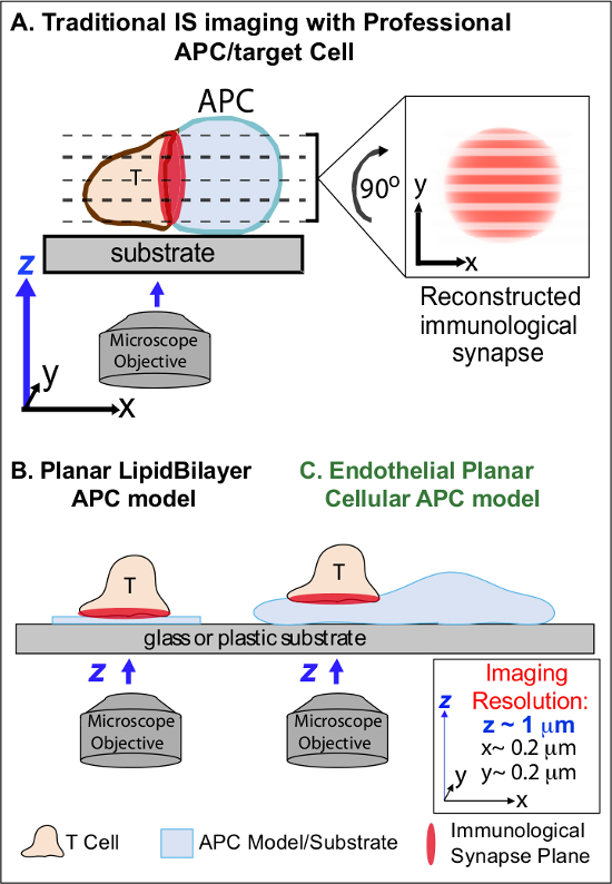
イメージング免疫学的シナプスのダイナミクスについては、図1のA生理学的平面内細胞APCモデル。概略的には、T細胞とprofessio間の免疫学的シナプスの伝統的なイメージングを示しています最終APC(A)とT細胞とこの新規内皮平面APCモデル(C)と比較して、従来の平面脂質二重層APCモデル(B)。プロフェッショナルAPCは、生理学的、免疫学的シナプスを提供するが、不十分指向細胞間のインタフェースを提供(最適なX-Yの撮像面に対してすなわち 、;解像度〜0.2μm)で、劇的空間(Zの撮像面〜1μmの分解能)および時間的( すなわち 、妥協します繰り返しイメージングのすべてのzイメージングプレーン)の解像度をスキャンする必要性に起因します。二層モデルは、最適な時空間分解能イメージングを提供する平面のトポロジーを持っているだけでなく、高度に簡略化された非生理学的な剛性です。この内皮細胞モデルは、生理学的な設定で最適な空間と時間的画像の解像度を提供するために、古典的なAPCの生理学的基質と脂質二重層の平面トポロジを兼ね備えています。M /ファイル/ ftp_upload / 53288 / 53288fig1large.jpg「ターゲット= "_空白">この図の拡大版をご覧になるにはこちらをクリックしてください。

以前の研究は、部分的に最適なXYイメージングに平行である、単一の計画に、T細胞活性化の面を固定経由で最適な時空間解像度( すなわちを提供し 、平面基板モデル( すなわち 、脂質二重層と抗体被覆表面)、開発することによって、これらの障害物を回避していますプレーン)11-15( 図1B)。これらのモデルは、動的なアクチン/ TCRシグナリングマイクロクラスター7,11-14の発見を含むT細胞における抗原性のシグナル伝達を制御する分子/細胞内動態に重要な洞察を容易にしました。しかし、このようなモデルは、本質的に単純化されるだけでなく、剛性( 図1B)(3次元位相幾何学的特徴の開発/研究を排除)。したがって、PHYに、このような調査結果を関連付ける方法を依然として不透明siologic細胞 – 細胞免疫監視。

まだunderstudiedものの、血管やリンパ管の内皮細胞は、「セミプロ」のAPC 16-18の大( すなわち 、〜1000倍により、すべてのプロフェッショナルAPCより数の大きい)周辺の区画として浮上しています。これらの細胞は、MHC-I、MHC-II-および共刺激分子の多数を表現例えば、CD40、LFA3、ICOSL、4-1BB、OX40L、TL1A、PD-L1;ではなく、CD80およびCD86)と戦略的です彼らは16-18特殊なセンチネル機能を果たし、血液-組織界面に位置します。以前の研究では、内皮細胞が効果的にエフェクター/メモリー、しかしナイーブではない、T細胞の19-25を再刺激することができることを実証しました。従って、内皮細胞は、T細胞の活性化、分化、メモリーと寛容16,17,26のローカル影響などの末梢組織中での適応免疫応答のエフェクター相に固有のAPCの役割を果たしている可能性があります。 CRIin vitroで増殖させた場合tically、 内皮細胞は、実質的に平面状のセル表面を形成し、(蛍光タンパク質レポーターを用いて、 例えば、)容易にトランスフェクトされています。これらの機能は、細胞間相互作用19,27の間の位相幾何学的力学の高時空間分解能イメージングに最適です。従って、内皮細胞は、抗原認識を駆動し、応答( 1C)19,20 を制御する細胞内分子/改造メカニズムの研究のための明らかに適した生理的な「平面携帯APC」モデルとなる恐れがあります。

接着および経内皮移動27中の白血球-内皮相互作用の詳細を研究するための(形質膜と細胞質ゾルの蛍光タンパク質メーカーと内皮細胞のトランスフェクションを含む)以前に確立された補完的なイメージング技術は、白血球が積極的にダイナミックによって内皮の表面を探査することを示しました挿入ANサブミクロンスケールのd後退は、アクチン豊富な円筒状の突起が(〜直径、深さ200〜1,000 nm)をinvadosome状突起( すなわち 、「ILPS ')27,28と呼ばれます 。これらのイメージング手法は、さらに19,20を報告し、本明細書でさらに説明するように、T細胞、内皮免疫学的シナプスの高時空間分解能イメージングのための最初の方法を開発する内皮APC機能を利用するためのプロトコルの作成 ​​に伴って拡張されました。この小説平面携帯APCモデル由来中央知見は、T細胞ILPSは、初期のAg検出を促進する上で、その後のシグナル伝達を維持するには、両方の機能することです。実際、(安定化およびカルシウム流入初期に応じて計上されていた)、TCRと活性なシグナルなどのPKC-Q、ZAP-70、ホスホチロシンとHS1の示唆に富む分子でshow濃縮を複数ILPSの配列。したがって、ILPSは、TCRシグナルマイクロへの3次元生理学的同等を表しているように見えます平面二重層モデルで見られたクラスタ。このアプローチは、このように、敏感に明らかに/レポート分子と建築(および生体力学的暗黙の)ダイナミクスそうでなければ検出できません。

本明細書に記載の方法は、適応免疫応答の基本的なメカニズムを調べるために我々の能力を向上させるために、プロのAPCと人工APC基板モデル間のギャップを埋めるに有用であるはずです。ここでの焦点は、CD4 + Th1型エフェクター/メモリー細胞の活性化にある間に、以下に説明するように、この基本的なアプローチは、容易に、T細胞型とのAgの広い範囲を研究するために改変することができます。

Protocol

このプロトコルで説明されているすべての実験は、初代ヒトT細胞および市販の一次ヒト内皮細胞(皮膚や肺の微小血管のEC)を用いて実施されているヒトを対象とする.ANY研究プロトコルは、治験審査委員会によって承認されなければならず、書面によるインフォームドコンセントから提供されなければなりません各血液ドナー。このプロトコルを使用して行った実験は、ベス・イスラエル・ディーコネス医…

Representative Results

内皮細胞を使用し、生理的な複雑さとプロフェッショナルAPCの変形能を有する平面脂質二重層モデルの解像度の利点を組み合わせた新たなイメージング手法は、( 図1)を開発しました。2本で観察された典型的な移行、カルシウムフラックスとトポロジカルダイナミクスの例を示します。図アプローチ。内皮上のサグ不在下でサ?…

Discussion

全体的に、このプロトコルは、ⅰ)understudied生理学的なAPCとして内皮細胞を調査するための方法を説明し、ii)の「平面携帯APCモデル」の新規タイプとして。前者に関しては、ますます適応免疫応答16-18を形成する上で(造血APCに比べて、 すなわち )を、周辺非造血(または「間質')APCは、重要な、非冗長な役割を果たしていることが理解されるようになりました。このよ…

Declarações

The authors have nothing to disclose.

Acknowledgements

We thank Dr. Peter T. Sage for his assistance in generating some of the representative images. This work was supported by an NIH R01 grant to C.V.C. (HL104006).

Materials

BD Vacutainer stretch latex free tourniquet BD Biosciences 367203
BD alcohol swabs BD Biosciences 326895
BD Vacutainer Safety-Lok BD Biosciences 367861 K2 EDTA
BD Vacutainer Push Button Blood Collection Set BD Biosciences 367335
RPMI-1640 Sigma-Aldrich R8758-1L
Ficoll-Paque  Sigma-Aldrich GE17-1440-02 Bring to RT before use
FCS-Optima Atlanta Biologics s12450 Heat inactivated
Penicillin-Streptomycin  Sigma-Aldrich  P4458-100ML  
Trypan blue Sigma-Aldrich T8154-20ML
staphylococcal enterotoxin B  Toxin Technology BT202RED Stock solution 1mg/ml in PBS
toxic shock syndrome toxin 1  Toxin Technology TT606RED Stock solution 1mg/ml in PBS
human IL-15 R&D Systems 247-IL-025 Stock solution 50ug/ml in PBS
PBS Life Technologies 10010-049
Fibronectin Life Technologies 33016-015 Stock solution 1mg/ml in H20
HMVEC-d Ad-Dermal MV Endo Cells Lonza CC-2543 Other Human Microvascular ECs can be used, i.e. HLMVECs
EGM-2 MV bullet kit Lonza CC-3202
Trypsin-EDTA Sigma-Aldrich T-4174 Stock solution 10x, dilute in PBS
amaxa-HMVEC-L Nucleofector Kit Lonza vpb1003 Required Kit for step 4
IFN-g Sigma-Aldrich I3265 Stock solution 1mg/ml in H20
TNF-alpha 10ug, human Life Technologies PHC3015 Stock solution 1mg/ml in H20
phenol Red-free HBSS  Life Technologies 14175-103
Hepes Fisher Scientific BP299-100
Calcium Chloride Sigma-Aldrich C1016-100G Stock solution 1M in H20
Magnesium chloride Sigma-Aldrich 208337 Stock solution 1M in H20
Human Serum albumin Sigma-Aldrich A6909-10ml
Immersol 518 F fluorescence free Immersion oil Fisher Scientific 12-624-66A
Fura-2 AM 20x50ug Life Technologies F1221 Stock solution 1mM in DMSO
pEYFP-Mem (Mem-YFP) Clontech 6917-1
pDsRed-Monomer (Soluble Cytoplasmic DsRed) Clontech 632466
pDsRed-Monomer Membrane (Mem-DsRed) Clontech 632512
pEGFP-Actin Clontech 6116-1
Alexa Fluor 488 Phalloidin Life Technologies A12379
Formaldehyde solution 37% Fisher Scientific BP531-500 Toxic, use fumehood
Triton X-100 Sigma-Aldrich X100-5ML
 Falcon 15mL Conical Centrifuge Tubes Fisher Scientific 14-959-70C
Falcon 50mL Conical Centrifuge Tubes Fisher Scientific 14-959-49A 
Falcon Tissue Culture Treated Flasks T25 Fisher Scientific 10-126-9
 Falcon Tissue Culture Treated Flasks T75 Fisher Scientific   13-680-65
 Corning Cell Culture Treated T175 Fisher Scientific 10-126-61 
Glass coverslips  Fisher Scientific 12-545-85  12 mm diameter
 Falcon Tissue Culture Plates 24-well Fisher Scientific 08-772-1
Delta-T plates Bioptechs 04200415B
Wheaton Disposable Pasteur Pipets Fisher Scientific 13-678-8D
1.5 ml Eppendorf tube  Fisher Scientific 05-402-25
 ICAM1 mouse anti-human BD Biosciences 555509
HS1 mouse anti-human BD Biosciences 610541
Anti-Human CD11a (LFA-1alpha) Purified ebioscience BMS102
Anti-Human CD3 Alexa Fluor® 488 ebioscience 53-0037-41
Anti-MHC Class II antibody  Abcam ab55152
Anti-Talin 1 antibody Abcam ab71333
Anti-PKC theta antibody  Abcam ab109481
phosphotyrosine (4G10 Platinum) Millipore 50-171-463
Nucleofector II Amaxa Biosystems Required electroporator for step 4
Zeiss Axiovert Carl Zeiss MicroImaging
Zeiss LSM510  Carl Zeiss MicroImaging
Zeiss Axiovison Software Carl Zeiss MicroImaging
NU-425 (Series 60) Biological Safety Cabinet NuAIRE Nu-425-600
 Forma STRCYCLE 37 °C, 5% CO2 Cell culture Incubator Fisher Scientific 202370
Centrifuge 5810 Eppendorf EW-02570-02
Hemocytometer Sigma-Aldrich  Z359629 Bright-Line Hemocytometer
Isotemp Waterbath model 202 Fisher Scientific 15-462-2

Referências

  1. von Andrian, U. H., Mackay, C. R. T-cell function and migration. Two sides of the same coin. N. Engl. J. Med. 343 (14), 1020-1034 (2000).
  2. Springer, T. A. Adhesion receptors of the immune system. Nature. 346 (6283), 425-434 (1990).
  3. Shaw, A. S., Dustin, M. L. Making the T cell receptor go the distance: a topological view of T cell activation. Immunity. 6 (4), 361-369 (1997).
  4. Monks, C. R., Freiberg, B. A., Kupfer, H., Sciaky, N., Kupfer, A. Three-dimensional segregation of supramolecular activation clusters in T cells. Nature. 395 (6697), 82-86 (1998).
  5. Delon, J., Stoll, S., Germain, R. N. Imaging of T-cell interactions with antigen presenting cells in culture and in intact lymphoid tissue. Immunol Rev. 189, 51-63 (2002).
  6. Brossard, C., et al. Multifocal structure of the T cell – dendritic cell synapse. Eur J Immunol. 35 (6), 1741-1753 (2005).
  7. Dustin, M. L. The cellular context of T cell signaling. Immunity. 30 (4), 482-492 (2009).
  8. Balagopalan, L., Sherman, E., Barr, V. A., Samelson, L. E. Imaging techniques for assaying lymphocyte activation in action. Nat Rev Immunol. 11 (1), 21-33 (2011).
  9. Cebecauer, M., Spitaler, M., Serge, A., Magee, A. I. Signalling complexes and clusters: functional advantages and methodological hurdles. J Cell Sci. 123, 309-320 (2010).
  10. Oddos, S., et al. High-speed high-resolution imaging of intercellular immune synapses using optical tweezers. Biophys J. 95 (10), L66-L68 (2008).
  11. Grakoui, A., et al. The immunological synapse: a molecular machine controlling T cell activation. Science. 285 (5425), 221-227 (1999).
  12. Bunnell, S. C., et al. T cell receptor ligation induces the formation of dynamically regulated signaling assemblies. J Cell Biol. 158 (7), 1263-1275 (2002).
  13. Yokosuka, T., et al. Newly generated T cell receptor microclusters initiate and sustain T cell activation by recruitment of Zap70 and SLP-76. Nat Immunol. 6 (12), 1253-1262 (2005).
  14. Seminario, M. C., Bunnell, S. C. Signal initiation in T-cell receptor microclusters. Immunol Rev. 221, 90-106 (2008).
  15. Dustin, M. L. Supported bilayers at the vanguard of immune cell activation studies. J Struct Biol. 168 (1), 152-160 (2009).
  16. Martinelli, R., Carman, C. V., Bradshaw, R. A., Stahl, P. Lymphocyte Endothelilal Interactions. Encyclopedia of Cell Biology. , (2015).
  17. Choi, J., Enis, D. R., Koh, K. P., Shiao, S. L., Pober, J. S. T lymphocyte-endothelial cell interactions. Annu Rev Immunol. 22, 683-709 (2004).
  18. Marelli-Berg, F. M., Jarmin, S. J. Antigen presentation by the endothelium: a green light for antigen-specific T cell trafficking?. Immunol Lett. 93 (2-3), 109-113 (2004).
  19. Sage, P. T., et al. Antigen recognition is facilitated by invadosome-like protrusions formed by memory/effector T cells. J Immunol. 188 (8), 3686-3699 (2012).
  20. Kumari, S., et al. Actin foci facilitate activation of the phospholipase C-gama in primary T lymphocytes via the WASP pathway . eLife. , (2015).
  21. Marelli-Berg, F. M., et al. Major histocompatibility complex class II-expressing endothelial cells induce allospecific nonresponsiveness in naive T cells. J Exp Med. 183 (4), 1603-1612 (1996).
  22. Ma, W., Pober, J. S. Human endothelial cells effectively costimulate cytokine production by, but not differentiation of, naive CD4+ T cells. J Immunol. 161 (5), 2158-2167 (1998).
  23. Perez, V. L., Henault, L., Lichtman, A. H. Endothelial antigen presentation: stimulation of previously activated but not naive TCR-transgenic mouse T cells. Cell Immunol. 189 (1), 31-40 (1998).
  24. Epperson, D. E., Pober, J. S. Antigen-presenting function of human endothelial cells. Direct activation of resting CD8 T cells. J Immunol. 153 (12), 5402-5412 (1994).
  25. Shiao, S. L., et al. Human effector memory CD4+ T cells directly recognize allogeneic endothelial cells in vitro and in vivo. J Immunol. 179 (7), 4397-4404 (2007).
  26. Marelli-Berg, F. M., Okkenhaug, K., Mirenda, V. A two-signal model for T cell trafficking. Trends Immunol. 28 (6), 267-273 (2007).
  27. Carman, C. V., et al. Transcellular diapedesis is initiated by invasive podosomes. Immunity. 26 (6), 784-797 (2007).
  28. Carman, C. V. Mechanisms for transcellular diapedesis: probing and pathfinding by ‘invadosome-like protrusions’. J Cell Sci. 122 ((Pt 17)), 3025-3035 (2009).
  29. Dustin, M. L., Tseng, S. Y., Varma, R., Campi, G. T. T cell-dendritic cell immunological synapses. Curr Opin Immunol. 18 (4), 512-516 (2006).
  30. Saito, T., Yokosuka, T. Immunological synapse and microclusters: the site for recognition and activation of T cells. Curr Opin Immunol. 18 (3), 305-313 (2006).
  31. Gomez, T. S., et al. HS1 functions as an essential actin-regulatory adaptor protein at the immune synapse. Immunity. 24 (6), 741-752 (2006).
  32. Burbach, B. J., Medeiros, R. B., Mueller, K. L., Shimizu, Y. T-cell receptor signaling to integrins. Immunol Rev. 218, 65-81 (2007).
  33. Vicente-Manzanares, M., Sanchez-Madrid, F. Role of the cytoskeleton during leukocyte responses. Nat Rev Immunol. 4 (2), 110-122 (2004).
  34. Ma, Z., Janmey, P. A., Finkel, T. H. The receptor deformation model of TCR triggering. Faseb J. 22 (4), 1002-1008 (2008).
  35. Ma, Z., Sharp, K. A., Janmey, P. A., Finkel, T. H. Surface-anchored monomeric agonist pMHCs alone trigger TCR with high sensitivity. PLoS Biol. 6 (2), e43 (2008).
  36. Groves, J. T. Bending mechanics and molecular organization in biological membranes. Annu Rev Phys Chem. 58, 697-717 (2007).
  37. Xu, C., et al. Regulation of T cell receptor activation by dynamic membrane binding of the CD3epsilon cytoplasmic tyrosine-based motif. Cell. 135 (4), 702-713 (2008).
check_url/pt/53288?article_type=t

Play Video

Citar este artigo
Martinelli, R., Carman, C. V. An Endothelial Planar Cell Model for Imaging Immunological Synapse Dynamics. J. Vis. Exp. (106), e53288, doi:10.3791/53288 (2015).

View Video